. 2024 Nov 7:14:1480076.
doi: 10.3389/fcimb.2024.1480076. eCollection 2024.

Iron transport pathways in the human malaria parasite Plasmodium falciparum revealed by RNA-sequencing

Affiliations

Iron transport pathways in the human malaria parasite Plasmodium falciparum revealed by RNA-sequencing

Juliane Wunderlich et al. Front Cell Infect Microbiol. .

Abstract

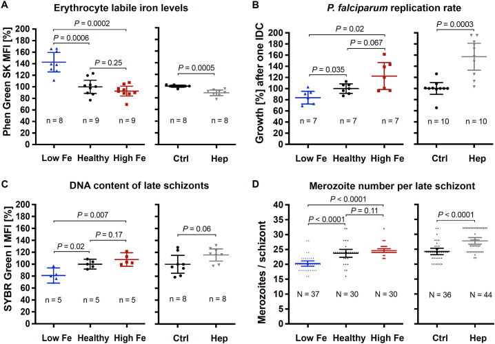
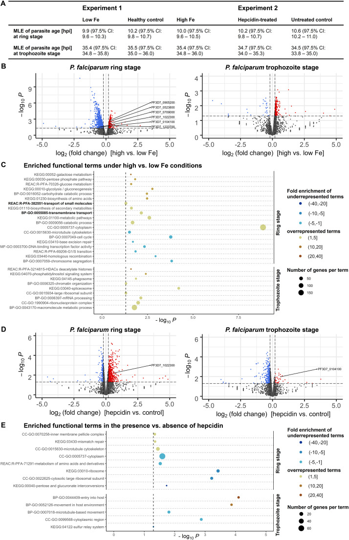
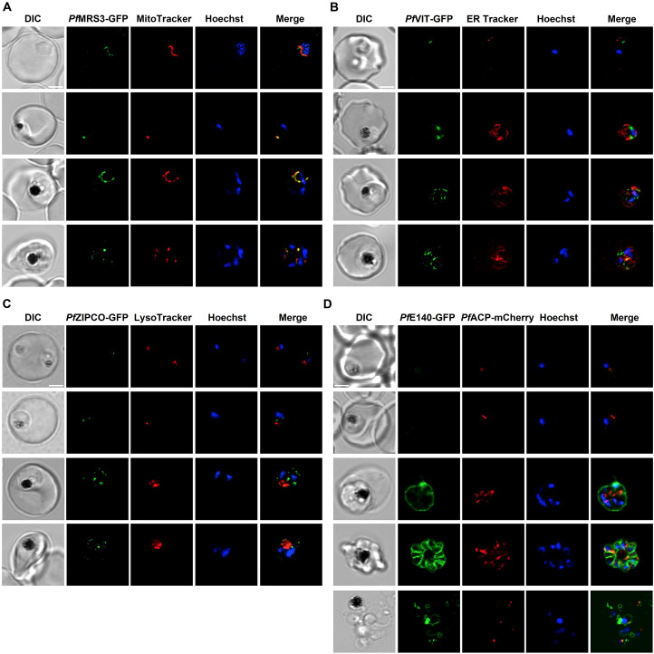
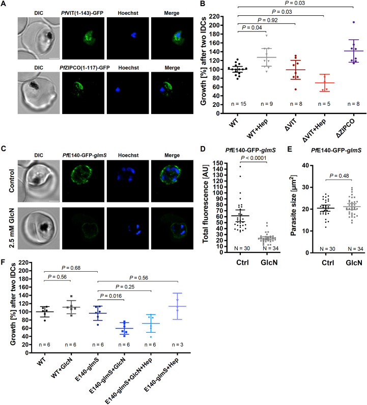
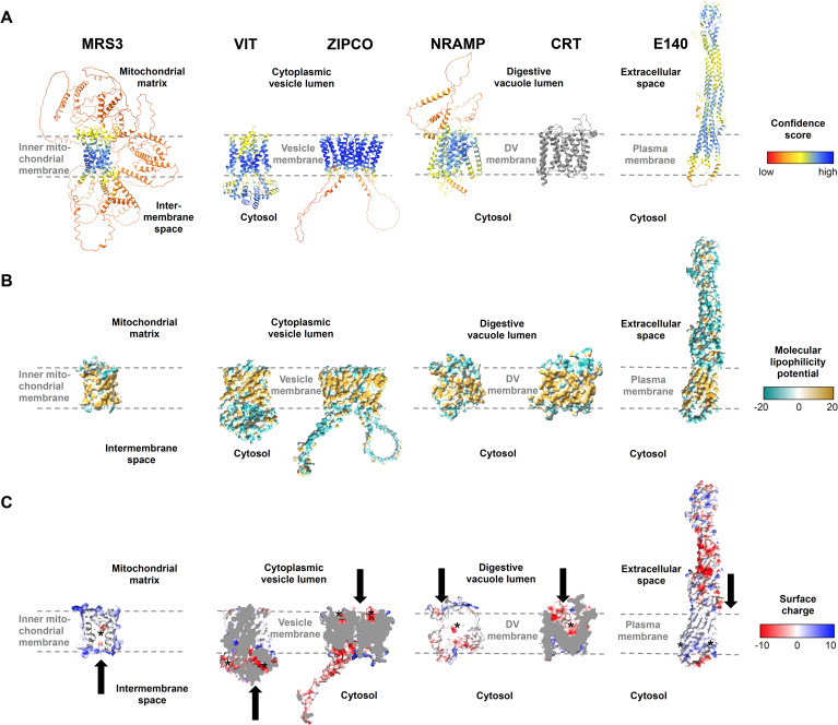
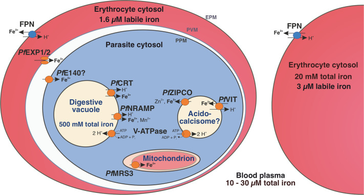
Host iron deficiency is protective against severe malaria as the human malaria parasite Plasmodium falciparum depends on bioavailable iron from its host to proliferate. The essential pathways of iron acquisition, storage, export, and detoxification in the parasite differ from those in humans, as orthologs of the mammalian transferrin receptor, ferritin, or ferroportin, and a functional heme oxygenase are absent in P. falciparum. Thus, the proteins involved in these processes may be excellent targets for therapeutic development, yet remain largely unknown. Here, we show that parasites cultured in erythrocytes from an iron-deficient donor displayed significantly reduced growth rates compared to those grown in red blood cells from healthy controls. Sequencing of parasite RNA revealed diminished expression of genes involved in overall metabolism, hemoglobin digestion, and metabolite transport under low-iron versus control conditions. Supplementation with hepcidin, a specific ferroportin inhibitor, resulted in increased labile iron levels in erythrocytes, enhanced parasite replication, and transcriptional upregulation of genes responsible for merozoite motility and host cell invasion. Through endogenous GFP tagging of differentially expressed putative transporter genes followed by confocal live-cell imaging, proliferation assays with knockout and knockdown lines, and protein structure predictions, we identified six proteins that are likely required for ferrous iron transport in P. falciparum. Of these, we localized PfVIT and PfZIPCO to cytoplasmic vesicles, PfMRS3 to the mitochondrion, and the novel putative iron transporter PfE140 to the plasma membrane for the first time in P. falciparum. PfNRAMP/PfDMT1 and PfCRT were previously reported to efflux Fe2+ from the digestive vacuole. Our data support a new model for parasite iron homeostasis, in which PfE140 is involved in iron uptake across the plasma membrane, PfMRS3 ensures non-redundant Fe2+ supply to the mitochondrion as the main site of iron utilization, PfVIT transports excess iron into cytoplasmic vesicles, and PfZIPCO exports Fe2+ from these organelles in case of iron scarcity. These results provide new insights into the parasite's response to differential iron availability in its environment and into the mechanisms of iron transport in P. falciparum as promising candidate targets for future antimalarial drugs.

Keywords: AlphaFold; Plasmodium falciparum; drug target; gene expression; iron deficiency; malaria; nutrient uptake; transporters.


Conflict of interest statement

The authors declare that the research was conducted in the absence of any commercial or financial relationships that could be construed as a potential conflict of interest.

Figures

Figure 1
Figure 1
Effects of the iron status of the blood donor and of hepcidin on (A) erythrocyte labile iron levels, (B) P. falciparum 3D7 growth rates, (C) DNA content per mature schizont, and (D) the number of merozoites per mature schizont. The relative labile iron level and DNA content per cell were assessed in the presence or absence of 0.7 µM hepcidin (Hep) by measuring the mean fluorescence intensity (MFI) of Phen Green SK or SYBR Green I compared to control (Ctrl, untreated, normal hemoglobin level) using flow cytometry. Therefore, 100,000 cells were analyzed per replicate. Parasite growth rates refer to the fold change in parasitemia after one intraerythrocytic developmental cycle in vitro relative to control as determined by flow cytometry with SYBR Green I (Malleret et al., 2011). Mature schizonts were obtained by treating schizonts at 40 hpi with 1 mM compound 2 (4-[7-[(dimethylamino)methyl]-2-(4-fluorphenyl)imidazo[1,2-α]pyridine-3-yl]pyrimidin-2-amine) for 8 h. To count the number of merozoites, Giemsa-stained blood smears were analyzed microscopically and only single-infected cells with one digestive vacuole were considered. Means and 95% confidence intervals (indicated by error bars) are shown. Statistical significance was calculated with two-tailed unpaired t tests with Welch’s correction for unequal variances and adjusted with the Holm-Šídák method for multiple comparisons except for merozoite numbers, which were compared using Mann-Whitney test. N represents the number of parasites and n the number of independent experiments.
Figure 2
Figure 2
Differential expression of P. falciparum 3D7 genes under various iron conditions. Parasites were cultured with erythrocytes from an individual with high, medium (healthy) or low iron status (experiment 1) or with red blood cells from another healthy donor in the presence or absence of 0.7 µM hepcidin (experiment 2). Samples were harvested at the ring and trophozoite stage (6 – 9 and 26 – 29 hours post invasion, hpi) with three biological replicates per time point and condition. The maximum likelihood estimation (MLE) of the average developmental age of the parasites for each condition and time point (A) was calculated using an algorithm developed by Avi Feller and Jacob Lemieux (Lemieux et al., 2009). CI, confidence interval. The volcano plots (B, D) show transcriptional changes of all parasite genes. Red dots indicate significantly (P < 0.05, exact test for negative binomial distribution) upregulated genes (log2 (fold change) ≥ 0.2), blue dots stand for significantly downregulated genes (log2 (fold change) ≤ -0.2), while grey dots represent genes that did not significantly differ in transcription under the conditions described (P ≥ 0.05 and/or -0.2 < log2 (fold change) < 0.2). Differentially expressed genes encoding putative iron transporters (see Table 1 ) are labeled. Panels (C, E) show the enrichment of Gene Ontology (GO), Kyoto Encyclopedia of Genes and Genomes (KEGG) and Reactome (REAC) terms among significantly regulated genes excluding var, stevor and rifin gene families at the two time points. The functional terms were summarized using REVIGO (Supek et al., 2011) to remove redundancies, represented by circles and plotted according to the significance of their enrichment (-log10 (adjusted P), hypergeometric test). The size of the circle is proportional to the number of differentially regulated genes in the dataset that are associated with the respective term, while the color stands for the fold enrichment. The gray dashed line indicates the threshold of the adjusted P value (-log10 0.05 = 1.3).
Figure 3
Figure 3
Subcellular localization of known and putative iron transport proteins. Representative erythrocytes infected with P. falciparum 3D7 parasites endogenously expressing GFP-tagged PfMRS3 (A), PfVIT (B), PfZIPCO (C) or PfE140 (D) were additionally stained with the fluorescent dyes Hoechst-33342, MitoTracker Red, ER Tracker Red and/or LysoTracker Deep Red. Co-transfection with a construct that encodes the 60 N-terminal amino acids of acyl carrier protein (PfACP) tagged with mCherry (Birnbaum et al., 2020) resulted in red fluorescence of the apicoplast. Live-cell images were taken under physiological conditions at 37°C using an SP8 confocal laser-scanning microscope (Leica). DIC, differential interference contrast. Scale bar, 2 µm.
Figure 4
Figure 4
PfVIT and PfE140 are important for P. falciparum growth and may be involved in intracellular iron homeostasis. (A) Representative erythrocytes infected with P. falciparum 3D7 parasites that endogenously express a truncated version of PfVIT or PfZIPCO tagged with GFP (green). (B) Growth rates of knockout parasite lines generated. (C) Representative live 3D7 parasites endogenously expressing PfE140-GFP whose green fluorescence was reduced by glmS-mediated knockdown induced by treatment with 2.5 mM glucosamine for 36 h (GlcN) compared to untreated control (Ctrl). (D) Total parasite fluorescence intensities were quantified as background-corrected integrated densities using ImageJ version 2.9.0/1.53t (Schindelin et al., 2012) and compared using Mann-Whitney test. Images to which no averaging or deconvolution software was applied were used for quantification. (E) The size of the parasites was measured as the area of the region of interest and compared using equal variance unpaired t test. (F) Conditional knockdown of PfE140 induced by treatment with 2.5 mM GlcN results in a growth defect during asexual blood stage development. Live parasites were stained with Hoechst-33342 (blue) and imaged under physiological conditions at 37°C using an SP8 confocal laser-scanning microscope (Leica). DIC, differential interference contrast. Scale bar, 2 µm. Error bars represent 95% confidence intervals of the mean, N the number of parasites analyzed, n the number of independent experiments and Hep treatment with 0.7 µM hepcidin. Growth rates refer to the fold change in parasitemia after two intraerythrocytic developmental cycles in vitro relative to untreated wild-type 3D7 parasites (WT) as determined by flow cytometry with SYBR Green I (Malleret et al., 2011). Statistical significance of growth differences was calculated with two-tailed unpaired t tests with Welch’s correction for unequal variances and adjusted with the Holm-Šídák method for multiple comparisons.
Figure 5
Figure 5
Structures of known and putative P. falciparum iron transporters as viewed from the membrane plane. (A) Predicted protein structures with per-residue pLDDT (predicted local distance difference test) confidence scores on a scale from 0 to 100, where blue represents high and red low confidence, respectively. The experimentally determined structure of PfCRT is shown in gray. (B) Molecular lipophilicity potential of the protein surfaces as implemented in UCSF ChimeraX; tan is hydrophobic and cyan hydrophilic. Dashed lines above and below the tan regions of all proteins indicate the respective membrane and disordered loops were removed for clarity. (C) Surface charge of the proteins with positively charged areas colored blue and negatively charged ones red. Putative cation-binding site are indicated with an asterisk and transport directions by arrows. PfE140 likely forms a dimer but is shown as a monomer, as no predicted dimer structure could be obtained using AlphaFold2-multimer. The putative cation-binding sites for this protein are based on DeepFRI gradCAM scores for the functional term GO:0015075 “monoatomic ion transmembrane transporter activity” ( Supplementary Figure S5 ).
Figure 6
Figure 6
Iron homeostasis in a P. falciparum-infected erythrocyte. The human blood plasma contains between 10 and 30 µM total iron and the erythrocyte cytosol approximately 20 mM (Egan et al., 2002). However, the labile iron pool is only 3 µM in an uninfected erythrocyte and 1.6 µM in a P. falciparum-infected one (Loyevsky et al., 1999). Human ferroportin (FPN) at the host cell surface (erythrocyte plasma membrane, EPM) exports ferrous iron from the erythrocyte (Ward and Kaplan, 2012) and the nutrient pore formed by PfEXP1 and PfEXP2 allows the passage of ions through the parasitophorous vacuole membrane (PVM) (Mesén-Ramírez et al., 2021). PfE140 at the parasite plasma membrane (PPM) may mediate iron uptake into the parasite cytosol and the mitochondrial carrier protein PfMRS3 likely translocates Fe2+ into the mitochondrion, a site of de novo heme biosynthesis (this study). We propose that the vacuolar iron transporter (PfVIT) is involved in iron detoxification by transporting excess Fe2+ from the cytosol into cytoplasmic vesicles that may be acidocalcisomes, whereas PfZIPCO releases Fe2+ from these organelles under low-iron conditions. The digestive vacuole (DV) contains a high amount of total iron as it is the site of hemoglobin degradation and hemozoin formation (Becker et al., 2004). The chloroquine resistance transporter (PfCRT) and the natural resistance-associated macrophage protein (PfNRAMP, also called PfDMT1 for divalent metal transporter 1) were suggested to mediate proton-coupled export of Fe2+ from the DV into the parasite cytosol (Martin et al., 2005; Bakouh et al., 2017). Both acidocalcisomes and the DV are likely acidified by the plant-like H+-pump V-ATPase, which can fuel secondary active transport processes (Wunderlich et al., 2012; de Oliveira et al., 2021). Parasite-encoded proteins are shown in orange and human-encoded transporters in blue.

Similar articles

Cited by

References

    1. Abrahamian M., Ah-Fong A. M., Davis C., Andreeva K., Judelson H. S. (2016). Gene expression and silencing studies in Phytophthora infestans reveal infection-specific nutrient transporters and a role for the nitrate reductase pathway in plant pathogenesis. PloS Pathog. 12, e1006097. doi: 10.1371/journal.ppat.1006097 - DOI - PMC - PubMed
    1. Aghabi D., Sloan M., Gill G., Hartmann E., Antipova O., Dou Z., et al. . (2023). The vacuolar iron transporter mediates iron detoxification in Toxoplasma gondii. Nat. Commun. 14, 3659. doi: 10.1038/s41467-023-39436-y - DOI - PMC - PubMed
    1. Agrawal S., Striepen B. (2010). More membranes, more proteins: complex protein import mechanisms into secondary plastids. Protist. 161, 672–687. doi: 10.1016/j.protis.2010.09.002 - DOI - PMC - PubMed
    1. Ahiya A. I., Bhatnagar S., Morrisey J. M., Beck J. R., Vaidya A. B. (2022). Dramatic consequences of reducing erythrocyte membrane cholesterol on Plasmodium falciparum . Microbiol. Spectr. 10, e0015822. doi: 10.1128/spectrum.00158-22 - DOI - PMC - PubMed
    1. Altschul S. F., Madden T. L., Schäffer A. A., Zhang J., Zhang Z., Miller W., et al. . (1997). Gapped BLAST and PSI-BLAST: a new generation of protein database search programs. Nucleic Acids Res. 25, 3389–3402. doi: 10.1093/nar/25.17.3389 - DOI - PMC - PubMed

MeSH terms

LinkOut - more resources