. 2024 Dec 3;13(23):e036872.
doi: 10.1161/JAHA.124.036872. Epub 2024 Nov 22.

Cytokine Adsorption During Ex Vivo Blood Perfusion Improves Contractility of Donation After Circulatory Death Hearts

Affiliations

Cytokine Adsorption During Ex Vivo Blood Perfusion Improves Contractility of Donation After Circulatory Death Hearts

Lars Saemann et al. J Am Heart Assoc. .

Abstract

Background: Donation after circulatory death (DCD) hearts have to withstand ischemia/reperfusion injury that is partially driven by proinflammatory cytokines and decreases ventricular contractility. We hypothesize that cytokine adsorption during normothermic ex vivo blood perfusion of DCD hearts reduces the cytokine levels and improves ventricular contractility.

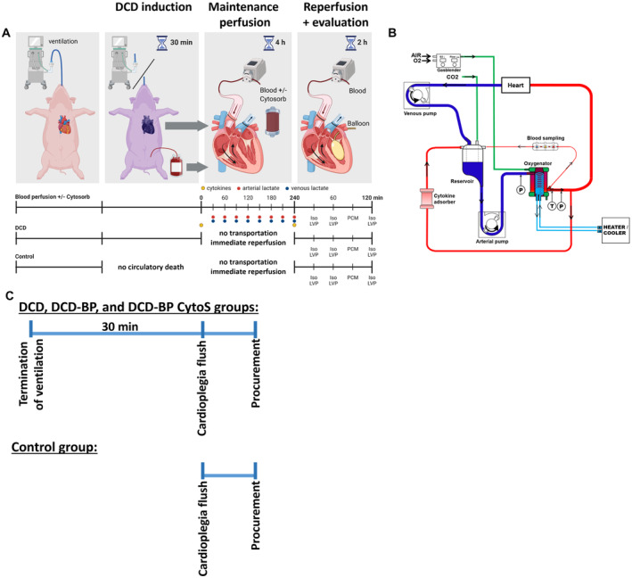
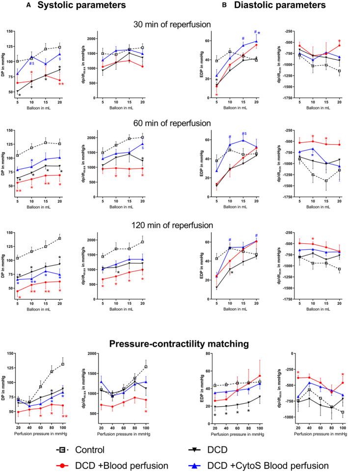
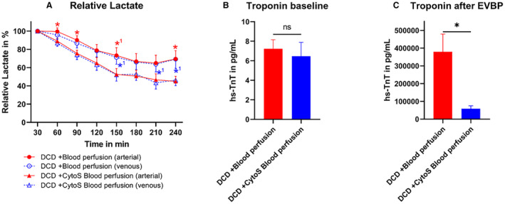
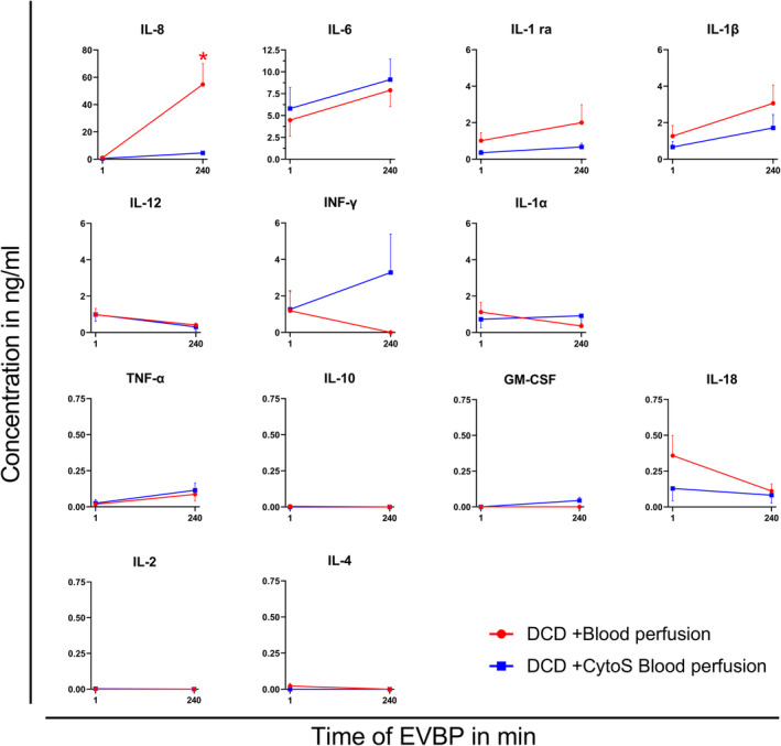
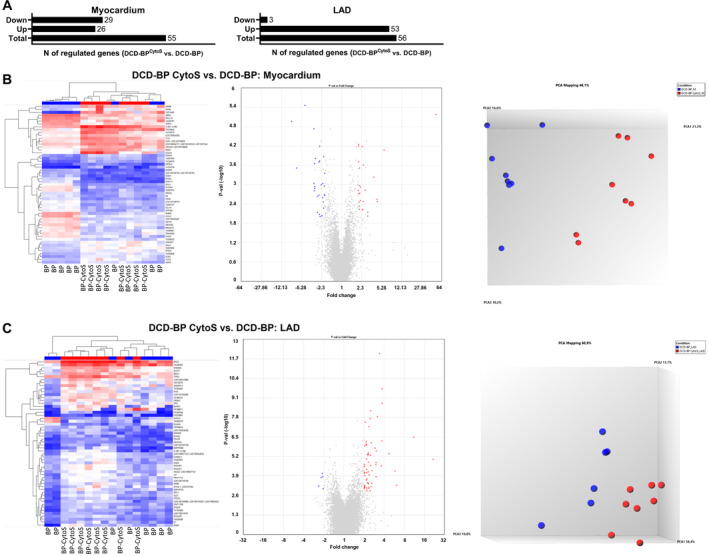
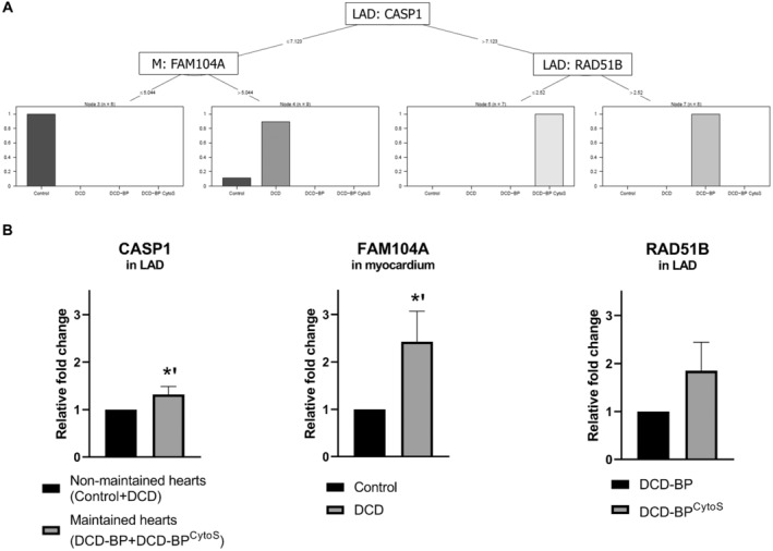
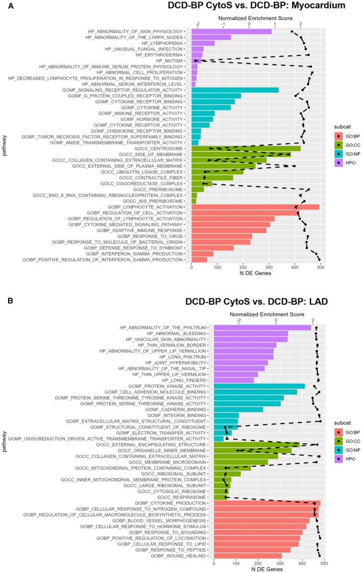
Methods and results: Porcine DCD hearts were maintained 4 hours by ex vivo blood perfusion with (DCD-BPCytoS, all groups: N=8) or without (DCD-BP) CytoSorb, followed by 2 hours reperfusion with fresh blood, including left ventricular functional analysis using a balloon catheter. In a control and a DCD group, hearts were evaluated after procurement. We determined lactate and cytokines after ex vivo blood perfusion and the myocardial and left anterior descending artery transcriptome using microarrays after reperfusion. In DCD-BPCytoS, the developed pressure (control: 124±7 mm Hg/s, DCD: 86±4 mm Hg/s, DCD-BP: 69±11 mm Hg/s, DCD-BPCytoS: 112±9 mm Hg/s; P<0.05) and maximal slope of pressure increment (control: 2010±39 mm Hg/s, DCD: 1219±164 mm Hg/s, DCD-BP: 964±163 mm Hg/s, DCD-BPCytoS: 1794±205 mm Hg/s; P<0.05) were higher compared with DCD-BP and DCD hearts. However, contractility decreased later during reperfusion without CytoSorb. After 4 hours, troponin, lactate (45±5% versus 69±9%, P<0.05), IL (interleukin)-1β, -1ra, and -8 were lower in DCD-BPCytoS hearts. In the myocardium of DCD-BPCytoS compared with DCD-BP hearts, inflammatory mediator receptor activity/binding pathways were enriched, and pathways for collagen-containing extracellular matrix and contractile fiber were underrepresented. In the left anterior descending artery of DCD-BPCytoS hearts, serine/threonine/tyrosine kinase activity and wound-healing pathways were enriched, and mitochondrial protein-containing complex and respiratome-associated pathways were underrepresented.

Conclusions: CytoSorb during ex vivo blood perfusion enhances the maintenance of DCD hearts and is likely to improve graft function after transplantation.

Keywords: CytoSorb; cytokine adsorption; cytokines; donation after circulatory death; heart transplantation; machine perfusion.


Figures

Figure 1
Figure 1. Workflow.
A, DCD model and blood sample collection (marked by blue, red, and yellow dots). B, Perfusion system. C, Ischemic period and procurement workflow. DCD indicates donation after circulatory death; DCD‐BP, donation after circulatory death hearts blood perfusion without CytoSorb; DCD‐BP CytoS, donation after circulatory death hearts blood perfusion with CytoSorb; LVP, left ventricular pressure; and PCM, pressure contractility matching.
Figure 2
Figure 2. Left ventricular contractility.
A, Systolic parameters. B, Diastolic parameters. *P<0.05 vs control. **P<0.001 vs control. # P<0.05 vs DCD. $ P<0.05 vs DCD+blood perfusion. N=8 per group. CytoS indicates CytoSorb; DCD, donation after circulatory death; DP, developed pressure; Dp/dtmax, slope of systolic pressure increment; Dp/dtmin, slope of diastolic pressure decrement; and EDP, end‐diastolic pressure.
Figure 3
Figure 3. Metabolic markers and cardiac enzymes during maintenance perfusion.
A, Lactate during maintenance perfusion. Lactate was analyzed in relation to a baseline measurement after 30 minutes of machine perfusion. At 210 minutes of perfusion, 1 lactate sample in the DCD‐BPCytoS group was missing. B, Baseline troponin concentration in the organ donor before DCD induction. C, Troponin concentration in the perfusate at the end of maintenance perfusion (after 240 minutes). *P<0.05 DCD‐blood perfusion vs DCD‐CytoS blood perfusion. *1 P<0.1 DCD‐blood perfusion vs DCD‐CytoS blood perfusion. N=8 per group. The error bars represent the standard error. CytoS indicates CytoSorb; DCD, donation after circulatory death; DCD‐BPCytoS, donation after circulatory death hearts blood perfusion with CytoSorb; EVBP, ex vivo blood perfusion; and hs‐TnT, high‐sensitivity troponin T.
Figure 4
Figure 4. Cytokine concentrations during machine perfusion.
*P<0.05 vs DCD+CytoS blood perfusion. N=8 per group. CytoS indicates CytoSorb; DCD, donation after circulatory death; DCD‐BP, donation after circulatory death hearts blood perfusion without CytoSorb; DCD‐BPCytoS, donation after circulatory death hearts blood perfusion with CytoSorb; EVBP, ex vivo blood perfusion; GM‐CSF, granulocyte‐monocyte colony‐stimulating factor; IL, interleukin; INF, interferon; and TNF, tumor necrosis factor.
Figure 5
Figure 5. Regulated transcripts.
A, Myocardium and LAD artery. B, DCD‐BPCytoS vs DCD‐BP; analyzed tissue: myocardium. C, DCD‐BPCytoS vs DCD‐BP; analyzed tissue: LAD artery. BP indicates blood perfusion; CytoS, CytoSorb; DCD, donation after circulatory death; DCD‐BP, donation after circulatory death hearts blood perfusion without CytoSorb; DCD‐BPCytoS, donation after circulatory death hearts blood perfusion with CytoSorb; LAD, left anterior descending; and PCA, principal component analysis.
Figure 6
Figure 6. Identified genes.
A, Decision tree analysis. B, Regulation of identified genes based on RT‐qPCR. *P<0.01. The error bars represent the standard error. BP indicates blood perfusion; CASP1, caspase 1; CytoS, CytoSorb; DCD, donation after circulatory death; DCD‐BP, donation after circulatory death hearts blood perfusion without CytoSorb; DCD‐BPCytoS, donation after circulatory death hearts blood perfusion with CytoSorb; FAM104A, Family With Sequence Similarity 104 Member A; LAD, left anterior descending; M, myocardium; and RT‐qPCR, quantitative real‐time polymerase chain reaction.
Figure 7
Figure 7. Pathway analysis for treatment comparison.
The dashed black line represents the normalized enrichment score. All plotted pathways are P<0.05. BP indicates biological process; CC, cellular component; DCD‐BP, donation after circulatory death hearts blood perfusion without CytoSorb; DCD‐BP CytoS, donation after circulatory death hearts blood perfusion with CytoSorb; DE, differentially expressed; GO, gene ontology; HPO, human phenotype ontology; and MF, molecular function.
Figure 8
Figure 8. Immunohistochemical staining of PARP and TUNEL.
The error bars represent the standard error. *P<0.01. DCD indicates donation after circulatory death; DCD‐BP, donation after circulatory death hearts blood perfusion without CytoSorb; DCD‐BPCytoS, donation after circulatory death hearts blood perfusion with CytoSorb; PARP, poly(adenosine diphosphate‐ribose)polymerase; and TUNEL, terminal deoxynucleotidyl transferase dUTP nick end labeling.

References

    1. Messer S, Cernic S, Page A, Berman M, Kaul P, Colah S, Ali J, Pavlushkov E, Baxter J, Quigley R, et al. A 5‐year single‐center early experience of heart transplantation from donation after circulatory‐determined death donors. J Heart Lung Transplant. 2020;39:1463–1475. doi: 10.1016/j.healun.2020.10.001 - DOI - PubMed
    1. Chew HC, Iyer A, Connellan M, Scheuer S, Villanueva J, Gao L, Hicks M, Harkness M, Soto C, Dinale A, et al. Outcomes of donation after circulatory death heart transplantation in Australia. J Am Coll Cardiol. 2019;73:1447–1459. doi: 10.1016/j.jacc.2018.12.067 - DOI - PubMed
    1. Miñambres E, Royo‐Villanova M, Pérez‐Redondo M, Coll E, Villar‐García S, Canovas SJ, Francisco Nistal J, Garrido IP, Gómez‐Bueno M, Cobo M, et al. Spanish experience with heart transplants from controlled donation after the circulatory determination of death using thoraco‐abdominal normothermic regional perfusion and cold storage. Am J Transplant. 2021;21:1597–1602. doi: 10.1111/ajt.16446 - DOI - PubMed
    1. Saemann L, Korkmaz‐Icöz S, Hoorn F, Veres G, Kraft P, Georgevici A‐I, Brune M, Guo Y, Loganathan S, Wenzel F, et al. Reconditioning of circulatory death hearts by ex‐vivo machine perfusion with a novel HTK‐N preservation solution. J Heart Lung Transplant. 2021;40:1135–1144. doi: 10.1016/j.healun.2021.07.009 - DOI - PubMed
    1. Moeslund N, Ertugrul IA, Hu MA, Dalsgaard FF, Ilkjaer LB, Ryhammer P, Pedersen M, Erasmus ME, Eiskjaer H. Ex‐situ oxygenated hypothermic machine perfusion in donation after circulatory death heart transplantation following either direct procurement or in‐situ normothermic regional perfusion. J Heart Lung Transplant. 2023;42:730–740. doi: 10.1016/j.healun.2023.01.014 - DOI - PubMed

MeSH terms

LinkOut - more resources