

Classics in abdominal radiology: the jumping deer sign
- PMID: 39576316
- PMCID: PMC11991963
- DOI: 10.1007/s00261-024-04699-6
Classics in abdominal radiology: the jumping deer sign
Abstract
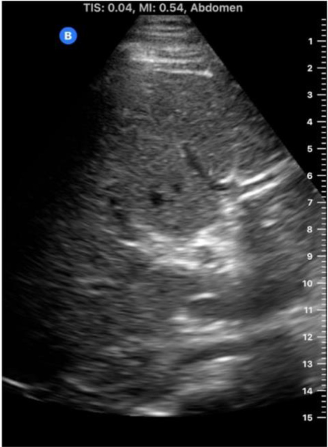
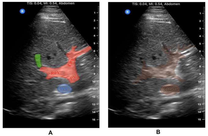
The "jumping deer sign" is an ultrasonographic pattern that aids in identifying normal liver anatomy and distinguishing it from pathology. It includes the portal vein (deer's head and body), the gallbladder or cystic duct (tail), and the inferior vena cava (obstacle). This sign helps differentiate portal veins from intrahepatic ducts, crucial for diagnosing conditions like portal hypertension. It also assists in identifying gallbladder pathologies and assessing the IVC for hydration status. The jumping deer sign provides a clear reference for clinicians, improving diagnostic accuracy, especially for those with limited ultrasound experience.
Keywords: Classics in abdominal radiology; Hepatobiliary; Jumping deer; Jumping stag; Liver; Normal anatomy; Radiology signs; Signs; Ultrasound.
© 2024. The Author(s).
Conflict of interest statement
Declarations. Competing interests: The authors declare no competing interests.
Figures
References
-
- Laing FC, Linda McKay London, Filly RA. Ultrasonographic Identification of Dilated Intrahepatic Bile Ducts and their Differentiation from Portal Venous Structures. Springer eBooks. Published online January 1, 1978:173–176. doi: 10.1007/978-1-4613-4021-8_46 - PubMed
-
- Bennett GL. Evaluating Patients with Right Upper Quadrant Pain. Radiologic Clinics of North America. 2015;53(6):1093–1130. doi: 10.1016/j.rcl.2015.06.002 - PubMed
-
- L Garcia-Sancho Tellez, Rodriguez-Montes JA, Fernandez S, L Garcia-Sancho Martin. Acute emphysematous cholecystitis. Report of twenty cases. PubMed. 1999;46(28):2144–2148 - PubMed
-
- Ali AA, Salem OM, Elghonimy MM, Sharaf MS. Ultrasound guided measurement of inferior vena cava diameter, common carotid artery diameter versus central venous pressure for estimation of intravascular volume status in Septic Shock Patients. The Scientific Journal of Medical Scholar. 2024;3(3). doi:10.55675/sjms.v3i3.91
Publication types
MeSH terms
LinkOut - more resources
Full Text Sources
