. 2025 Jan;14(1):153-167.
doi: 10.1007/s40123-024-01074-y. Epub 2024 Nov 22.

One-Year Comparison of Efficacy and Safety of PreserFlo MicroShunt with Mitomycin C Applied by Sub-Tenon Injection Versus Sponge

Affiliations

One-Year Comparison of Efficacy and Safety of PreserFlo MicroShunt with Mitomycin C Applied by Sub-Tenon Injection Versus Sponge

Nora Majtanova et al. Ophthalmol Ther. 2025 Jan.

Abstract

Introduction: This study was performed to compare the efficacy and safety of PreserFlo MicroShunt (PMS) implantation with mitomycin C (MMC) applied by sub-tenon injection versus conventional application by MMC-soaked sponges.

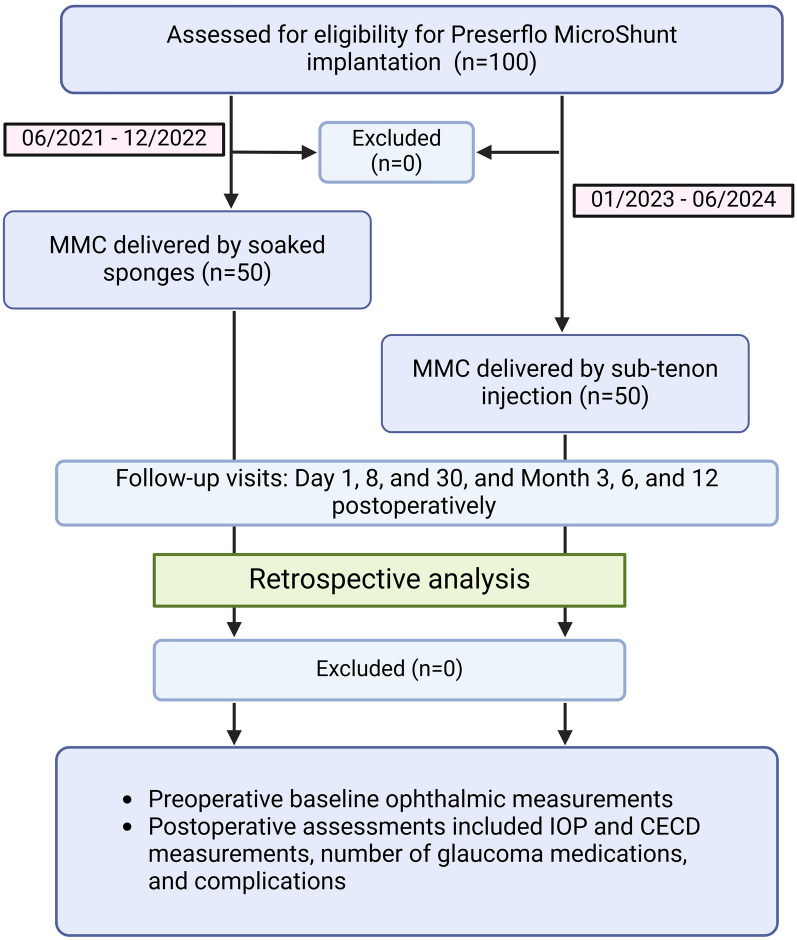
Methods: This retrospective, 1-year cohort study included 100 eyes of 100 patients with glaucoma who underwent PMS implantation with MMC (0.4 mg/ml) delivered either by sub-tenon injection (50 eyes) or via soaked sponges (50 eyes). The primary outcome measure at 1 year was intraocular pressure (IOP) reduction, with complete success defined as an IOP reduction of ≥ 20% and achieving a target IOP of ≤ 21 or 18 mmHg without the use of medication. Secondary outcomes, including corneal endothelial cell density (CECD) loss, the number of medications, and complications, were assessed and compared between the groups.

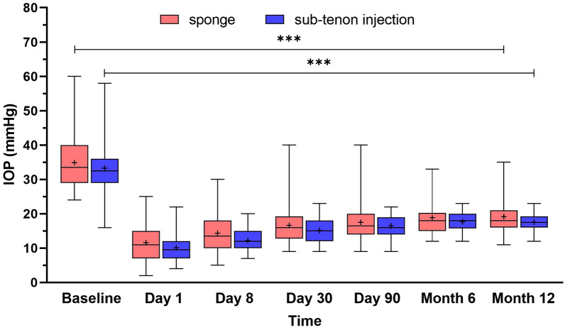
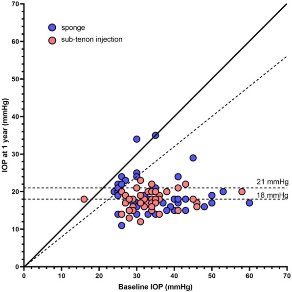
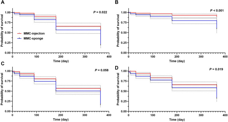
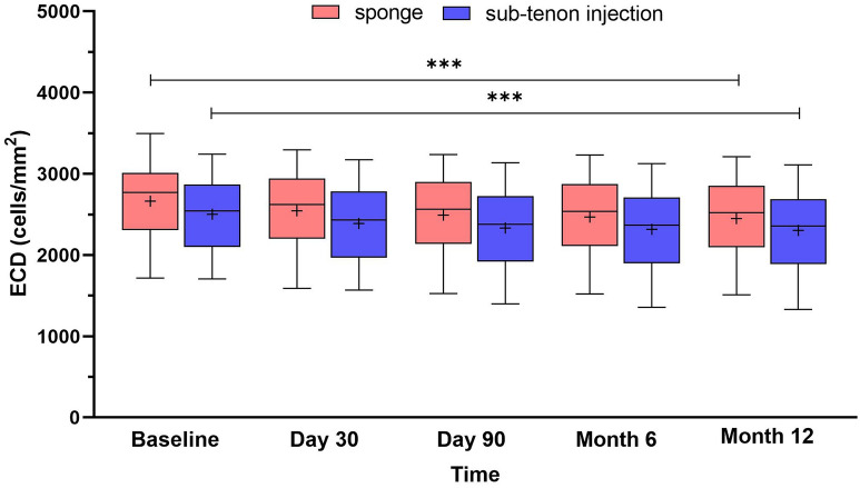
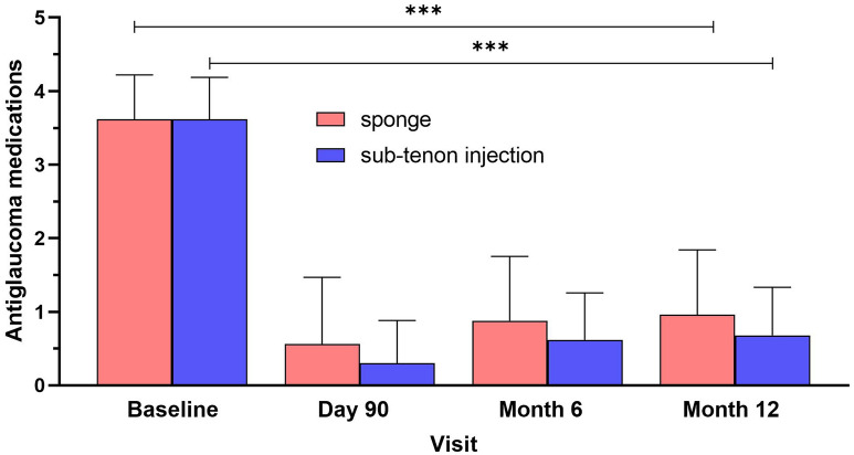
Results: Sustained reductions in mean IOP were observed in both groups over the 1-year follow-up, with no significant differences between the groups. The complete success rate, with a target IOP of ≤ 21 mmHg after 1 year, was 19.3% in the sponge group and 26.4% in the injection group. The qualified success rate was 59.0% and 87.4% in the sponge and injection groups, respectively. A longer survival rate was observed in the injection group than in the sponge group when IOP was below 21 mmHg. The mean CECD significantly decreased (P < 0.01) from baseline to each postoperative follow-up time point in both groups. At 1 year postoperatively, the percentage of total CECD loss was 8.1% in the sponge group and 8.0% in the injection group. However, no significant differences in mean CECD values, the number of medications, or adverse events were found between the groups.

Conclusions: PMS implantation with sub-tenon injection of MMC was comparable in terms of efficacy and safety to traditional MMC delivery via soaked sponges. However, the injection group demonstrated a significantly higher success rate than the sponge group.

Keywords: Corneal endothelial cells; Glaucoma; Glaucoma surgery; Intraocular pressure; MIBS.


Conflict of interest statement

Declarations. Conflict of Interest: Nora Majtanova and Petr Kolar have the following financial disclosures: Alcon (honorarium); Santen (consultancy and honorarium) and Bayer (honorarium). Adriana Takacova has the following financial disclosures: Alcon (honorarium) and Santen (honorarium). Veronika Kurilova, Libor Hejsek, and Juraj Majtan have no competing interests. Ethical Approval: The study was conducted according to the guidelines of the Declaration of Helsinki and approved by the Institutional Review Board of St. Cyril and Method University Hospital No. 1/9/20/24. Informed consent for surgical procedure was obtained from all subjects involved in the study.

Figures

Fig. 1
Fig. 1
Flow chart of the selection process of the study participants in the retrospective cohort study. MMC mitomycin C
Fig. 2
Fig. 2
Graph showing intraocular pressure (IOP) at baseline and during follow-up. Data are expressed as mean values with standard deviation. ***P < 0.001 for baseline versus 12-month comparison within the groups of patients who underwent PreserFlo MicroShunt implantation with different mitomycin C (MMC) delivery methods (sponge vs. sub-tenon injection), based on the paired Wilcoxon signed-rank test
Fig. 3
Fig. 3
Scatter plot of postoperative intraocular pressure (IOP) values compared with preoperative IOP at the 1-year follow-up. Dots are color-coded according to subgroups. The solid diagonal line represents matching baseline IOP and IOP at the 1-year follow-up. The dashed diagonal line represents a 20% reduction in mean IOP. The dashed horizontal lines indicate postoperative IOP thresholds of 18 and 21 mmHg
Fig. 4
Fig. 4
Kaplan–Meier plots comparing the success rates of the two subgroups of patients who underwent PreserFlo MicroShunt implantation with different mitomycin C (MMC) delivery methods (sponge vs. sub-tenon injection). A Complete success, defined as IOP ≤ 21 mmHg, ≥ 20% IOP reduction, and no medication allowed. B Qualified success, defined as IOP ≤ 21 mmHg, ≥ 20% IOP reduction, and medication allowed. C Complete success with IOP ≤ 18 mmHg, ≥ 20% IOP reduction, and no medication allowed. D Qualified success with IOP ≤ 18 mmHg, ≥ 20% IOP reduction, and medication allowed. The dashed lines represent the 95% confidence interval curves. P values was calculated by log-rank test
Fig. 5
Fig. 5
Time course of postoperative corneal endothelial cell density (CECD) over 1 year of follow-up. Data are expressed as mean values with standard deviation. ***P < 0.001
Fig. 6
Fig. 6
Graphical representation of the relationship between corneal endothelial cell density (CECD) loss and tube–endothelium (TE) distance in patients who underwent PreserFlo MicroShunt implantation with different mitomycin C (MMC) delivery methods: A sponge soaked with MMC and B sub-tenon injection of MMC. A Spearman correlation test was used for the correlation analysis
Fig. 7
Fig. 7
Postoperative change in the number of antiglaucoma medications over 1 year compared with preoperative values. Data are expressed as mean values with standard deviation. ***P < 0.001

Similar articles

References

    1. Vision Loss Expert Group of the Global Burden of Disease Study. Global estimates on the number of people blind or visually impaired by glaucoma: a meta-analysis from 2000 to 2020. Eye. 2024;38:2036. - PMC - PubMed
    1. Chen W, Xu Y, Liu Z, Zhao J. Global, regional and national burden of Glaucoma: an update analysis from the Global Burden of Disease Study 2019. Int Ophthalmol. 2024;44:234. - PubMed
    1. Jayaram H, Kolko M, Friedman DS, Gazzard G. Glaucoma: now and beyond. Lancet. 2023;402:1788–801. - PubMed
    1. Chan L, Moster MR, Bicket AK, et al. New devices in glaucoma. Ophthalmol Ther. 2023;12:2381–95. - PMC - PubMed
    1. Saeed E, Gołaszewska K, Dmuchowska DA, Zalewska R, Konopińska J. The PreserFlo MicroShunt in the context of minimally invasive glaucoma surgery: a narrative review. Int J Env Res Public Health. 2023;20:2904. - PMC - PubMed

LinkOut - more resources