. 2024 Nov 22;10(47):eadq9960.
doi: 10.1126/sciadv.adq9960. Epub 2024 Nov 22.

Enhanced dual-mode imaging: Superior photoacoustic and ultrasound endoscopy in live pigs using a transparent ultrasound transducer

Affiliations

Enhanced dual-mode imaging: Superior photoacoustic and ultrasound endoscopy in live pigs using a transparent ultrasound transducer

Jaewoo Kim et al. Sci Adv. .

Abstract

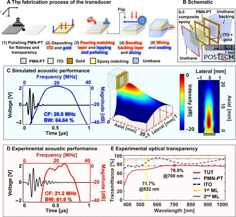
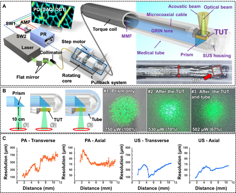
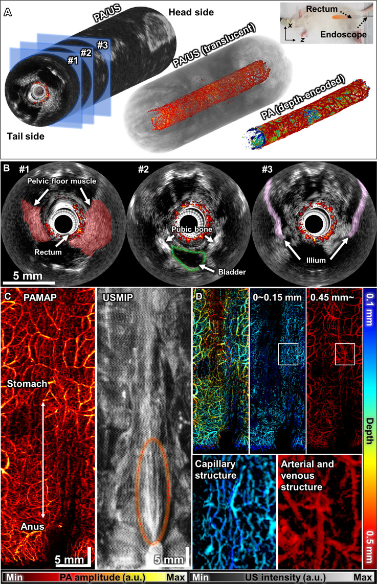
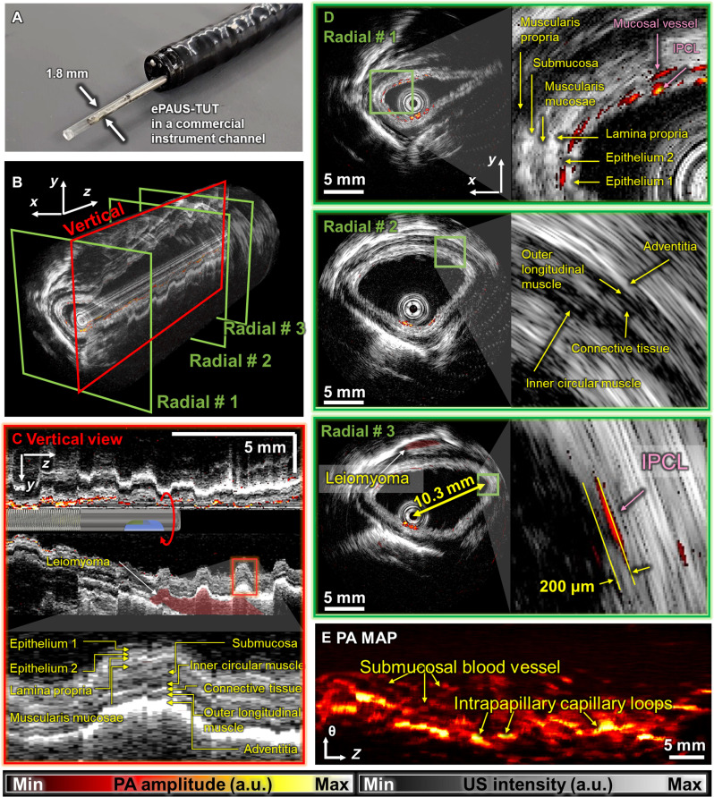
Dual-mode photoacoustic/ultrasound endoscopy (ePAUS) is a promising tool for preclinical and clinical interventions. To be clinically useful, ePAUS must deliver high-performance ultrasound imaging comparable to commercial systems and maintain high photoacoustic imaging performance at long working distances. This requires a transducer with an intact physical aperture and coaxial alignment of acoustic and optical beams within the probe, a challenging integration task. We present a high-performance ePAUS probe with a miniaturized, optically transparent ultrasonic transducer (TUT) called ePAUS-TUT. The 1.8-mm-diameter probe, fitting into standard endoscopic channels, aligns acoustic and optical beams efficiently, achieving commercial-level ultrasound and high-resolution photoacoustic imaging over long distances. These imaging capabilities were validated through in vivo imaging of a rat's rectum and a pig's esophagus. The ePAUS-TUT system substantially enhances feasibility and potential for clinical applications.


Figures

Fig. 1.
Fig. 1.. Fabrication and operating characteristics of a TUT.
(A) Fabrication of the TUT. (B) Schematic of the layer structure and (inset) photograph of the TUT. (C) Simulated and (D) Experimental acoustic performances. (E) Experimental optical transmittance. CF, center frequency; BW, bandwidth; and ML, matching layer.
Fig. 2.
Fig. 2.. System schematic and performance of the TUT-based ePAUS system.
(A) Schematic of the overall ePAUS-TUT system and a photograph of the probe. (B) Optical beam shapes after each component: #1 immediately after the prism, #2 after passing the TUT, and #3 after passing through the TUT and tube. (C) PA and US transverse and axial resolutions as a function of distance. MMF, multimode fiber; SUS, steel use stainless; DAQ, data acquisition device; DGT, digitizer; PR, pulser/receiver; SW, switch; HSR, hollow-shaft slip ring; ORJ, optical rotary joint; and AMP, amplifier.
Fig. 3.
Fig. 3.. Endoscopic PA/USI of a rat’s rectum in vivo.
(A) 3D-rendered overlaid PA/US, translucent PA/US, and depth-encoded PA-only images (movie S1). (B) Cross-sectional overlaid B-mode PA/US images indicated by numbers in (A). (C) PA MAP and US MIP images. (D) Three depth-encoded PA MAP images: The entire depth, from 0- to 0.15-mm deep, and below 0.45 mm. Insets are magnified images from the boxes in (D). All scale bars, 5 mm.
Fig. 4.
Fig. 4.. Endoscopic PA/USI of a pig’s esophagus in vivo.
(A) Photograph of the ePAUS probe inserted into a commercial endoscope. (B) 3D-rendered overlaid PA/US image of a pig’s esophagus. (C) Cross-sectional PA/US image cut along the vertical direction in (B). (D) Cross-sectional PA/US images (#1 to #3) cut along the radial direction in (B). (E) PA MAP image of the pig’s esophagus. IPCL, intrapapillary capillary loops. All scale bars, 5 mm.

Similar articles

Cited by

References

    1. Weber J., Beard P. C., Bohndiek S. E., Contrast agents for molecular photoacoustic imaging. Nat. Methods 13, 639–650 (2016). - PubMed
    1. Kim J., Kim J. Y., Jeon S., Baik J. W., Cho S. H., Kim C., Super-resolution localization photoacoustic microscopy using intrinsic red blood cells as contrast absorbers. Light Sci. Appl. 8, 103 (2019). - PMC - PubMed
    1. Baik J. W., Kim H., Son M., Choi J., Kim K. G., Baek J. H., Park Y. H., An J., Choi H. Y., Ryu S. Y., Intraoperative label-free photoacoustic histopathology of clinical specimens. Laser Photon. Rev. 15, 2100124 (2021).
    1. Kim J., Park B., Ha J., Steinberg I., Hooper S. M., Jeong C., Park E.-Y., Choi W., Liang T., Bae J. S., Multiparametric photoacoustic analysis of human thyroid cancers in vivo. Cancer Res. 81, 4849–4860 (2021). - PubMed
    1. Kim J., Kim G., Li L., Zhang P., Kim J. Y., Kim Y., Kim H. H., Wang L. V., Lee S., Kim C., Deep learning acceleration of multiscale superresolution localization photoacoustic imaging. Light Sci. Appl. 11, 131 (2022). - PMC - PubMed

LinkOut - more resources