. 2024 Nov 22;15(1):10150.
doi: 10.1038/s41467-024-54584-5.

An inhibitory segment within G-patch activators tunes Prp43-ATPase activity during ribosome assembly

Affiliations

An inhibitory segment within G-patch activators tunes Prp43-ATPase activity during ribosome assembly

Daniela Portugal-Calisto et al. Nat Commun. .

Abstract

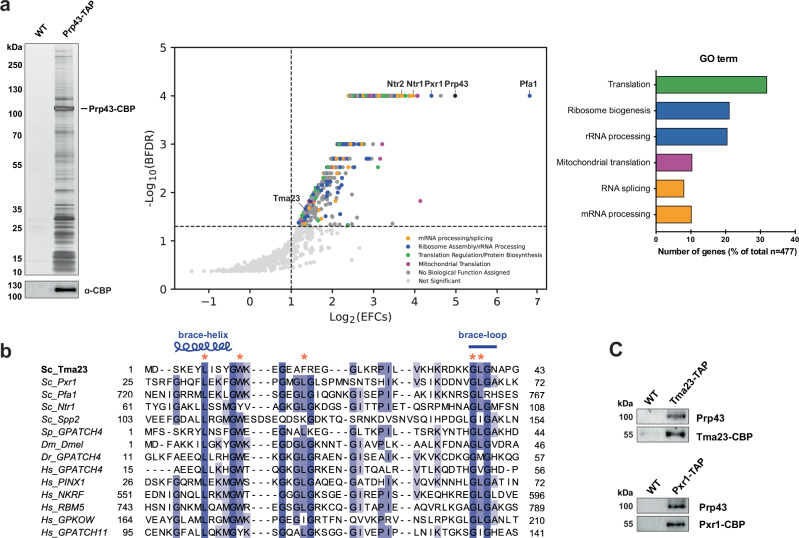
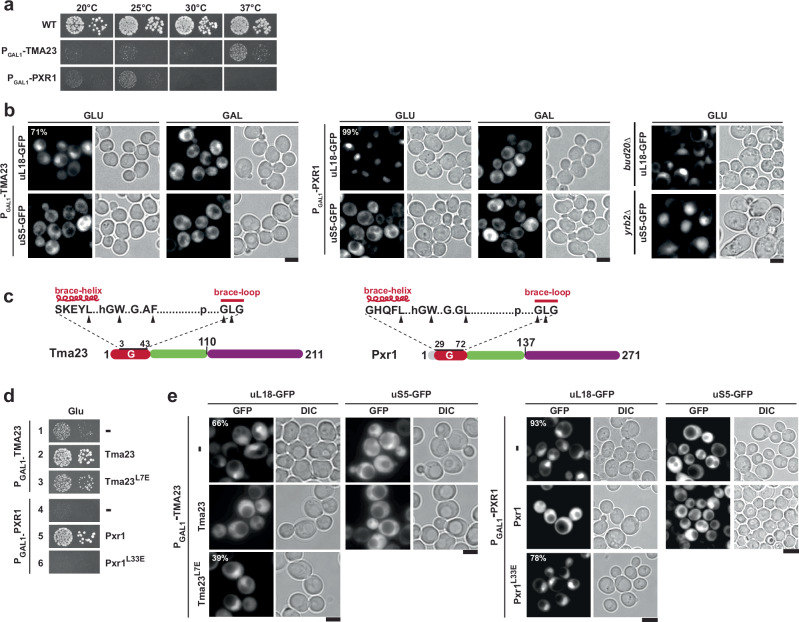
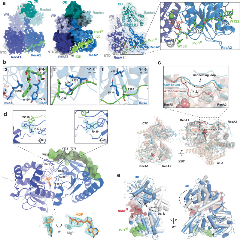
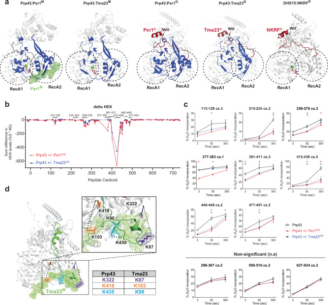
Mechanisms by which G-patch activators tune the processive multi-tasking ATP-dependent RNA helicase Prp43 (DHX15 in humans) to productively remodel diverse RNA:protein complexes remain elusive. Here, a comparative study between a herein and previously characterized activators, Tma23 and Pxr1, respectively, defines segments that organize Prp43 function during ribosome assembly. In addition to the activating G-patch, we discover an inhibitory segment within Tma23 and Pxr1, I-patch, that restrains Prp43 ATPase activity. Cryo-electron microscopy and hydrogen-deuterium exchange mass spectrometry show how I-patch binds to the catalytic RecA-like domains to allosterically inhibit Prp43 ATPase activity. Tma23 and Pxr1 contain dimerization segments that organize Prp43 into higher-order complexes. We posit that Prp43 function at discrete locations on pre-ribosomal RNA is coordinated through toggling interactions with G-patch and I-patch segments. This could guarantee measured and timely Prp43 activation, enabling precise control over multiple RNA remodelling events occurring concurrently during ribosome formation.


Conflict of interest statement

Competing interests: The authors declare no competing interests.

Figures

Fig. 1
Fig. 1. Tma23 is a G-patch containing Prp43 activator.
a Left panel: Prp43-TAP eluate was separated on a NuPAGE 4–12% Bis-Tris gradient gel and analysed by Silver staining or Western blotting using α-CBP antibody. WT (BY4741 untagged strain) was used as negative control. Middle panel: Prp43-TAP proteome was identified by label-free quantitative mass spectrometry analysis. The data was analysed with the R package prolfqua. The plot shows a Bayesian false discovery rate (BFDR) of less than 10% as a function of empirical fold change (EFC) score >2. Right panel: Statistically significant proteins (p-value < 0.001) were grouped based on the DAVID functional GO clustering analysis. This experiment was performed independently three times with similar results. b Sequence alignments of Tma23 with known G-patch proteins across species: (Sc) Saccharomyces cerevisiae; (Sp) Schizosaccharomyces pombe; (Dm) Drosophila melanogaster; (Dr) Danio rerio; (Hs) Homo sapiens. Brace-helix and brace-loop as defined previously are indicated on top. The alignments were made with MAFFT and visualized with Jalview (version 2.11.2.5). Asterisks indicate residues substituted for functional studies. c, Tma23-TAP and Pxr1-TAP eluates were separated on a NuPAGE 4-12% Bis-Tris gradient gel and subjected to Western blotting using antibodies directed against Tma23, Pxr1, and Prp43. This experiment was performed independently three times with similar results. Source data are provided as a Source Data file.
Fig. 2
Fig. 2. Tma23M and Pxr1M mediates binding to Prp43.
a Upper panel: Domain organization of Tma23 and Pxr1. Lower panel: Plasmids encoding indicated LexA DNA-binding domain (DBD) and GAL4 activation domain (AD) fusion proteins were transformed into NMY32. Transformants were spotted in serial 10-fold dilutions on the indicated selective media and incubated at 30 °C for 2–4 days. LaminC and LargeT served as negative controls. b GST tagged variants of Tma23 and Pxr1 were co-expressed with His6-Prp43 in E. coli cells and affinity-purified using Glutathione Sepharose 4 Fast Flow beads (Cytiva). The bound proteins were eluted using PierceTM LDS Sample buffer at 70 °C, separated on a SurePAGE 4-20% BIS-TRIS gradient gel and visualized by Coomassie Blue staining. Pfa1 G-patch (Pfa1G) and GST-alone were used as positive and negative controls, respectively. Asterisks indicate the baits. cf ATP hydrolysis rates at 2.5 mM ATP normalized for enzyme concentration (Prp43). Initial ATP hydrolysis rates were calculated by applying a linear regression to the NADH oxidation over time and normalized to the enzyme concentration for Prp43. Each independent experiment is shown (dots) and error bars indicate mean values ± SD. cd Tma23G or Pxr1G was added to Prp43 in a 1:1 or 10:1 molar ratio, as indicated. Pfa1G was used as control. ef Tma23G or Pxr1G were fused to Prp43 (Prp43-TG and Prp43-PG, respectively). ATPase activities of the fusions were compared to Tma23G or Pxr1G added to Prp43 in a 1:1 ratio. Source data are provided as a Source Data file.
Fig. 3
Fig. 3. Tma23G and Pxr1G mutants are impaired in 60S pre-ribosome export.
a Wild type (BY4741), PGAL1-TMA23, and PGAL1-PXR1 strains were spotted in serial 10-fold dilutions on glucose-repressive medium and incubated at the indicated temperature for 3 – 7 days. b PGAL1-TMA23, PGAL1-PXR1 expressing uL18-GFP or uS5-GFP, nuclear export reporters for 60S and 40S respectively, were grown to mid-log phase in glucose-(GLU) and galactose- (GAL) containing media at 37 °C. The location of the reporters was monitored by fluorescence microscopy and images were processed using ImageJ (version 1.50e; NIH and LOCI, USA). Scale bar = 5 μm. Percentage of cells exhibiting nuclear accumulation of uL18-GFP is indicated. bud20Δ and yrb2Δ were used as a positive control for nuclear accumulation of the reporters. This experiment was performed independently three times with similar results. c Domain organization of Tma23 and Pxr1 showing partial sequence of the G-patch with mutated residues indicated by black arrowheads. d PGAL1-TMA23 strain expressing empty vector (-), Tma23 or Tma23L7E and PGAL1-PXR1 strain expressing empty vector (-), Pxr1, or Pxr1L33E were spotted in 10-fold serial dilutions on glucose-containing medium and incubated at 37 °C for 2–7 days. e, The indicated yeast strains expressing uL18-GFP or uS5-GFP reporters, were grown to mid-log phase in glucose-containing medium at 37 °C. Cellular localization of the reporters was visualized by fluorescence microscopy. The percentage of cells exhibiting nuclear accumulation of uL18-GFP reporter is indicated. This experiment was performed independently three times with similar results. Images were processed using ImageJ (version 1.50e; NIH and LOCI, USA). Scale bar = 5 μm.
Fig. 4
Fig. 4. I-Patch restrains Prp43-ATPase activity.
ah Initial hydrolysis rates at 2.5 mM ATP were normalized to Prp43 concentration. Initial rates were calculated by applying a linear regression to the NADH oxidation over time and normalized to Prp43 concentration. Each independent experiment is shown (dots) and error bars indicate mean values ± SD. a Tma23G was added to Prp43 in a 10:1 molar ratio. Tma23GM, Tma23GM-L7E, and Tma23M were co-expressed with Prp43. b ATPase activity of Prp43-TG was compared with Prp43-TG:Tma23M or Prp43-TG:Tma23M44. c Pxr1G was added to Prp43 in a 10:1 molar ratio. Pxr1GM, Pxr1GM-L33E, and Pxr1M were co-expressed with Prp43. d ATPase activity of Prp43-TG was compared to either Prp43-TG:Pxr1M or Prp43-TG:Pxr1M14. e ATPase activities of Prp43:Tma23GM and Prp43:Tma23M complexes in presence/absence of U20 RNA. f ATPase activities of Prp43-TG and Prp43-TG:Tma23M, in presence/absence of U20 RNA. g ATPase activity of Prp43:Pxr1GM and Prp43:Pxr1M complexes in presence/absence of U20 RNA. Pxr1GM and Pxr1M were co-expressed with Prp43. h ATPase activity of Prp43-TG and Prp43-TG:Pxr1M, in presence and absence of U20 RNA. i KD determination for Prp43 binding to Cy5-labelled U12-RNA by fluorescence polarization. Cy5-labelled U12-RNA was titrated with Prp43 (circles), Prp43:Pxr1M (triangle up) or Prp43:Tma23M (triangle down) and the change in polarization was fitted to a single-site binding model (solid lines). An average of four independent measurements is plotted, and error bars indicate mean ± SD. j Kinetic parameters for ATPase activities of Prp43 (square, diamond), Prp43:Tma23M (triangle down, circle; left panel) and Prp43:Pxr1M (triangle down, circle; right panel), in the presence and absence of U20, respectively. This experiment was performed independently three times with similar results. A representative measurement in triplicate is presented, and error bars indicate mean values ± SD. k Sequence alignments of Tma23M and Pxr1M across yeasts and higher eukaryotes. Light green lines indicate the M domain of Tma23 (Tma23M) and Pxr1 (Pxr1M). Dark green lines mark the segments that inhibit Prp43-ATPase activity (I-patch; Tma23M44, Pxr1M14). The conserved FVKGE motif is indicated. Alignments were made with MAFFT and visualized with Jalview (version 2.11.2.5). Source data are provided as a Source Data file.
Fig. 5
Fig. 5. Cryo-EM structure of a Prp43-Pxr1GM complex.
a Cryo-EM structure of Prp43:Pxr1GM complex at 3.3 Å resolution shows binding of Pxr1 peptide 122-136 to Prp43. Surface (left panel) and cartoon (right panel) representation of Prp43:Pxr1GM complex. The individual domains of Prp43 are indicated and colour-coded in shades of blue. Pxr1 122-136 peptide is shown in green. Left panel: Pxr1M peptide (green) contacts Prp43 RecA2 (blue) and the connecting loop between RecA2 and RecA1 (dark blue). The ADP and the magnesium ion are shown in pink/orange and green, respectively. b Hydrophobic contacts between Pxr1GM and Prp43. Residues are depicted as sticks. c Prp43:Pxr1GM complex, bound to ADP (blue), shows a 7 Å displacement of the connecting loop between RecA1 and RecA2 domains, superposed with an ATP-bound Prp43 structure of C. thermophilum (light red, PDB 5LTA). DHX15:NKRFG (beige) complex was used as representative of an ADP-bound structure (PDB 6SH6). d Upper panel: The density map of Prp43:Pxr1GM complex shows two different conformations of Prp43 R430 and R270. When R430 is distant from the ADP, it contacts R270 and consequently, D386 does not interact with ADP (compared to other ADP-bound structures [PDB 2XAU]). Lower panel: The active site of Prp43 shows a density compatible with ADP. ADP is depicted in orange stick representation. e Structural comparison of Prp43:Pxr1GM complex (blue and green) with DHX15:NKRFG complex (grey and red; PDB 6SH6) shows an open RNA-binding tunnel. The CTD of Prp43:Pxr1GM complex is rotated outwards as compared to DHX15-NKRFG complex by 24 Å as indicated by the arrows.
Fig. 6
Fig. 6. A conserved Phe anchors I-patch to Prp43.
a Upper panel: Domain organization of Pxr1 and Tma23. The star indicates the substituted F126/F96 within the M-domains. Lower panel: NMY32 or a modified NMY32 Pxr1GM-CoV2C strain transformed with plasmids encoding for the indicated LexA DNA-binding domain (DBD) and GAL4 activation domain (AD) fusion proteins were spotted on selective SD-Leu-Trp and SD-His media and incubated at 30 °C for 2–4 days. LaminC and LargeT were used as negative controls. b Prp43 was co-expressed in E. coli with the indicated GST-fused wild-type proteins and mutants of Pxr1M and Tma23M, or with GST, and affinity-purified using Glutathione Sepharose 4 Fast Flow beads (Cytiva). The bound proteins were eluted using PierceTM LDS Sample buffer at 70 °C separated by SDS-PAGE and visualized by Coomassie Blue staining. GST was used as a negative control. Asterisks indicate the baits. This experiment was performed independently three times with similar results. c PGAL1-PXR1 and PGAL1-TMA23 strains expressing empty vector (-), the mutants Pxr1F126A and Tma23F96A, or C-terminal fusions of Tma23GM and Pxr1GM to CoV2 dimerization motif (CoV2C) were transformed in the PGAL1-TMA23 and PGAL1-PXR1 strains were spotted in 10-fold serial dilutions on glucose-containing medium and incubated at 37 °C for 2–7 days. d PGAL1-TMA23 and PGAL1-PXR1 strains expressing empty vector (-), wild type Tma23 or Pxr1, or the indicated chimeric constructs were spotted in 10-fold serial dilutions on selective medium and incubated at 37 °C for 2–7 days. e PGAL1-PXR1 and PGAL1-TMA23 expressing the 60S nuclear export reporter uL18-GFP and empty vector (−), wild type protein or mutants (indicated) were grown to mid-log phase in glucose-containing media at 37 °C. The location of the reporter was analysed by fluorescence microscopy and images were processed using ImageJ (version 1.50e; NIH and LOCI, USA). The percentage of cells exhibiting nuclear accumulation of uL18-GFP reporter is indicated. This experiment was performed independently three times with similar results. Scale bar = 5 μm.
Fig. 7
Fig. 7. HDX-MS and XL-MS of Pxr1GM:Prp43 and Tma23GM:Prp43 complexes.
a Binding interface between Prp43 and either Pxr1GM or Tma23GM shows extensive protection of both RecA-like domains. Regions of Prp43 showing reduced H/D exchange rates upon complex formation are highlighted in blue. Regions in green indicate parts of Pxr1 present in the cryo-EM structure (left). The Pxr1 and Tma23 predicted G-patch position is displayed in red. The x-ray structure of DHX15:NKRFG (PDB 6SH6) is shown for comparison and the winged-helix domain (WH) is marked. b Difference in H/D exchange rate between Prp43 alone or when in complex with either Pxr1GM (red) or Tma23GM (blue). Each point represents a peptide, plotted according to its centroid value. The Y axis shows the H/D difference calculated as the product of the difference in percentage deuteration multiplied by the difference calculated in number of deuterons (%D * #D). Peptides shown in the uptake plots (c) are indicated above the plot (the length of the lines is not in scale). c Uptake plots for Prp43 peptides representing the different regions (indicated above each plot) that show significant differences in H/D exchange rates in different conditions. Peptides 296-307, 505-516, and 627-634 represent statistically non-significant (n.s.) H/D exchange rates. H/D exchange rates for Prp43, Prp43-Pxr1GM, and Prp43-Tma23GM are shown in black, red, and blue, respectively. Stars mark the conditions for which H/D exchange of Prp43 was statistically significant (p < 0.05) in presence of Pxr1GM (red) or Tma23GM (blue). A two-sided t-test student was applied for data analysis; three technical replicates are presented with mean values ± SD (See also Supplementary Data. 1). d Crosslinks of Prp43-Tma23GM complex were mapped on the AlphaFold2 predicted model. The crosslinked amino acids are shown in sticks. The cross-linked pairs are indicated in the same colour, connected with a dashed lined, and summarized in the table. The I-patch is shown in mesh representation. See also Supplementary Data. 2.

References

    1. Bohnsack, K. E., Yi, S., Venus, S., Jankowsky, E. & Bohnsack, M. T. Cellular functions of eukaryotic RNA helicases and their links to human diseases. Nat. Rev. Mol. Cell Biol.24, 749–769 (2023). - PubMed
    1. Khreiss, A., Bohnsack, K. E. & Bohnsack, M. T. Molecular functions of RNA helicases during ribosomal subunit assembly. Biol. Chem.404, 781–789 (2023). - PubMed
    1. Ozgur, S. et al. The conformational plasticity of eukaryotic RNA-dependent ATPases. FEBS J.282, 850–863 (2015). - PubMed
    1. Fairman-Williams, M. E., Guenther, U. P. & Jankowsky, E. SF1 and SF2 helicases: family matters. Curr. Opin. Struct. Biol.20, 313–324 (2010). - PMC - PubMed
    1. Jarmoskaite, I. & Russell, R. RNA helicase proteins as chaperones and remodelers. Annu Rev. Biochem83, 697–725 (2014). - PMC - PubMed

Publication types

MeSH terms

Associated data

LinkOut - more resources