. 2024 Nov 22;9(1):232.
doi: 10.1038/s41541-024-01031-7.

The PvRBP2b-TfR1 interaction is not essential for reticulocytes invasion by Plasmodium vivax isolates from Cambodia

Affiliations

The PvRBP2b-TfR1 interaction is not essential for reticulocytes invasion by Plasmodium vivax isolates from Cambodia

Lionel B Feufack-Donfack et al. NPJ Vaccines. .

Abstract

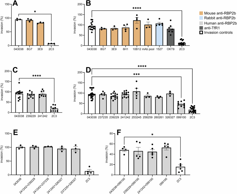
Plasmodium vivax is the most widespread of the different Plasmodium species able to infect humans and is responsible for most malaria cases outside Africa. An effective, strain-transcending vaccine that alleviates or suppresses erythrocyte invasion would be a game-changer in eliminating vivax malaria. Recently, the binding of P. vivax Reticulocyte Binding Protein 2b (PvRBP2b) to human Transferrin receptor (TfR1) has been described as essential for reticulocyte invasion, making this parasite protein an appealing vaccine candidate. Here, using P. vivax Cambodian clinical isolates in robust ex vivo invasion assays, we show that anti-PvRBP2b polyclonal and monoclonal antibodies that inhibit binding of PvRBP2b to TfR1 do not block P. vivax invasion into reticulocytes even at high concentrations. Anti-TfR1 antibodies do not inhibit P. vivax invasion either. Combinations at high concentrations of human monoclonal antibodies targeting different PvRBP2b epitopes do not inhibit invasion. Combinations of anti-PvRBP2b with anti-PvDBP do not enhance invasion inhibition caused by anti-PvDBP alone. We also show that the invasion of Cambodian P. vivax is trypsin-resistant while TfR1 is trypsin-sensitive, and we demonstrate that TfR1 is not recycled following trypsin treatment. We determined the PvRBP2b sequence of all isolates used in the invasion assays and analyzed polymorphism within epitopes recognized by anti-PvRBP2b antibodies. We show that polymorphism does not explain the absence of neutralization. Anti-PvRBP2b polyclonal antibodies recognized all four isolates tested in immunofluorescence assays while not inhibiting P. vivax invasion. Overall, our results demonstrate that PvRBP2b binding to TfR1 is not essential for invasion into reticulocytes of P. vivax Cambodian strains questioning the relevance of PvRBP2b as vaccine candidate.


Conflict of interest statement

Competing interests: The authors declare no competing interests.

Figures

Fig. 1
Fig. 1. Anti-PvRBP2b antibodies do not inhibit the invasion of Cambodian clinical isolates.
The mean percentage (± SEM) of reticulocyte invasion by P. vivax clinical isolates in the presence of different anti-PvRBP2b antibodies tested at 100, 250, and 500 μg/ml is represented. All invasions are normalized to no-antibody controls. HumAbs 043038 specific of tetanus toxin C terminal fragment and the anti-Duffy mouse mAbs 2C3 were used as negative and positive invasion inhibition controls for each experiment. Each dot represents the invasion for a different clinical isolate with a single technical replicate per isolate. Differences are assessed by the Kruskal-Wallis test with Dunn’s post-hoc correction (*p < 0.05; ***p = 0.0007; ****p < 0.0001). A Invasion of P. vivax in presence of 100 μg/ml of mouse anti-PvRBP2b mAbs 3E9 and 8G7. B Invasion of P. vivax in presence of 500 μg/ml (100 μg/ml for the 2C3 control) mouse anti-PvRBP2b mAbs 8G7, 3E9, 6H1, 10B12, mAbs pool (each mAb at one-third of the final concentration) and rabbit polyclonal mAbs (1527). C Invasion of P. vivax in presence of 100 μg/ml of anti-PvRBP2b humAbs 239229 and 241242. D Invasion inhibition of P. vivax in presence of 500 μg/ml (100 μg/ml for the 2C3 control) of humAbs 237235, 239229, 241242, 253245, 258259, 260261, 326327. E Invasion inhibition of P. vivax in the presence of 500 μg/ml (100 μg/ml for the 2C3 control) anti-PvRBP2b humAbs combinations (250 μg/ml each huAbs). F Invasion inhibition of P. vivax in presence of anti-PvRBP2b humAbs (2392209 or 241242 each at 250 μg/ml) or 043038 control in combination with anti-PvDBP 099100 at 250 μg/ml (final concentration of humAbs of 500 μg/ml) compared to invasion in presence of 099100 alone at 250 μg/ml or 2C3 control at 100 μg/ml.
Fig. 2
Fig. 2. The absence of invasion inhibition by anti-PvRBP2b Abs is not dependent on PvRBP2b sequence polymorphism of P. vivax isolates.
A Epitopes recognized by anti-PvRBP2b mAbs,. B Invasion in the presence of 500 μg/ml of mouse anti-PvRBP2b mAbs is not affected by mutations in residues recognized by 6H1, 3E9, and 10B12 compared to the wild-type (WT). C Invasion in the presence of 500 μg/ml of humAbs is not affected by mutations in residues recognized by 237235, 239229, 253245, 258259 and 260261 compared to WT. For 241242 and 326327, all isolates had at least one mutation in the epitopes recognized by these humAbs. HumAbs 043038 specific of tetanus toxin C terminal fragment and the anti-Duffy mouse mAbs 2C3 were used as negative and positive invasion inhibition controls for each experiment. All invasions are normalized to no-antibody controls.
Fig. 3
Fig. 3. Polyclonal anti-PvRBP2b 1527 Abs binds to merozoites from all four P. vivax clinical isolates tested by immunofluorescence.
Each row shows representative images of segmented schizonts for a different clinical isolate. A: bright field, B: Hoechst 33342, C: 1527, D: merged.
Fig. 4
Fig. 4. P. vivax Cambodian isolates are trypsin-resistant.
A Inhibition of P. vivax invasion after chymotrypsin, neuraminidase or trypsin treatment of reticulocytes. Only chymotrypsin treatment inhibits P. vivax merozoite invasion. B The absence of recycling of TfR1/CD71 on trypsin-treated reticulocytes. Trypsin treatment removed > 80% of TfR1 molecules on the surface of reticulocytes and the TfR1 levels remain below 10% to the original level. Data expressed are the % of the control (% of non-treated TfR1+ reticulocytes at Time 0). This experiment is representative of 4 separate experiments with 4 different batches of cord blood reticulocytes. All invasions are normalized to no-antibody controls.

References

    1. Battle, K. E. et al. Mapping the global endemicity and clinical burden of Plasmodium vivax, 2000–17: a spatial and temporal modelling study. Lancet394, 332–343 (2019). - PMC - PubMed
    1. Baird, J. K. Evidence and implications of mortality associated with acute Plasmodium vivax malaria. Clin. Microbiol. Rev.26, 36–57 (2013). - PMC - PubMed
    1. Douglas, N. M. et al. Major burden of severe anemia from non-falciparum malaria species in Southern Papua: a hospital-based surveillance study. PLos Med.10, e1001575 (2013). - PMC - PubMed
    1. Douglas, N. et al. Mortality attributable to Plasmodium vivax malaria: a clinical audit from Papua, Indonesia. BMC Med.12, 217 (2014). - PMC - PubMed
    1. Mueller, I. et al. Key gaps in the knowledge of Plasmodium vivax, a neglected human malaria parasite. Lancet Infect. Dis.9, 555–566 (2009). - PubMed

LinkOut - more resources