. 2024 Nov 22;15(1):10126.
doi: 10.1038/s41467-024-54378-9.

Viral sequence determines HLA-E-restricted T cell recognition of hepatitis B surface antigen

Affiliations

Viral sequence determines HLA-E-restricted T cell recognition of hepatitis B surface antigen

Gavuthami Murugesan et al. Nat Commun. .

Abstract

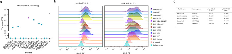
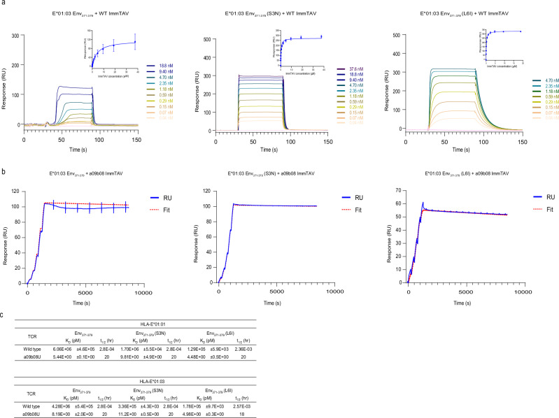
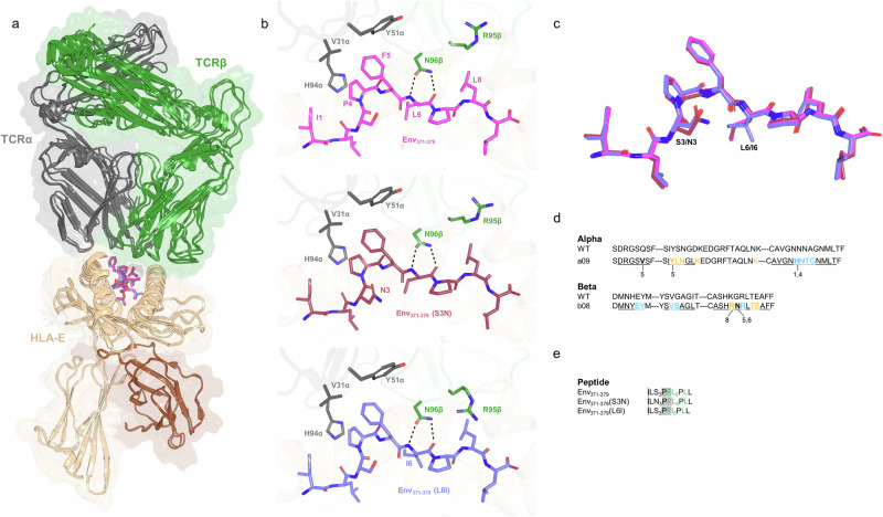
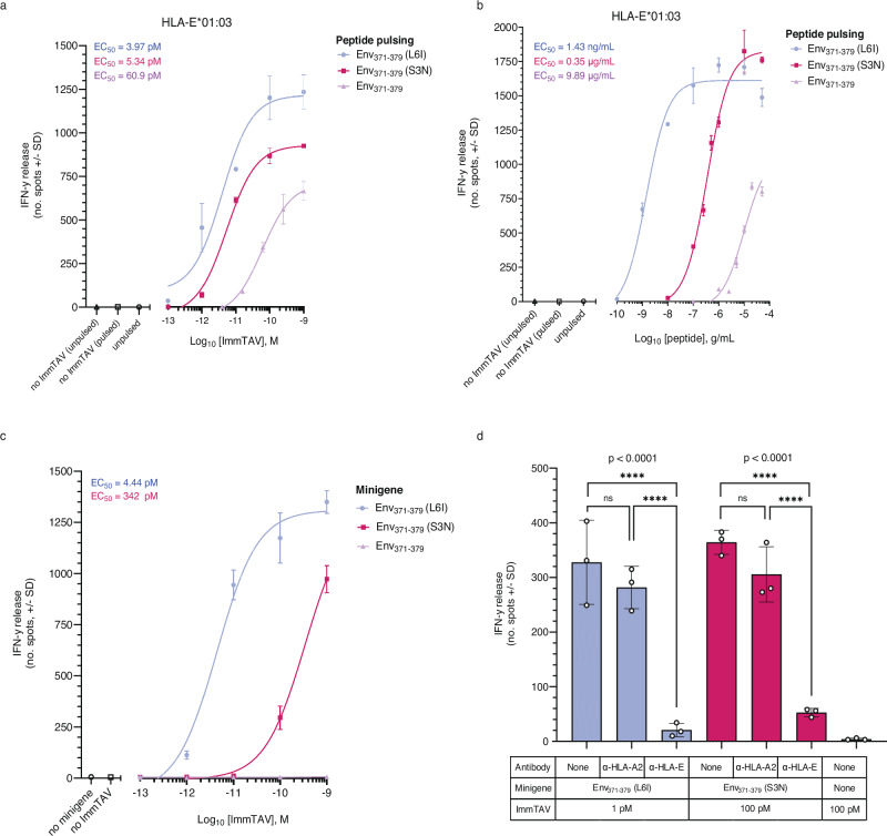
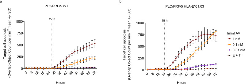
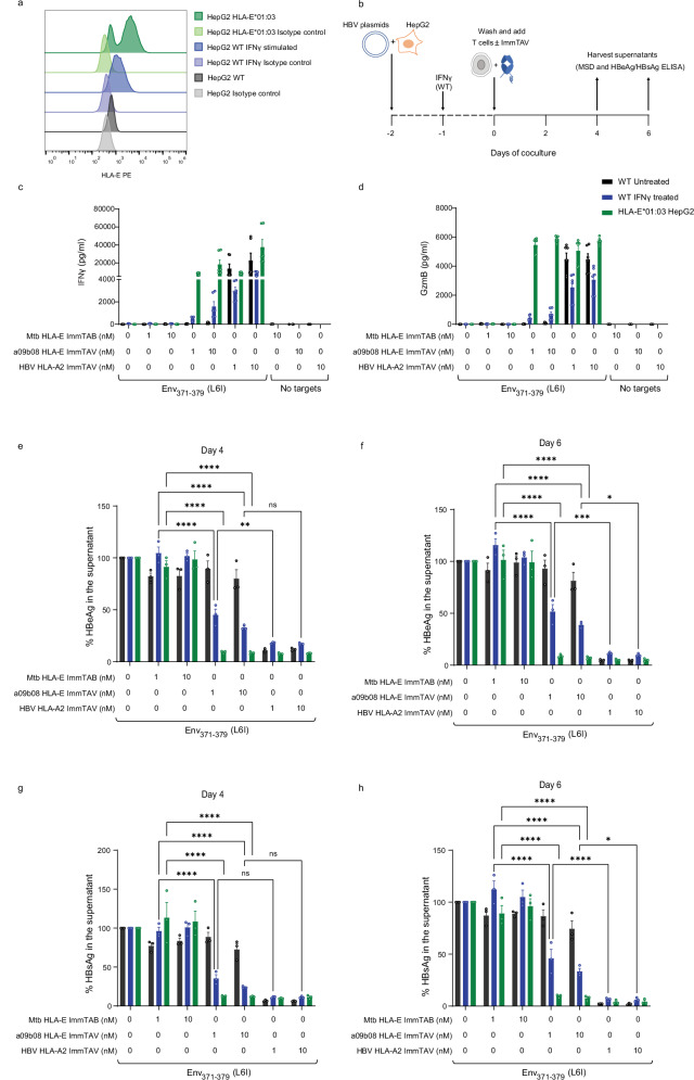
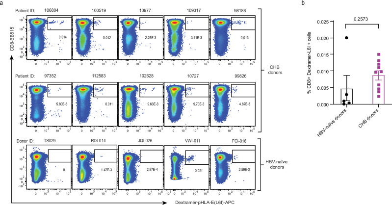
The non-polymorphic HLA-E molecule offers opportunities for new universal immunotherapeutic approaches to chronic infectious diseases. Chronic Hepatitis B virus (HBV) infection is driven in part by T cell dysfunction due to elevated levels of the HBV envelope (Env) protein hepatitis B surface antigen (HBsAg). Here we report the characterization of three genotypic variants of an HLA-E-binding HBsAg peptide, Env371-379, identified through bioinformatic predictions and verified by biochemical and cellular assays. Using a soluble affinity-enhanced T cell receptor (TCR) (a09b08)-anti-CD3 bispecific molecule to probe HLA-E presentation of the Env371-379 peptides, we demonstrate that only the most stable Env371-379 variant, L6I, elicits functional responses to a09b08-anti-CD3-redirected polyclonal T cells co-cultured with targets expressing endogenous HBsAg. Furthermore, HLA-E-Env371-379 L6I-specific CD8+ T cells are detectable in HBV-naïve donors and people with chronic HBV after in vitro priming. In conclusion, we provide evidence for HLA-E-mediated HBV Env peptide presentation, and highlight the effect of viral mutations on the stability and targetability of pHLA-E molecules.


Conflict of interest statement

Competing interests: G.M., R.L.P., R.K., V.I., R.J.S., M.M.C., V.K., R.P., A.J., J.D., T.H., W.B., G.P., K.O., D.K., A.S., C.B., R.R., C.P., T.G., A.T., A.W., M.D., D.G., M.H., R.J.C., K.E.A., L.D., A.K., S.L., M.S., and L.F.G. are or were employees of Immunocore Ltd.

Figures

Fig. 1
Fig. 1. Identification and validation of HBV peptides binding HLA-E.
a Thermal melting point (Tm) assessed by thermal shift assay screen of HBV peptides (Env371-379 peptide, magenta triangle) and Mtb RLPA (positive control, gray circle). Peptides were added to refolded and purified HIV Gag275-283-HLA-E*01:03 complexes (in PBS at 0.25 mg/mL) at a 60:1 molar ratio. Data were presented as mean values ± SD of triplicates. b HLA-E binding of HBV Env371-379 variants, Mtb RLPA, and signal peptides (A80, B7, and Cw3) was measured by HLA-E upregulation at the cell surface of K562 cells transduced with single chain β2m-HLA-E*01:01 or β2m-HLA-E*01:03. Plotted are flow cytometry histograms after staining with 3D12 antibody (HLA-E antibody). Histograms are representative of two independent experiments, each conducted in triplicates (full dataset and gating strategy available in Supplementary Fig. 1). c Summary of t1/2 and Tm of HBV Env371-379 variants and signal peptides (Cw3 and A1) pHLA-E*01:03 complexes as determined by SPR and thermal shift assays in triplicate. All SPR experiments were performed at 25 °C. Data were presented as mean values ± SD of triplicates. Source data are provided as a Source Data file.
Fig. 2
Fig. 2. Binding kinetics of wildtype and a09b08 ImmTAVs to HBV Env371-379 pHLA-E*01:03 complexes.
a Binding curves for wildtype ImmTAV and all three HBV Env371-379 pHLA-E*01:03 complexes. Binding was determined over a range of analyte concentrations from 37 nM to 37.6 µM. Insets: calculation of steady-state affinity, data were presented as mean values ± SD. b Binding kinetics for a09b08 ImmTAV and all three HBV Env371-379 pHLA-E*01:03 complexes. Graphs show the mean of the raw data (blue) and the 1:1 fit (dotted red line). For the binding kinetic graphs, the ImmTAV molecule was flown over the chip as the analyte, at concentrations ranging from 0.313 to 5 nM. Kinetic constants were determined using a 1:1 Langmuir model. c Summary of KD values and t1/2 values of the wild-type and a09b08 ImmTAV. All experiments were performed at 25 °C in triplicate. Data were presented as mean values ± SD. Source data are provided as a Source Data file.
Fig. 3
Fig. 3. Structural overview of the a09b08 TCR in complex with three HBV Env371-379 pHLA-E complexes.
a Cartoon overview of the three TCR-pHLA complexes aligned on HLA-E*01:03 (TCR alpha chain in gray, TCR beta chain in green, HLA-E in wheat, B2M is brown, peptides: Env371-379 in magenta, Env371-379 (S3N) in red and Env371-379 (L6I) in blue). b Close-up view of the three TCR-peptide interactions. TCR residues within 4 Å of the peptide are shown as sticks. Dotted lines indicate polar contacts. c Overlay of the three peptides, aligned on HLA-E, showing highly similar conformation in the complexes. d a09b08 TCR contacts to pHLA-E mapped onto the truncated TCR sequence. A wild-type TCR sequence is provided to show the mutations introduced by affinity maturation. CDR residues are underlined and residues within 4.1 Å of the peptide are highlighted in bold with peptide positions indicated below. Residues highlighted in blue and orange indicate positions that are within 4.1 Å of the HLA-E helix 1 and HLA-E helix 2, respectively. e a09b08 TCR contacts to peptide mapped onto the peptide sequences. Peptide residues within 4.1 Å of the TCR alpha chain are highlighted gray, and those of the TCR beta chain are colored green.
Fig. 4
Fig. 4. The a09b08 ImmTAV molecule elicits T cell responses against cells displaying HBV Env371-379 pHLA-E complexes.
a ELISPOT assay measuring dose-dependent IFN-γ release induced by a09b08 ImmTAV in co-cultures of PBMC from 3 HBV-naïve donors and THP-1-E*01:03 cells pulsed with Env371-379 (purple line), Env371-379 (S3N) (red line) and Env371-379 (L6I) (blue line) peptides (10 μg/mL). b IFN-γ ELISPOT assays showing titratable activation of PBMC from 3 HBV-naïve donors by a09b08 ImmTAV (1 nM) in the presence of THP-1-E*01:03 cells pulsed with the indicated amounts of peptide. Controls (a, b clear symbols) include PBMC + target cells (no ImmTAV; unpulsed or pulsed with 10 μg/mL peptide), and PBMC + ImmTAV + target cells (unpulsed). c ELISPOT assay measuring dose-dependent IFN-γ release from HBV-naïve donor PBMC in the presence of HepG2 targets stably transfected with minigenes encoding the indicated peptides Env371-379, Env371-379 (S3N), and Env371-379 (L6I). Untransfected HepG2 (no minigene) and PBMC + minigene target cells (no ImmTAV) were included as controls (clear symbols). d Cumulative IFN-γ responses induced by a09b08 ImmTAV in PBMC cocultured with HepG2 Env371-379 (L6I) (blue) or Env371-379 (S3N) (red) minigene targets in the presence or absence of blocking mAbs against HLA-E or HLA-A*02:01 (10 µg/mL); ****p = <0.0001, two-way ANOVA. Results (ad) are representative of one of three PBMC donors tested in triplicate. Data were presented as mean values ± SD. Average EC50 are indicated in the inset at the top left corner of the figures (ac). All donor EC50 values and averages are displayed in Supplementary Table 5. Source data are provided as a Source Data file.
Fig. 5
Fig. 5. The a09b08 ImmTAV elicits antigen-dependent T cell killing of target cells.
Apoptosis (measured by IncuCyte assay, see methods) of Env371-379 (L6I) Ag+ PLC/PRF/5 target cells wildtype (a) or HLA-E*01:03 lentiviral transduced (b), cocultured with HBV-naïve donor PBMC and a titration of a09b08 ImmTAV. Dashed vertical lines indicate timepoints from which target cell apoptosis begins (at 1 nM ImmTAV). Results (a, b) are representative of one of three PBMC donors, each tested in triplicate. Data were presented as mean values ± SD. Source data are provided as a Source Data file.
Fig. 6
Fig. 6. The a09b08 ImmTAV activates T cells to eliminate HBV-transfected HepG2 cells.
a Surface expression levels of HLA-E on HepG2 cells were analysed by flow cytometry. HepG2 wildtype and HLA-E*01:03 overexpressing cells were transfected with a replication-competent HBV plasmid encoding genotype A2 (Env371-379 (L6I)). b 24 h post-transfection, some wells of HepG2 wildtype cells were stimulated with IFN-γ (1 ng/mL) for 16 h. Cells were washed and cocultured with pan T cells from three donors (1:1 ratio) with or without a09b08 ImmTAV, HBV HLA-A*02:01 ImmTAV or Mtb RLPA HLA-E ImmTAB (negative control) at 1 and 10 nM. Culture supernatants were harvested on day 4 and day 6 according to the schematics shown (modified from ref. ). Panel b is released under a Creative Commons Attribution-Non Commercial 4.0 International license (https://creativecommons.org/licenses/by-nc/4.0/deed.en). Levels of IFN-γ (c) and granzyme B (d) in the culture supernatants at day 4 were quantified using MSD. Data represents the mean ± SEM of triplicates (n = 3). HBeAg (e, f) and HBsAg (g, h) levels in the culture supernatant at day 4 and day 6 were quantified using ELISA. Data were represented as mean ± SEM of duplicates from three donors. Significant differences are *p < 0.05, **p < 0.001, ****p < 0.0001 by ANOVA followed by Tukey’s post hoc test. Flow cytometry gating strategy and additional datasets are shown in Supplementary Figs. 11, 12, 13. Source data are provided as a Source Data file.
Fig. 7
Fig. 7. Detection of HBV antigen-specific T cells by pHLA-E multimers.
a T cells from people with CHB (n = 10) and HBV-naïve donors (n = 5) were expanded in vitro by stimulation with streptavidin magnetic beads conjugated to Env371-379 (L6I)-pHLA-E and anti-CD28 for two weeks and stained with dextramer-E and antibody cocktail. Dot plots show the percentage of CD8+ dextramer-E+ cells (dextramer positive cells were gated as live CD3+CD4-CD8+ singlet cells). b Quantification of % of CD8+ dextramer-E positive cells. Statistical analyses were performed using two-tailed unpaired t-test in GraphPad Prism software (version 10.0.2), data represented as dot plot ± SEM (n = 5 for HBV-naïve donors (in black); n = 10 for people with CHB (in purple)). Source data are provided as a Source Data file.

Similar articles

Cited by

References

    1. WHO. Hepatitis B. https://www.who.int/news-room/fact-sheets/detail/hepatitis-b (2024).
    1. Jeng, W.-J., Papatheodoridis, G. V. & Lok, A. S. F. Hepatitis B. Lancet401, 1039–1052 (2023). - PubMed
    1. El-Serag, H. B. Epidemiology of viral hepatitis and hepatocellular carcinoma. Gastroenterology142, 1264–1273.e1 (2012). - PMC - PubMed
    1. Xie, Y. Infectious agents associated cancers: epidemiology and molecular biology. Adv. Exp. Med. Biol.1018, 11–21 (2017). - PubMed
    1. Bray, F. et al. Global cancer statistics 2018: GLOBOCAN estimates of incidence and mortality worldwide for 36 cancers in 185 countries. CA Cancer J. Clin.68, 394–424 (2018). - PubMed

MeSH terms

Substances