. 2024 Nov 22;14(1):29004.
doi: 10.1038/s41598-024-79114-7.

The effects of WWP1 overexpression on the golgi apparatus stress response and proteoglycan production in adipocytes

Affiliations

The effects of WWP1 overexpression on the golgi apparatus stress response and proteoglycan production in adipocytes

Yuka Nozaki et al. Sci Rep. .

Abstract

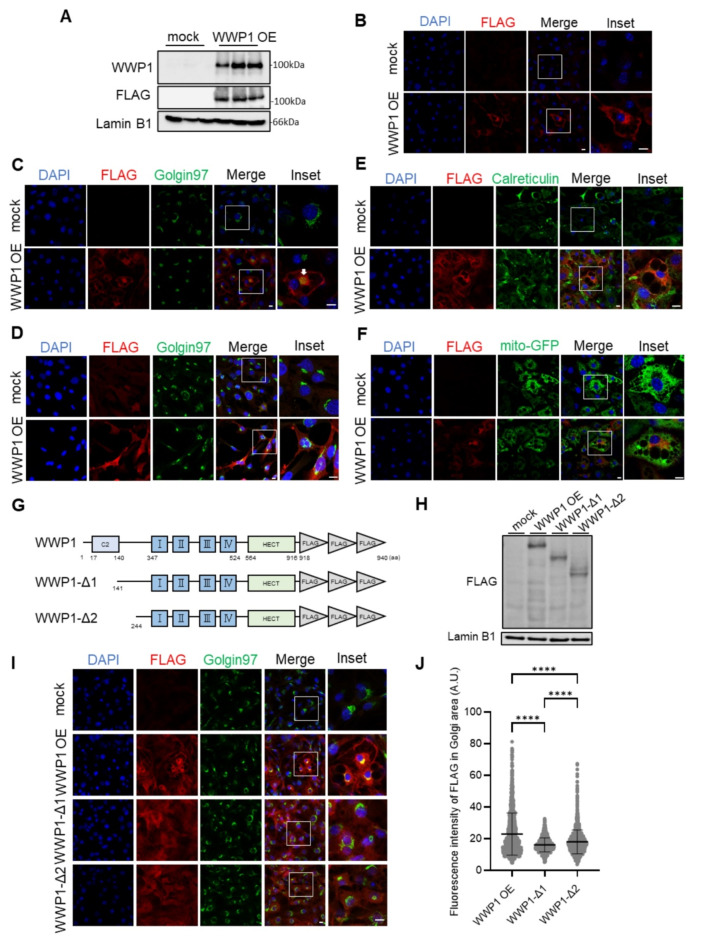
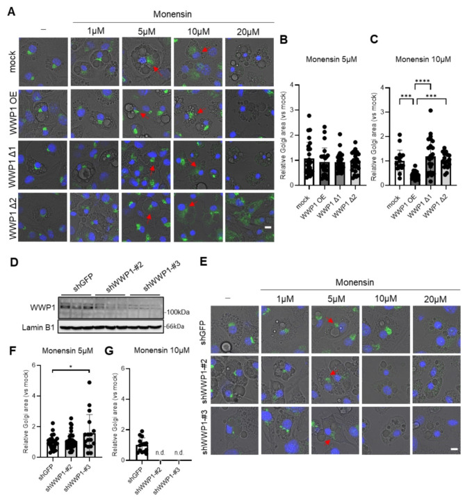
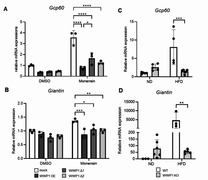
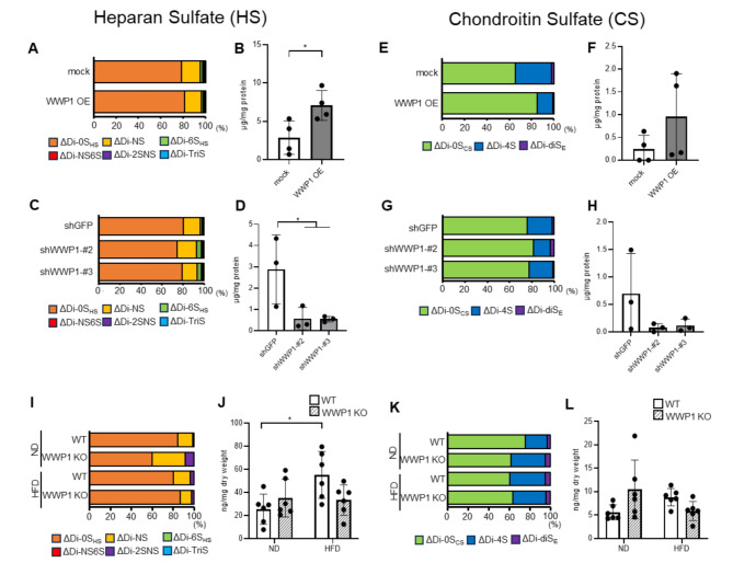
White adipocytes are a major component of white adipose tissue (WAT) and help to maintain systemic metabolic homeostasis by storing energy and secreting adipokines. In mice deficient in the protein WWP1 (WW domain-containing E3 ubiquitin protein ligase 1), oxidative stress in adipocytes increases but insulin resistance induced by obesity improves. However, the specific roles of WWP1 in adipocytes remain unclear. Here, we show that in 3T3L1 adipocytes, WWP1 localized in the Golgi apparatus via its C2 domain, where it protected the Golgi apparatus from monensin-induced disruption. By contrast, WWP1 knockdown by short hairpin RNA failed to protect the Golgi apparatus and enhanced Golgi apparatus disruption by monensin. The Golgi apparatus acts as a central organelle to establish accurate protein glycosylation of proteoglycans containing glycosaminoglycans, including chondroitin sulfate and heparan sulfate (HS). WWP1 overexpression increased chondroitin sulfate and HS levels, whereas WWP1 knockdown decreased them. Furthermore, obesity-related increases in HS were prevented by WWP1 knockout in adipose tissue. In summary, our results demonstrate a novel role for WWP1 in maintaining Golgi apparatus morphology and proteoglycan synthesis in adipocytes.

Keywords: Adipose tissue; Glycosaminoglycan; Golgi apparatus; WWP1.


Conflict of interest statement

Declarations. Competing interests: The authors declare no competing interests.

Figures

Fig. 1
Fig. 1
Subcellular localization of WWP1 in the Golgi apparatus in 3T3L1 adipocytes. (A) WWP1 protein abundance in WWP1 overexpressing (OE) 3T3L1 cells was analyzed by immunoblotting with an anti-WWP1 and FLAG antibody (n = 3). Lamin B1 was used as a loading control. (B) Confocal immunofluorescence images of the differentiated mock-transfected and WWP1 OE 3T3L1 adipocytes stained for FLAG (red) and DAPI (blue). (CD) Confocal immunofluorescence images of the differentiated (C) or undifferentiated (D) mock-transfected and WWP1 OE 3T3L1 adipocytes stained for DAPI (blue), FLAG (red), and Golgin97 (green). (E) Confocal immunofluorescence images of the differentiated mock-transfected and WWP1 OE 3T3L1 adipocytes stained for DAPI (blue), FLAG antibody (red), and calreticulin (green), indicating the endoplasmic reticulum. (F) Confocal immunofluorescence images of the differentiated mock-transfected and WWP1 OE 3T3L1 adipocytes with expression of mito-GFP (green), indicating the mitochondria, and stained for DAPI (blue) and FLAG antibody (red). (G) Schematic representation of mock-transfected, WWP1 overexpressing (OE), WWP1-Δ1, and WWP1-Δ2 protein structure. (H) Verification of expression of mock-transfected, WWP1 OE, WWP1-Δ1, and WWP1-Δ2 differentiated 3T3L1 adipocytes using a FLAG antibody analyzed by western blot. Lamin B was used as an internal control. (I) Representative confocal immunofluorescence images of the differentiated mock-transfected, WWP1 OE, WWP1-Δ1 and WWP1-Δ2 3T3L1 adipocytes stained for DAPI (blue), FLAG antibody (red), and Golgin97 (green). (J) The fluorescence intensity of WWP1 within the Golgi area was determined based on the overlap of FLAG and Golgin97 fluorescence. Each dot represents a single cell. N = 1356–1731 cells. Data are means ± SD. Differences between each value were analyzed by one-way ANOVA with Tukey’s multiple comparisons test (****: p < 0.0001). Scale bar is 10 μm.
Fig. 2
Fig. 2
WWP1 is required for maintaining the structure of the Golgi apparatus in differentiated 3T3L1 adipocytes. (A) Representative images of the differentiated mock-transfected, WWP1 OE, WWP1-Δ1, and WWP1-Δ2 3T3L1 cells stained for Golgin97 (green) and DAPI (blue). Cells were treated with each concentration of monensin for 6 h. Scale bar is 10 μm. (B–C) Relative Golgi size was determined based on Golgin97 staining. Each dot represents a single cell. N = 17–42 cells. Data are means ± SD. Differences between each value were analyzed by one-way ANOVA with Tukey’s multiple comparisons test (***: p < 0.001, ****: p < 0.0001). (D) WWP1 protein abundance in WWP1 knockdown (KD) 3T3L1 cells was analyzed by immunoblotting with anti-WWP1 (n = 3). Lamin B1 was used as a loading control. (E) Representative images of the differentiated mock-transfected and WWP1 KD differentiated 3T3L1 stained for Golgin97 (green) and DAPI (blue). Cells were treated with each concentration of monensin for 6 h. Scale bar is 10 μm. (F–G) Relative Golgi size was determined based on Golgin97 staining. Each dot represents a single cell. N = 17–42 cells. Data are means ± SD. Differences between each value were analyzed by one-way ANOVA with Tukey multiple comparisons test (*: p < 0.05).
Fig. 3
Fig. 3
WWP1 suppresses monensin-induced increases in Golgi stress markers. (A–B) The mRNA level of Gcp60 and Giantin in differentiated mock-transfected, WWP1 OE, WWP1-Δ1, and WWP1-Δ2 overexpressing (OE) 3T3L1 cells were treated with DMSO or 10 µM monensin for 6 h and analyzed by real-time PCR. Rps18 was used as a reference control. (C–D) The mRNA level of Gcp60 and Giantin in subcutaneous white adipose tissue (WAT) derived from normal diet (ND)-fed or high-fat diet (HFD)-fed Wwp1 KO mice of 13–15 weeks of age analyzed by real-time PCR. Rps18 was used as a reference control. Differences between each value were analyzed by one-way ANOVA with Tukey’s multiple comparisons test (*: p < 0.05, **: p < 0.01, ***: p < 0.001, ***: p < 0.0001).
Fig. 4
Fig. 4
WWP1 contributes to the synthesis of heparan sulfate (HS) and chondroitin sulfate (CS) in 3T3L1 adipocytes and white adipose tissue (WAT). (A, B) Effect of WWP1 overexpression (OE) on the disaccharide composition and amount of HS in differentiated 3T3L1 cells. (C, D) Effect of WWP1 knockdown (KD) on the disaccharide composition and amount of HS in differentiated 3T3L1 cells. (E, F) Effect of WWP1 OE on the disaccharide composition and amount of CS in differentiated 3T3L1 cells. (G, H) Effect of WWP1 KD on the disaccharide composition and amount of CS in differentiated 3T3L1 cells. (I, J) Disaccharide composition and levels of HS in subcutaneous WAT derived from normal diet (ND)-fed or high-fat diet (HFD)-fed Wwp1 KO mice of 13–15 weeks of age. (K, L) Disaccharide composition and levels of CS in subcutaneous WAT derived from ND-fed or HFD-fed Wwp1 KO mice of 13–15 weeks of age. Extraction of glycosaminoglycans and analysis of their unsaturated disaccharides was performed as described. Differences between each value were analyzed by one-way ANOVA with Tukey’s multiple comparisons test (*: p < 0.05, **: p < 0.01).

Similar articles

Cited by

References

    1. Ouchi, N., Parker, J. L., Lugus, J. J. & Walsh, K. Adipokines in inflammation and metabolic disease. Nat. Rev. Immunol.11, 85–97. 10.1038/nri2921 (2011). - PMC - PubMed
    1. Hotamisligil, G. S. Inflammation and metabolic disorders. Nature. 444, 860–867. 10.1038/nature05485 (2006). - PubMed
    1. Seo, S. R. et al. The novel E3 ubiquitin ligase Tiul1 associates with TGIF to target Smad2 for degradation. Embo j.23, 3780–3792. 10.1038/sj.emboj.7600398 (2004). - PMC - PubMed
    1. Zhi, X. & Chen, C. WWP1: A versatile ubiquitin E3 ligase in signaling and diseases. Cell. Mol. Life Sci.69, 1425–1434. 10.1007/s00018-011-0871-7 (2012). - PMC - PubMed
    1. Hu, X. et al. The emerging role of WWP1 in cancer development and progression. Cell. Death Discov.7, 163. 10.1038/s41420-021-00532-x (2021). - PMC - PubMed

LinkOut - more resources