. 2025 Nov;43(11):1813-1820.
doi: 10.1038/s41587-024-02470-2. Epub 2024 Nov 22.

Lipid nanoparticle-mediated mRNA delivery to CD34+ cells in rhesus monkeys

Affiliations

Lipid nanoparticle-mediated mRNA delivery to CD34+ cells in rhesus monkeys

Hyejin Kim et al. Nat Biotechnol. 2025 Nov.

Abstract

Transplantation of ex vivo engineered hematopoietic stem cells (HSCs) can lead to robust clinical responses but carries risks of adverse events from bone marrow mobilization, chemotherapy conditioning and other factors. HSCs have been modified in vivo using lipid nanoparticles (LNPs) decorated with targeting moieties, which increases manufacturing complexity. Here we screen 105 LNPs without targeting ligands for effective homing to the bone marrow in mouse. We report an LNP named LNP67 that delivers mRNA to murine HSCs in vivo, primary human HSCs ex vivo and CD34+ cells in rhesus monkeys (Macaca mulatta) in vivo at doses of 0.25 and 0.4 mg kg-1. Without mobilization and conditioning, LNP67 can mediate delivery of mRNA to HSCs and their progenitor cells (HSPCs), as well as to the liver in rhesus monkeys, without serum cytokine activation. These data support the hypothesis that in vivo delivery to HSCs and HSPCs in nonhuman primates is feasible without targeting ligands.


Conflict of interest statement

Competing interests: M.Z.C.H., H.N. and J.E.D. have filed a provisional patent related to this manuscript (US patent application number 63/632,354). J.E.D. is an advisor to GV, Readout, Edge Animal Health and Nava Therapeutics and P.J.S. is a cofounder of Tether Therapeutics. None of these companies provided any financial support for this work. The other authors declare no competing interests.

Figures
Figure 1.
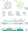
Figure 1.. Barcoded LNPs deliver mRNA to transcriptionally defined HSCs in vivo.
a, Chemical composition of the 128 formulated LNPs. Of these, 105 LNPs met quality control (QC) criteria. b, The hydrodynamic diameter and polydispersity of the LNP pool were contained within the range of the LNPs composing the pool. c, After injecting 105 LNPs, we isolated the lineage (Ter119, CD45, CD3e, ITGAM, and Gr-1)-negative (Lin) population from bone marrow and quantified LNP (barcode), functional delivery (aVHH protein), and the cell identity (transcriptome) in single cells, then overlaid the data. Functional delivery to liver endothelial (CD31+CD45) and immune cells (CD31CD45+) was also analyzed by DNA barcode sequencing. This figure was generated in part using graphics created by Biorender.com. d, Bone marrow cells were subdivided as MPP, multipotent progenitor; CMP, common myeloid progenitor; CLP, common lymphoid progenitor; MEP, megakaryocyte-erythrocyte progenitor; and GMP, granulocyte-monocyte progenitor. e, UMAP representation of 21,618 single cells within the bone marrow. f, Barcode counts and aVHH density distribution overlaid on the UMAP. g, Delivery mediated by the top 20 LNPs in aVHH+ cells in the transcriptionally defined Lin cell subtypes in bone marrow. Off-target delivery to liver endothelial cells (ECs) is also shown in gray.
Figure 2.
Figure 2.. LNP67 delivers mRNA to cells in mouse bone marrow.
a, Radar plot showing the percentage of aVHH-positive cells after PBS, or LNP67, LNP95, and LNP108, or LP01 LNP carrying aVHH mRNA were administered to C57BL/6J mice. LSK for bone marrow (BM), immune cells (CD45+CD31) for spleen (Sp), and endothelial cells (CD31+CD45) for the liver (Li), lung (Lu), heart (He), and kidney (Ki) are shown. Axis scales are 0% to 25% (BM, Ki, and He), 50% (Sp and Li), or 100% (Lu) toward corresponding directions. Data are presented as mean (black line) ± SD (gray) (N=3, biological replicates). b, LNP67 comprises an ionizable lipid, cholesterol, C14PEG2000, and DOTAP. c-e, Characteristics of LNP67. c, TEM image, d, hydrodynamic diameter, and e, zeta potential. f, TNS fluorescence intensity between pH 4 and 10 reveals the pKa of LNP67. g, Representative hematoxylin and eosin (H&E) images of liver from PBS- or LNP67-treated mice. N=3 (biological replicates) for both groups were analyzed, and the representative images are shown. h, Chemokines (CCL1, CCL2, CCL3, CCL4, CCL5, CCL11, CCL12, CCL17, CXCL1, CXCL2, CXCL9, CXCL10, CXCL11, CXCL12, CXCL13), cytokines (TNF-α, IL-6, IL-12p70, IL-1β, IFN-γ, IL-2, IL-17, IL-5, IL-16, TIMP-1, TREM-1, IL-23, IL-10, IL-1ra, IL-13, IL-4), colony-stimulating factors and growth factors (CSF and GF; G-CSF, GM-CSF, M-CSF, IL-3, IL-7), and soluble cell adhesion molecules (CD54 and complement component 5a) were analyzed in serum from PBS-, LNP67-, or lipopolysaccharide (LPS, 1 mg/kg)-treated mice at 6- or 24-hour timepoints. Data show mean fold-change compared to PBS group (N=3, biological replicates were pooled for measurement). Raw values are shown in Supplementary Fig. 12. i, The percentage of aVHH-positive cells in bone marrow cell subsets at 16 hours after administration of LNP67 at 0.5, 1.0, and 2.0 mg/kg. Data are presented as mean ± SD (N=3, biological replicates). **** p < 0.0001, analyzed by two-way ANOVA with Tukey post-hoc test for multiple comparisons.
Figure 3.
Figure 3.. LNP67 delivers mRNA to transcriptionally defined HSCs in vivo.
a, t-SNE clustering of 21 cell types in the bone marrow and their aVHH protein expression with single-cell resolution, overlaid on the t-SNE (36,142 cells). b, aVHH protein expression in for all 21 clusters. * p < 0.05, ** p < 0.01, and *** p < 0.001 analyzed by two-tailed unpaired student’s t-test (2 mg/kg LNP67 compared to PBS, N=3). c, aVHH protein, CD48 mRNA, and cKit (CD117) mRNA expression levels in LSK cells with or without CD48 expression.
Figure 4.
Figure 4.. LNP67 delivers mRNA to CD34+ cells in rhesus monkeys.
a, Hydrodynamic diameter (Dh) and polydispersity index (PD index) of LNP67 before (N=3, independent batches) and after freeze-thawing (N=2, independent batches). Data are presented as mean ± SD. ns = not significant, analyzed by two-tailed unpaired student’s t-test. b, aVHH expression did not show significant differences in mice treated with fresh LNP67 or freeze-thawed LNP67 in bone marrow. Data were analyzed by two-way ANOVA with Tukey post-hoc test for multiple comparisons. All data are presented as mean ± SD (N=3, biological replicates). c, aNLuc mRNA-carrying LNP67 was administered intravenously to young rhesus monkeys at 0.25 or 0.4 mg/kg. Bone marrow cells were immunoselected (CD34 and CD34+ fractions) and liver cell suspensions prepared. Luminescence intensities were evaluated after adding the luciferase substrate luciferin to address the dose response. This figure was generated in part using graphics created by Biorender.com. d-e, IVIS imaging of CD34 and CD34+ fractions from the bone marrow and liver cell suspension from untreated (N=1) or LNP67 (0.25 mg/kg, N=4; or 0.4 mg/kg, N=3)-treated rhesus. d, raw images after adding luciferin and e, the signal quantification. Data are presented as mean ± SEM. f-g, z-projection of Fourier light-field fluorescence microscopy images of f, rhesus bone marrow CD34+ and g, liver cell suspensions from animals administered LNP67 (0.4 mg/kg). The cells were stained with anti-NanoLuc and anti-CD34 (bone marrow, f) or anti-NanoLuc and anti-ASGPR (liver, g). h, 23 rhesus monkey serum cytokines were analyzed from untreated (N=1) or LNP67 (0.25 mg/kg, N=4; or 0.4 mg/kg, N=3)-treated rhesus collected at 24 hours post administration. Raw values and qualified assay ranges are shown in Supplementary Fig. 18.
Figure 5.
Figure 5.. LNP67 delivers mRNA to HSCs to primary human HSCs ex vivo.
aVHH expression from LNP67-treated primary human stem and progenitor cells. MLP, multilymphoid progenitor. * p < 0.05, ** p < 0.01, *** p < 0.001, **** p < 0.0001, and ns = not significant, compared to matched untreated group, analyzed by two-way ANOVA with Tukey post-hoc test. All data are presented as mean ± SEM (n=3).

References

    1. Polack FP et al. Safety and efficacy of the BNT162b2 mRNA Covid-19 vaccine. N. Engl. J. Med. 383, 2603–2615 (2020). - PMC - PubMed
    1. Baden LR et al. Efficacy and safety of the mRNA-1273 SARS-CoV-2 vaccine. N. Engl. J. Med. 384, 403–416 (2020). - PMC - PubMed
    1. Adams D et al. Patisiran, an RNAi therapeutic, for hereditary transthyretin amyloidosis. N. Engl. J. Med. 379, 11–21 (2018). - PubMed
    1. Gillmore JD et al. CRISPR-Cas9 in vivo gene editing for transthyretin amyloidosis. N. Engl. J. Med. 385, 493–502 (2021). - PubMed
    1. Verve Therapeutics doses first human with an investigational in vivo base editing medicine, VERVE-101, as a potential treatment for heterozygous familial hypercholesterolemia. https://ir.vervetx.com/news-releases/news-release-details/verve-therapeu.... (2022).

Methods-only references

    1. Lindsay KE et al. Aerosol delivery of synthetic mRNA to vaginal mucosa leads to durable expression of broadly neutralizing antibodies against HIV. Mol. Ther. 28, 805–819 (2020). - PMC - PubMed
    1. Sago CD et al. High-throughput in vivo screen of functional mRNA delivery identifies nanoparticles for endothelial cell gene editing. Proc. Natl. Acad. Sci. 115, E9944–E9952 (2018). - PMC - PubMed
    1. Chen D et al. Rapid discovery of potent siRNA-containing lipid nanoparticles enabled by controlled microfluidic formulation. J. Am. Chem. Soc. 134, 6948–6951 (2012). - PubMed
    1. Belliveau NM et al. Microfluidic synthesis of highly potent limit-size lipid nanoparticles for in vivo delivery of siRNA. Mol. Ther. Nucleic Acids 1, e37 (2012). - PMC - PubMed
    1. Mastronarde DN Automated electron microscope tomography using robust prediction of specimen movements. J. Struct. Biol. 152, 36–51 (2005). - PubMed

LinkOut - more resources