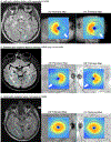
Homonymous hemi-macular atrophy in multiple sclerosis
- PMID: 39579046
- PMCID: PMC11617262
- DOI: 10.1177/13524585241297816
Homonymous hemi-macular atrophy in multiple sclerosis
Abstract
Background: Retrograde trans-synaptic degeneration (TSD) following retro-chiasmal pathology, typically retro-geniculate in multiple sclerosis (MS), may manifest as homonymous hemi-macular atrophy (HHMA) of the ganglion cell/inner plexiform layer (GCIPL).
Objective: To determine the frequency, association with clinical outcomes, and retinal and radiological features of HHMA in people with MS (PwMS).
Methods: In this cross-sectional study, healthy controls (HC) and PwMS underwent retinal optical coherence tomography scanning. For quantitative identification of HHMA, a normalized asymmetry ratio was used, and its normative cutoffs were established from the HC. HHMA PwMS were propensity score matched 1:2 to non-HHMA PwMS. Mixed-effects linear regression models were used in analyses.
Results: Based on normative data from 238 HC (466 eyes), 79 out of 942 PwMS exhibited HHMA (8.4%; 143 eyes). Compared to non-HHMA eyes from matched PwMS (158 PwMS; 308 eyes), HHMA eyes had lower average GCIPL (diff: -5.7 μm (95% CI -7.6 to -3.8); p < 0.001) but also inner nuclear layer (diff: -0.9 μm (95% CI -1.6 to -0.1); p = 0.02), and outer nuclear layer (diff: -1.9 μm (95% CI -3.4 to -0.4); p = 0.02) thicknesses; in further analyses, these differences were exclusive to the homonymous side of HHMA. HHMA participants also exhibited higher expanded disability status scale scores (diff: 0.5 (95% CI 0.1 to 0.9); p = 0.02), worse 100% and 2.5% visual acuity scores (diff: -3.2 (95% CI -4.1 to -1.0); p = 0.002, -5.4 (95% CI -7.5 to -3.5); p < 0.001), and higher frequency of microcystoid macular changes (10.1% vs. 3.2%; p = 0.03) compared to non-HHMA participants.
Conclusions: HHMA, possibly as a marker of TSD, may signify higher disability in MS.
Keywords: Trans-synaptic degeneration; ganglion cell/inner plexiform layer; homonymous hemi-macular atrophy; microcystoid macular pathology; multiple sclerosis; neurodegeneration; optical coherence tomography.
Conflict of interest statement
Declaration of Conflicting InterestsThe author(s) declared the following potential conflicts of interest with respect to the research, authorship, and/or publication of this article: OCM receives funding from the Race to Erase MS Foundation.KCF reports research funding from NIH, NMSS, and the DoD. She has served on Data and Safety Monitoring Boards for trials funded by the NIH and NMSS, and has received honorarium for serving as an external thesis committee reviewer.SDN reports grants or contracts from Biogen, Roche, Genentech, National Multiple Sclerosis Society, Department of Defense, and Patient Centered Outcomes Research Institute; personal compensation for consulting from Biogen, Roche, Genentech, Bristol Myers Squibb, EMD Serono, Greenwich Biosciences, Novartis, and Horizon Therapeutics; and participation on a Data Safety Monitoring Board or Advisory Board for MedDay Pharmaceuticals. SDN also reports support from the Stiff Person Syndrome Research Foundation.ESS reports scientific advisory boards and/or consulting for Alexion, Viela Bio, Horizon Therapeutics, Genentech, and Ad Scientiam; speaking honoraria from Alexion, Viela Bio, and Biogen.BN has received research funding from the NMSS, DoD, NIH, PCORI, and Genentech.JP is a founder of Sonovex, Inc. and serves on its Board of Directors, and has received consulting fees from JuneBrain LLC and is PI on research grants to Johns Hopkins from Genentech and Biogen.SS has received consulting fees from Medical Logix for the development of CME programs in neurology and has served on scientific advisory boards for Biogen, Genzyme, Genentech Corporation, EMD Serono, and Celgene. He is the PI of investigator-initiated studies funded by Genentech and Biogen, was the site investigator of a trial sponsored by MedDay Pharmaceuticals and received support from the Race to Erase MS foundation. He has consulted for Carl Zeiss Meditec, and has received equity compensation for consulting from JuneBrain LLC, a retinal imaging device developer.PAC has received consulting fees from Disarm, Nervgen, and Biogen and is Principal Investigator on grants to JHU from Biogen, Genentech, Prinicpia, and Annexon.
Figures


References
MeSH terms
Grants and funding
LinkOut - more resources
Full Text Sources
Medical

