. 2024 Nov 23;148(1):68.
doi: 10.1007/s00401-024-02836-5.

Genome-wide methylation profiling differentiates benign from aggressive and metastatic pituitary neuroendocrine tumors

Affiliations

Genome-wide methylation profiling differentiates benign from aggressive and metastatic pituitary neuroendocrine tumors

Jelena Jotanovic et al. Acta Neuropathol. .

Erratum in

  • Correction: Genome‑wide methylation profiling differentiates benign from aggressive and metastatic pituitary neuroendocrine tumors.
    Jotanovic J, Boldt HB, Burton M, Andersen MS, Bengtsson D, Bontell TO, Ekman B, Engström BE, Feldt-Rasmussen U, Heck A, Jakovcevic A, Jørgensen JOL, Kraljevic I, Kunicki J, Lindsay JR, Losa M, Loughrey PB, Maiter D, Maksymowicz M, Manojlovic-Gacic E, Pahnke J, Petersenn S, Petersson M, Popovic V, Ragnarsson O, Rasmussen ÅK, Reisz Z, Saeger W, Schalin-Jäntti C, Scheie D, Terreni MR, Tynninen O, Whitelaw B, Burman P, Casar-Borota O. Jotanovic J, et al. Acta Neuropathol. 2025 Jan 2;149(1):4. doi: 10.1007/s00401-024-02838-3. Acta Neuropathol. 2025. PMID: 39747763 Free PMC article. No abstract available.

Abstract

Aggressive pituitary neuroendocrine tumors (PitNETs)/adenomas are characterized by progressive growth despite surgery and all standard medical therapies and radiotherapy. A subset will metastasize to the brain and/or distant locations and are termed metastatic PitNETs (pituitary carcinomas). Studies of potential prognostic markers have been limited due to the rarity of these tumors. A few recurrent somatic mutations have been identified, and epigenetic alterations and chromosomal rearrangements have not been explored in larger cohorts of aggressive and metastatic PitNETs. In this study, we performed genome-wide methylation analysis, including copy-number variation (CNV) calculations, on tumor tissue specimens from a large international cohort of 64 patients with aggressive (48) and metastatic (16) pituitary tumors. Twelve patients with non-invasive pituitary tumors (Knosp 0-2) exhibiting an indolent course over a 5 year follow-up served as controls. In an unsupervised hierarchical cluster analysis, aggressive/metastatic PitNETs clustered separately from benign pituitary tumors, and, when only specimens from the first surgery were analyzed, three separate clusters were identified: aggressive, metastatic, and benign PitNETs. Numerous CNV events affecting chromosomal arms and whole chromosomes were frequent in aggressive and metastatic, whereas benign tumors had normal chromosomal copy numbers with only few alterations. Genome-wide methylation analysis revealed different CNV profiles and a clear separation between aggressive/metastatic and benign pituitary tumors, potentially providing biomarkers for identification of these tumors with a worse prognosis at the time of first surgery. The data may refine follow-up routines and contribute to the timely introduction of adjuvant therapy in patients harboring, or at risk of developing, aggressive or metastatic pituitary tumors.

Keywords: Aggressive pituitary tumor; Methylation analysis; Pituitary adenoma; Pituitary carcinoma; Pituitary neuroendocrine tumor.


Figures

Fig. 1
Fig. 1
Chart showing the structure of the APT/PC cohort and the benign tumors
Fig. 2
Fig. 2
Heatmap showing the degree of methylation (beta value) of the top 5000 most variable CpG sites and the associated hierarchical clustering of specimens from the entire cohort (n = 81) (a) and the subset of first surgery specimens (n = 50) (b). Blue and yellow shadings indicate hypomethylation and hypermethylation, respectively. The annotation bar shows the tumor type (benign—blue, APT—orange, and PC—red), transcription factors, and functional status of the tumors. Paired samples are marked with the connecting arrows. Unsupervised principal component analysis (PCA) illustrates clustering of the c) specimens from the entire cohort (n = 81) and d) the subset of first surgery specimens (n = 50) based on the CpG sites with the top 5,000 variable β values. The blue diamonds, orange triangles, and red boxes represent the benign, APT, and PC specimens, respectively. PC1, PC2, and PC3 are the axes of the 1st, 2nd, and 3rd principal components (PCs), respectively. In the PCA of the entire cohort, the first three PCs explain 20.2%, 9.2%, and 4.8% of the total variance, while in the PCA of the first surgery specimens, the 1st, 2nd, and 3rd PCs explained 38.6%, 8.4% and 6.8% of the variance, respectively
Fig. 3
Fig. 3
Volcano plots showing differential methylation analysis results in APT/PCs vs. benign PitNETs. The X-axis shows the ∆β-value (difference in beta value), and the Y-axis shows the associated –log10 p values. CpGs with ∆β ≤ − 0.2 or ∆β ≥ 0.2 are shown in blue (hypomethylated), red (hypermethylated), and gray (not deviating from the threshold), respectively. The dotted line shows the global significance threshold (p = 1.3 × 10–7). a All specimens comparing APT/PC (n = 69) vs. benign tumors (n = 12); b all specimens comparing PC (n = 18) vs APT (n = 51); c the first surgery specimens comparing APT/PC (n = 38) vs. benign tumors (n = 12); and d the first surgery specimens comparing PC (n = 7) vs. APT (n = 31)
Fig. 4
Fig. 4
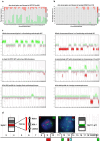
Cumulative CNV profiles showing abundant chromosomal alterations with a number of arm-level gains (green) and losses (red) in APT/PCs (a) in comparison to benign PitNETs, which were dominated by balanced chromosomes (gray) (b). The distribution of CNV alterations on chromosomal arms is shown on the X-axis, while the percentage of tumors with CNV alterations is shown on the Y-axis. c Examples showing a variation of distinct CNV profiles between APT/PCs and benign PitNETs. Chromosomes with a normal copy number (n = 2) exhibit an even distribution of data points around the calculated baseline, while gains (n > 2) and losses (n < 2) are depicted in green and red, respectively. Full vertical lines separate individual chromosomes, while stippled lines indicate the separation of the p and q arms. Chromosome numbers are shown on the X-axis of each CNV. d) Diagram showing the positions of FISH probes targeting chromosomes 9, 12, 13, and 22 with detailed information on probe locations for 9q arm and 12 centromere. Left FISH picture insert shows tumor cell with tetraploidy of chromosome 9 in red and normal ploidy of chromosome 22 in green, while right FISH picture insert shows tetraploidy of chromosome 12 in green and normal ploidy of chromosome 13 in red/cyan, respectively
Fig. 5
Fig. 5
Heatmap showing unsupervised CNV clustering in specimens from the entire cohort. Losses, gains, and balanced CNVs are shown in blue, yellow, and gray, respectively. Chromosome regions are ordered by number and displayed in rows, while the specimens are organized by hierarchical clustering and presented in columns. The annotations of the specimens with respect to tumor group (APT/PC/benign), transcription factor, and functional status are shown in the bars above the heatmap. Paired samples are marked with connecting arrows

References

    1. Andonegui-Elguera S, Silva-Román G, Peña-Martínez E, Taniguchi-Ponciano K, Vela-Patiño S, Remba-Shapiro I et al (2022) The genomic landscape of corticotroph tumors: from silent adenomas to ACTH-secreting carcinomas. Int J Mol Sci 23:4861. 10.3390/ijms23094861 - PMC - PubMed
    1. Bi WL, Greenwald NF, Ramkissoon SH, Abedalthagafi M, Coy SM, Ligon KL et al (2017) Clinical identification of oncogenic drivers and copy-number alterations in pituitary tumors. Endocrinology 158:2284–2291. 10.1210/en.2016-1967 - PMC - PubMed
    1. Bi WL, Horowitz P, Greenwald NF, Abedalthagafi M, Agarwalla PK, Gibson WJ et al (2017) Landscape of genomic alterations in pituitary adenomas. Clin Cancer Res 23:1841–1851. 10.1158/1078-0432.CCR-16-0790 - PMC - PubMed
    1. Burman P, Casar-Borota O, Perez-Rivas LG, Dekkers OM (2023) Aggressive pituitary tumors and pituitary carcinomas: from pathology to treatment. J Clin Endocrinol Metab 108:1585–1601. 10.1210/clinem/dgad098 - PMC - PubMed
    1. Caimari F, Korbonits M (2016) Novel genetic causes of pituitary adenomas. Clin Cancer Res 22:5030–5042. 10.1158/1078-0432.CCR-16-0452 - PubMed

Publication types

LinkOut - more resources