. 2024 Nov 23;24(1):389.
doi: 10.1186/s12935-024-03569-x.

Enhancing antitumor immunity in Lewis lung cancer through plasma-treated medium-induced activation of dendritic cells

Affiliations

Enhancing antitumor immunity in Lewis lung cancer through plasma-treated medium-induced activation of dendritic cells

Chae Bok Lee et al. Cancer Cell Int. .

Abstract

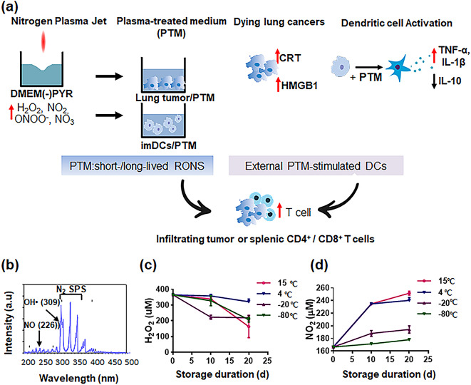
Background: Recently, atmospheric non-thermal plasma jet-treated medium (PTM) has been recognized as a novel strategy in cancer therapy and lymphocyte activation. However, PTM has limitations in inducing a robust antitumor-immune response. This study demonstrated that PTM treatment inhibited tumor progression by activating dendritic cells (DCs).

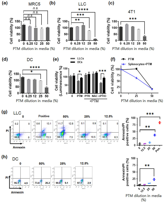
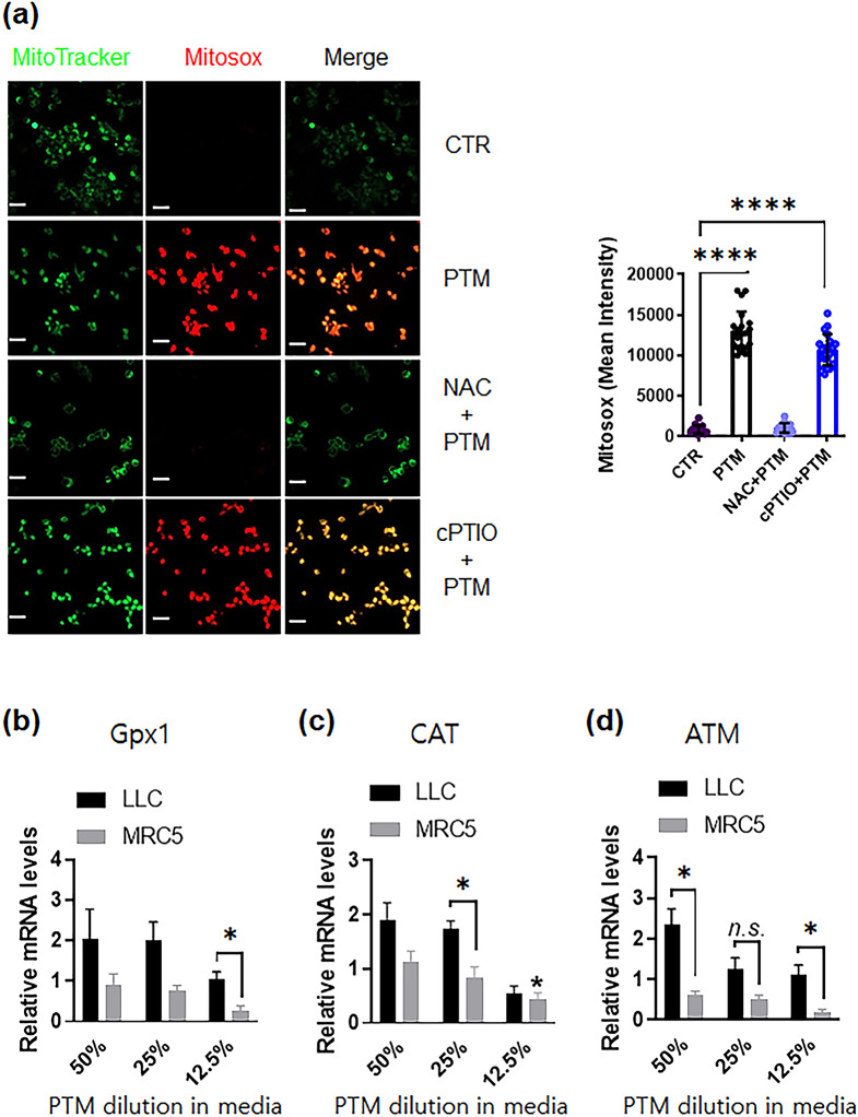
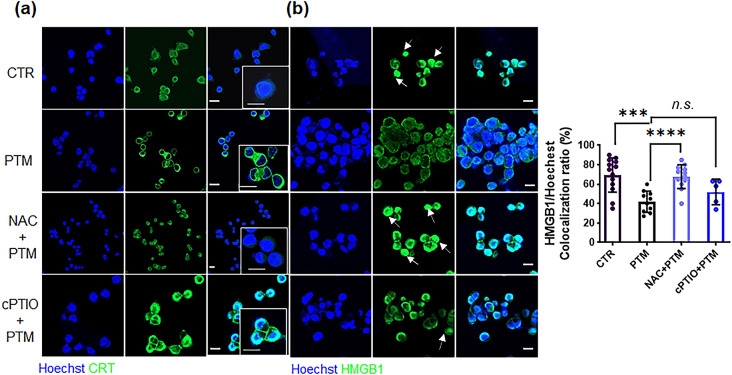
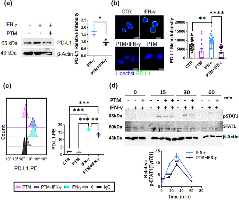
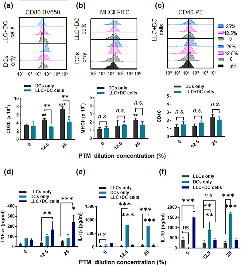
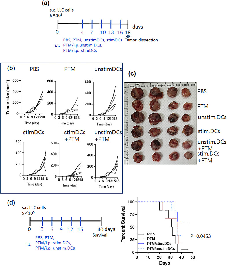
Method: In this study, we investigated the effects of PTM on selective cytotoxicity and intracellular reactive oxygen species (ROS) generation and oxidative stress-mediated signaling (e.g., glutathione peroxidase, catalase) using respective fluorescence probes in Lewis lung cancer (LLC) cells. Then, the PTM affects the expression of interferon-gamma (IFN)-γ-induced programmed death-ligand 1 (PD-L1) and inhibition of signal transducer and activator of transcription 1 (STAT1) in LLC cells using immunoblotting. Additionally, PTM effects on the tumor cell's death and activation of DCs were done by co-culturing DCs with or without tumor cells. Further, a mouse model was used to evaluate the synergistic antitumor effects of PTM and DCs where tumors are grown under the skin.

Results: PTM-exposed tumor cells increase intracellular superoxide production, enhancing ROS generation and leading to cancer immunogenic cell death. In addition, PTM suppresses IFN-γ-induced PD-L1 expression and STAT1 activation in tumor cells. The activation of DCs induced by PTM is downregulated when these cells are co-cultured with tumor cells. In vivo, intraperitoneal injection of PTM-activated DCs, as a synergistic agent to intertumoral PTM treatment, led to increased CD4+ and CD8+ T cell infiltration into the tumor and spleen and eventually decreased tumor growth.

Conclusion: Overall, this research introduces a promising avenue for improving lung cancer treatment using PTM to stimulate an immune response and induce cell death in tumor cells. Further studies will be essential to validate these findings and explore clinical applications.

Keywords: Cancer immunogenic cell death; Dendritic cell activation; Lewis Lung cancer cells; Non-thermal plasma-treated medium; Programmed death-ligand 1; Reactive oxygen species.


Conflict of interest statement

Declarations. Ethics approval and consent to participate: The Institutional Research and Ethics Committee of Chungnam National University approved all animal experiments (approval number: 202003 A-CNU-064), which complied with the relevant guidelines of the Korean Food and Drug Administration. Competing interests: The authors declare no competing interests.

Figures

Fig. 1
Fig. 1
PTM-stimulated DCs combined with PTM enhance antigen-specific T cells in cancer immune responses. (a) A scheme to increase the number of tumor-infiltrating lymphocytes involves the activation of extracellular DCs while concurrently introducing plasma-treated medium (PTM) treatment to inhibit tumor progression and activate immune T cells. (b) Optical emission spectrum (OES) of a non-thermal plasma jet with nitrogen at 200–500 nm. (c-d) H2O2 and NO2 concentrations corresponding to PTM stored at 15 ℃, 4 ℃, − 20 ℃, and − 80 ℃ for 10 or 20 d, determined using Amplex™ Red Reagent and Griess reagent assay, respectively. Results presented as the mean ± SEM of three replicates. These experiments were analyzed using a two-tailed unpaired Student’s t-test. *p < 0.01 (15 ℃), 20 d vs. PTM immediately following the preparation (0 d); **p < 0.01 (4 ℃ or 20 ℃), 10 or 20 d vs. 0 d. **p < 0.01 (− 80 ℃), 20 d vs. 0 d
Fig. 2
Fig. 2
Plasma-treated medium (PTM) induces a cytotoxic effect and alterations in LLC cells. (a) Lung normal MRC5 cells, (b-c) tumor cells (LLC or 4T1 cells), and (d) primary cells (DC) were incubated with various concentrations of PTM (6.25, 12.5, 25, and 50%) for 24 h. Cell viability was determined using CCK-8 assays. This experiment was repeated thrice. Statistical significance was analyzed via one-way ANOVA; *p < 0.05, **p < 0.01, ***p < 0.001, and ****p < 0.0001 compared to the control. n.s., not significant. (e) On day 1, intracellular ATP in LLC or DCs were assayed using luciferase-based viability following treatment with 25% PTM alone or pre-incubation with 2 mM PYR, 10 mM NAC, and 0.1 mM cPTIO for 1 h before PTM treatment. The mean ± SEM represents data; *p < 0.05 and ***p < 0.001; two-way analysis of variance (ANOVA). (f) LLCs were incubated alone (25% or 50% PTM) or co-cultured with splenocytes (1 × 105/well of 96-well plates) in the presence of PTM. Cell viability was assessed using CCK-8 assays. (g-h) LLC and DC cell death incubated in various PTM was evaluated via Annexin/PI staining and flow cytometry, respectively. The positive sample inactivates heated at 100℃ for 10 min. Results are represented by the means ± SD from three independent experiments. Significant differences were determined via a two-way ANOVA; **p < 0.01 and ***p < 0.001 compared to the control
Fig. 3
Fig. 3
Plasma-treated medium detoxifies antioxidant enzymes in LLC or MRC5 cells. (a) (Left) Representative confocal microscopy images indicate MitoTracker (green) and Mitosox (red) staining of LLCs incubated in different conditions: the control, 25% PTM, and 25% PTM pre-incubated with 10 mM NAC or 0.1 mM cPTIO for 5 h; scale bar, 10 µM. (Right) The intensity of red fluorescence was quantified using Zen blue software; n = 20, 25, 19, and 27 single cells for control, PTM alone, and PTM incubation with pre-incubated NAC or cPTIO, respectively. The mean ± SED represents results from three independent experiments. Unpaired Student’s t-test was used to calculate statistical significance; ****p < 0.0001. (b-d) PTM stimulates anticancer signals in LLCs or normal lung MRC5 cells. The mRNA expression of Gpx1 (glutathione peroxidase), CAT (catalase), and ATM gene was quantified by qRT-PCR. Fold changes of the three target genes were normalized using the 18 S mRNA or GAPDH control. n = 3, mean ± sem. *p < 0.05, n.s., not significant
Fig. 4
Fig. 4
PTM increases CRT exposure and cytoplasmic translocation of HMGB1 from the nucleus of LLCs. (a) Surface-exposed CRT (green) and (b) translocated HMGB1 (green) in LLCs treated with medium (CTR), 25% PTM, pretreated 10 mM NAC, or 0.1 mM 2-(4-carboxyphenyl)-4,4,5,5-tetramethylimidazoline-1-oxyl-3-oxide(cPTIO) and subsequently with 25% PTM for 12 h. Confocal images indicate stained nuclei (blue), and CRT or HMGB1 (green); scale bar, 10 μm. Arrows indicate increased fluorescence intensity of HMGB1 in the nuclei. Quantitative co-localization of HMGB1 expressed as nuclear (Hoechst) (n) per cell. HMGB1 staining intensity was analyzed using Zen blue lite. Data are represented by the mean ± standard error of the difference between two means (SED) and calculated using a two-tailed unpaired Student’s t-test. This experiment was repeated thrice; ***p < 0.001, and ****p < 0.0001.n.s., not significant
Fig. 5
Fig. 5
PTM inhibits IFN-γ-induced PD-L1 expression and attenuates STAT1 phosphorylation in LLCs. (a-c) LLCs were incubated with medium, and pretreated-PTM (12.5%) for 4 h and subsequently treated with IFN-γ (10 ng/mL) for 24 h. A positive sample was used for IFN-γ treatment (10 ng/mL). (a) PD-L1 expression was detected via western blot. The PD-L1 (relative to β-actin) expression was calculated using Image J. Statistical significances were estimated using a two-way analysis of variance (ANOVA); mean ± SEM; n = 3, *p < 0.05. (b) Confocal images indicate stained nuclei (blue) and mouse PD-L1 (green) being merged; scale bar, 10 µM. Mean fluorescence intensities were subsequently quantified using Zen blue; n = 20, 18, 20, and 18 single cells were used for control, PTM alone, IFN-γ, and PTM/IFN-γ, respectively; **p < 0.01, and ****p < 0.0001. n.s., not significant. (c) Flow cytometric analysis of PD-L1-PE was observed using Flowzo (n = 3). The graph indicates PD-L1 levels (clone 9A11) normalized by the lgG control. Statistical significance was determined via one-way ANOVA. Data are represented by the mean ± SEM of four representative experiments: **p < 0.01, and ***p < 0.001. (d) PTM markedly reduced IFN-γ-induced STAT1 phosphorylation at the indicated times. Cells were incubated with IFN-γ alone (positive control), and 12.5% PTM for 4 h, and subsequently treated with IFN-γ (10 ng/mL) at the indicated times; “–“, before indicated times (0, 15, 30, 60 min) indicates negative control. STAT1 phosphorylation (pSTAT1) at Tyr701, as well as at total STAT1 (relative to β-actin), was detected using western blot analysis. Line charts illustrate quantification data about pSTAT1 levels at indicated time points compared to levels induced by IFN-γ stimulation from the positive control group. Statistical differences between IFN-γ and IFN-γ/PTM were determined via a two-way ANOVA. Data are represented by the mean ± SEM of three independent experiments: ***p < 0.001 (15 min) and **p < 0.01 (30 min)
Fig. 6
Fig. 6
Plasma-treated medium (PTM) stimulates dendritic cells (DCs) alone and in a co-culture system. DCs were incubated alone (12.5 or 25% PTM) or co-cultured with Lewis lung cancer (LLC) cells in the presence of PTM. The DCs were cultured either alone or separated by a transwell system. After 24 h, surface marker expression was determined. (a-c). Representative histograms and bar plots demonstrate CD80-BV650, MHCII-FITC, and CD40-PE levels in the DCs only or in DCs with the co-culture, using flow cytometry, respectively. The data were analyzed and gated using FlowJo. The statistical significance of the difference between DC-alone and tumor cell co-culture groups was determined via a two-way ANOVA; *p < 0.05 and **p < 0.01; n.s., insignificant. Concurrently, data compared to the untreated cells using a two-tailed unpaired Student’s t-test are shown; *p < 0.05 and **p < 0.01, mean ± SEM; n = 4–5. (d-f) The TNF-α, IL-1β, and IL-10 levels in the supernatants from LLC cells, DCs cultured alone, DCs incubated with tumor cells, and both treated with 12.5% or 25% PTM, were measured. Comparison between LLCs, DCs, and LLC + DC cells using a two-way ANOVA; **p < 0.01, and ***p < 0.001. n.s., not significant. Statistical significance was determined using a two-tailed unpaired Student’s t-test; *p < 0.05, **p < 0.01, and ***p < 0.001; n = 4
Fig. 7
Fig. 7
PTM combined with PTM-stimulated DCs decreases tumor size and increases immune responses. (a) The treatment schedule for a mouse tumor model injected with Lewis lung cancer (LLC) cells (n = 4 per group). Unstimulated DCs or 6.25% stimulated DCs were injected intraperitoneally at 3-d intervals after LLC tumor formation. (b) Tumor growth curves. Each line indicates tumor growth in an individual mouse. n = 2. (c) On day 18, photographs of isolated tumors in the six groups were taken. (d) Mouse survival throughout the predetermined study period of 40 d was assessed, and the survival curves were plotted using the Log-rank with Mantel-Cox test (6 mice per group); *p = 0.0453. n = 2

Similar articles

Cited by

References

    1. Wu F, Fan J, He Y, Xiong A, Yu J, Li Y, et al. Single-cell profiling of tumor heterogeneity and the microenvironment in advanced non-small cell lung cancer. Nat Commun. 2021;12:2540. - PMC - PubMed
    1. Chen Z, Fillmore CM, Hammerman PS, Kim CF, Wong K-K. Non-small-cell lung cancers: a heterogeneous set of diseases. Nat Rev Cancer. 2014;14:535–46. - PMC - PubMed
    1. Domagala-Kulawik J. The role of the immune system in non-small cell lung carcinoma and potential for therapeutic intervention. Translational lung cancer Res. 2015;4(2):177. - PMC - PubMed
    1. Katopodi T, Petanidis S, Charalampidis C, Chatziprodromidou I, Eskitzis P, Tsavlis D, et al. Tumor-infiltrating dendritic cells: decisive roles in cancer immunosurveillance, immunoediting, and tumor T cell tolerance. Cells. 2022;11:3183. - PMC - PubMed
    1. Gardner A, Ruffell B. Dendritic cells and cancer immunity. Trends Immunol. 2016;37(12):855–65. - PMC - PubMed

LinkOut - more resources