. 2024 Nov 23;22(1):563.
doi: 10.1186/s12964-024-01931-0.

The cell adhesion molecule CD44 acts as a modulator of 5-HT7 receptor functions

Affiliations

The cell adhesion molecule CD44 acts as a modulator of 5-HT7 receptor functions

Saskia Borsdorf et al. Cell Commun Signal. .

Abstract

Background: Homo- and heteromerization of G protein-coupled receptors (GPCRs) plays an important role in the regulation of receptor functions. Recently, we demonstrated an interaction between the serotonin receptor 7 (5-HT7R), a class A GPCR, and the cell adhesion molecule CD44. However, the functional consequences of this interaction on 5-HT7R-mediated signaling remained enigmatic.

Methods: Using a quantitative FRET (Förster resonance energy transfer) approach, we determined the affinities for the formation of homo- and heteromeric complexes of 5-HT7R and CD44. The impact of heteromerization on 5-HT7R-mediated cAMP signaling was assessed using a cAMP responsive luciferase assay and a FRET-based cAMP biosensor under basal conditions as well as upon pharmacological modulation of the 5-HT7R and/or CD44 with specific ligands. We also investigated receptor-mediated G protein activation using BRET (bioluminescence resonance energy transfer)-based biosensors in both, homo- and heteromeric conditions. Finally, we analyzed expression profiles for 5-HT7R and CD44 in the brain during development.

Results: We found that homo- and heteromerization of the 5-HT7R and CD44 occur at similar extent. Functionally, heteromerization increased 5-HT7R-mediated cAMP production under basal conditions. In contrast, agonist-mediated cAMP production was decreased in the presence of CD44. Mechanistically, this might be explained by increased Gαs and decreased GαoB activation by 5-HT7R/CD44 heteromers. Unexpectedly, treatment of the heteromeric complex with the CD44 ligand hyaluronic acid boosted constitutive 5-HT7R-mediated cAMP signaling and receptor-mediated transcription, suggesting the existence of a transactivation mechanism.

Conclusions: Interaction with the hyaluronan receptor CD44 modulates both the constitutive activity of 5-HT7R as well as its agonist-mediated signaling. Heteromerization also results in the transactivation of 5-HT7R-mediated signaling via CD44 ligand.

Keywords: Bioluminescence Resonance Energy Transfer (BRET); Fluorescence Resonance Energy Transfer (FRET); G protein-coupled receptor (GPCR); Hyaluronan receptor CD44; Receptor oligomerization; Serotonin receptor 7 (5-HT7R).


Conflict of interest statement

Declarations. Ethics approval and consent to participate: Not applicable. Consent for publication: Not applicable. Competing interests: The authors declare no competing interests.

Figures

Fig. 1
Fig. 1
5-HT7R and CD44 form homo- and heteromeric complexes with similar probabilities. A Schematic representation of the experimental design. B-D Representative images of N1E-115 cells co-expressing 5-HT7R-eCFP/5-HT7R-eYFP (B, upper panel) or CD44-eCFP/CD44-eYFP (B, lower panel), CD44-eCFP/5-HT7R-eYFP (C) or CD86-eCFP/CD86-eYFP (D) depicted in eCFP: green, eYFP: red, merge: yellow. The right images show apparent FRET efficiencies (EfD) between eCFP and eYFP fluorophores. Scale bar: 20 µm. E Apparent FRET efficiencies (EfD) for 5-HT7R- and CD44-homomers, 5-HT7R/CD44-heteromers, and CD86 monomers. Data are means ± SEM (N = 5 experiment days, 41–45 cells). Statistical significance was assessed by one-way ANOVA with post hoc Tukey’s multiple comparison test (E). * p < 0.05, ** p < 0.01, *** p < 0.001. F Dimerization model of 5-HT7R and CD44. Relative dissociation constants K were computed from lux-FRET measurements (see Supp. Fig. S1 C-E) using a dynamic oligomerization model [7]
Fig. 2
Fig. 2
Constitutive 5-HT7R activity is regulated by its heteromerization with CD44. A Schematic presentation of the FRET-based cAMP biosensor that consists of EPAC1 (exchange factor directly activated by cAMP 1) connected to the fluorophores Citrine (acceptor) and mCerulean (donor). An increase in intracellular cAMP correlates with a decrease in the FRET efficiency and the fluorescence ratio of Citrine/mCerulean. B, C Representative images (B) and quantification (C) of the baseline cAMP levels as measured with the cAMP biosensor in N1E-115 cells transiently expressing 5-HT7R-eGFP/5-HT7R-mCherry or 5-HT7R-eGFP/CD44-mCherry without (Ctrl.) and with pre-blocking of the 5-HT7R with the inverse agonist SB-269970 (100 nM). Data are normalized to values obtained for 5-HT7R/5-HT7R. Data are means ± SEM (N = 21 experiment days, ≥ 61 coverslips, ≥ 1502 cells). Statistical significance was assessed by Kruskal–Wallis test with post hoc Dunn's multiple comparisons test. * p < 0.05, *** p < 0.001. D Changes in the Citrine/mCerulean ratio of the cAMP biosensor in response to blockage of cAMP degradation by phosphodiesterase inhibitor 3-Isobutyl-1-methylxanthine (IBMX, 50 µM). Responses to IBMX after pre-blocking with the 5-HT7R selective inverse agonist SB-2669970 (100 nM) are also shown. Basal values of the fluorescence ratio were normalized to one. Data are means ± SEM (N ≥ 3 experiment days, ≥ 6 coverslips, ≥ 131 cells). E, F Amplitude (E) and response time (F) of the cAMP biosensor response upon application of IBMX to cells expressing 5-HT7R-homomers and 5-HT7R/CD44-heteromers. Values were calculated by fitting the response-time curves to an exponential fit model (see Methods section). Data are shown relative to 5-HT7R homomers. Data are means ± SEM (N = 5 experiment days, 8 coverslips, ≥ 166 cells). Statistical significance was assessed by one sample t-test. * p < 0.05
Fig. 3
Fig. 3
Heteromerization affects agonist-induced 5-HT7R-mediated cAMP production. A Traces showing changes in the fluorescence ratio (Citrine/mCerulean) of the cAMP biosensor in response to treatment with the 5-HT7R agonist 5-Carboxamidotryptamine (5-CT, 10 µM). Results are presented for N1E-115 cells co-expressing 5-HT7R-eGFP/5-HT7R-mCherry, CD44-eGFP/CD44-mCherry, 5-HT7R-eGFP/CD44-mCherry, or pcDNA. Basal values of the fluorescence ratio were normalized to one. Data are means ± SEM ( ≥ 3 experiment days, ≥ 8 coverslips, ≥ 138 cells). B, C cAMP biosensor response amplitudes (B) and response times (C) after treatment with the 5-HT7R agonist 5-CT, compared between 5-HT7R-homodimers (5-HT7R/5-HT7R) and 5-HT7R/CD44-heteromers (5-HT7R/CD44). Amplitude and response time values were calculated after fitting the response-time curves to an exponential fit model (see Methods section). Data are shown relative to 5-HT7R homomerization. Data are means ± SEM (N = 6 experiment days, ≥ 12 coverslips, ≥ 278 cells). Statistical significance was assessed by one sample t-test. * p < 0.05. D cAMP production presented as the percentage change in the cAMP biosensor ratio compared to the basal levels in cells transfected with an empty vector. Changes in response to the expression of 5-HT7R-homomers and 5-HT7R/CD44-heteromers as well as stimulation with 5-CT are depicted. Data presented here are a combination of Figs. 2C and 3B. Data are means ± SEM (N = 6 experiment days, ≥ 12 coverslips, ≥ 278 cells). Statistical significance was assessed by two-way ANOVA testing for interaction between the effects of oligomer type (i.e., homo- vs. heteromers) and condition (i.e., basal vs. agonist-induced) on cAMP production. * p < 0.05
Fig. 4
Fig. 4
The CD44 agonist hyaluronic acid transactivates the 5-HT7R. A-C Time-course (A) of the cAMP biosensor response upon treatment with the CD44 ligand hyaluronic acid (20 µg/mL). Data are means ± SEM (N ≥ 4 experiment days, ≥ 8 coverslips, ≥ 160 cells). Response amplitudes (B) and response times (C) were calculated from the respective response-time curves. N1E-115 cells were transfected as indicated. Data are means ± SEM (N = 6 experiment days, 8 coverslips, ≥ 225 cells). Statistical significance was assessed by one sample t-test. * p < 0.05. D cAMP response after application of hyaluronic acid (20 µg/mL) in the presence or absence of SB-269970, measured in N1E-115 cells expressing 5-HT7R-eGFP/CD44-mCherry. Data are means ± SEM (N ≥ 4 experiment days, 8 coverslips, ≥ 141 cells). EG Traces of the cAMP biosensor ratio (E) as well as the calculated response amplitudes (F) and times (G) after simultaneous application of 5-CT and hyaluronic acid. For response-time curves, basal values were normalized to one. Amplitudes and response times are shown relative to 5-HT7R homomers. Data are weighted means ± SEM ( ≥ 3 experiment days, ≥ 6 coverslips, ≥ 82 cells (E), N = 5 experiment days, 8 coverslips, ≥ 134 cells (F, G)). The square root of the number of measured cells per experimental day was used as the weighting factor. Statistical significance was assessed by one sample t-test. * p < 0.05
Fig. 5
Fig. 5
Heteromerization modulates 5-HT7R-mediated gene transcription. A Scheme of the luciferase gene reporter assay. Upon production of cAMP, protein kinase A (PKA) gets activated and phosphorylates the cAMP response element binding protein (CREB). Phosphorylated CREB binds to the cAMP response element (CRE) and induces the expression of a luciferase. B-D Basal (B), 5-CT induced (C), and hyaluronic acid induced (D) activity of CRE-dependent luciferase in N1E-115 cells expressing 5-HT7R-, CD44-homomers, and 5-HT7R/CD44-heteromers. Data are means ± SEM (N = 5 experiment days, 15 technical replicates (B, D), N = 3 experiment days, 9 technical replicates (C)). Statistical significance was assessed by one-way ANOVA with post hoc Tukey’s multiple comparison test (B) or by two-way ANOVA with post hoc Šídák's multiple comparisons test (C, D; basal vs. treatment). * p < 0.05, ** p < 0.01, **** p < 0.0001
Fig. 6
Fig. 6
5-HT7R-mediated activation of Gα proteins is distinctly modified in the presence of CD44. A-B Schema of the BRET2-based G protein Effector Membrane Translocation Assay or GEMTA [40] that was used to study Gα protein activation. Upon activation, RlucII-linked Gαs leaves the plasma membrane, which results in a decreased enhanced bystander BRET (ebBRET) signal between the RlucII and the membrane-anchored rGFP-CAAX (A). Activation of Gαi/o proteins lead to the recruitment of RlucII-tagged Rap1GAP leading to an increased ebBRET (B). C-D Dose–response curves of the BRET2 ratio upon treatment with increasing concentrations of 5-CT in HEK-293 cells expressing pcDNA (grey), 5-HT7R (green), or a positive control (black) combined with the Gαs (C) or Gαi/o family (D) specific biosensor components (see Methods section). Cells expressing β2 adrenergic receptor (β2AR) or dopamine D2 receptor (D2R) were treated with epinephrine or dopamine, respectively. Data are means ± SEM (N = 3 experiment days). E Baseline BRET2 ratios estimated for HEK-293 cells expressing the 5-HT7R alone or in combination with CD44 assessed with the Gαs, or GαoB, or Gαz specific biosensors. Values are calculated relative to 5-HT7R/5-HT7R. Data are means ± SEM (N = 3 experiment days). Statistical significance was assessed by one sample t-test. * p < 0.05, ** p < 0.01, ns: not significant
Fig. 7
Fig. 7
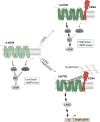
Schematic overview of the effects of CD44 co-expression on 5-HT7R-mediated signaling. Upon the formation of heteromeric complexes with CD44, the constitutive activity of the 5-HT7R towards cAMP signaling is increased, whereas the agonist-induced activity is decreased. The modulated constitutive activity results from an increased Gαs and a decreased GαoB activation under basal conditions (upper panel). Stimulation of heteromeric complexes with the CD44 ligand hyaluronic acid leads to the 5-HT7R-mediated production of cAMP and the subsequent activation of CRE-mediated gene expression (lower panel)

References

    1. Sriram K, Insel PA. G Protein-coupled receptors as targets for approved drugs: how many targets and how many drugs? Mol Pharmacol. 2018;93(4):251–8. - PMC - PubMed
    1. Rosenbaum DM, Rasmussen SGF, Kobilka BK. The structure and function of G-protein-coupled receptors. Nature. 2009;459(7245):356–63. - PMC - PubMed
    1. Rios CD, Jordan BA, Gomes I, Devi LA. G-protein-coupled receptor dimerization: modulation of receptor function. Pharmacol Ther. 2001;92(2):71–87. - PubMed
    1. Terrillon S, Bouvier M. Roles of G-protein-coupled receptor dimerization. EMBO Rep. 2004;5(1):30–4. - PMC - PubMed
    1. Herrick-Davis K. Functional significance of serotonin receptor dimerization. Exp Brain Res. 2013;230(4):375–86. - PMC - PubMed

LinkOut - more resources