. 2024 Nov 23;15(1):10165.
doi: 10.1038/s41467-024-54500-x.

Crystal structures of DCAF1-PROTAC-WDR5 ternary complexes provide insight into DCAF1 substrate specificity

Affiliations

Crystal structures of DCAF1-PROTAC-WDR5 ternary complexes provide insight into DCAF1 substrate specificity

Mark F Mabanglo et al. Nat Commun. .

Abstract

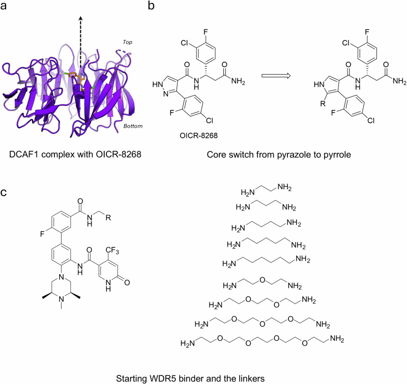
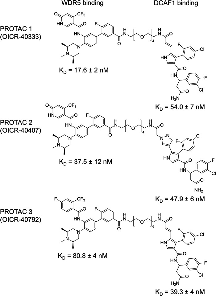
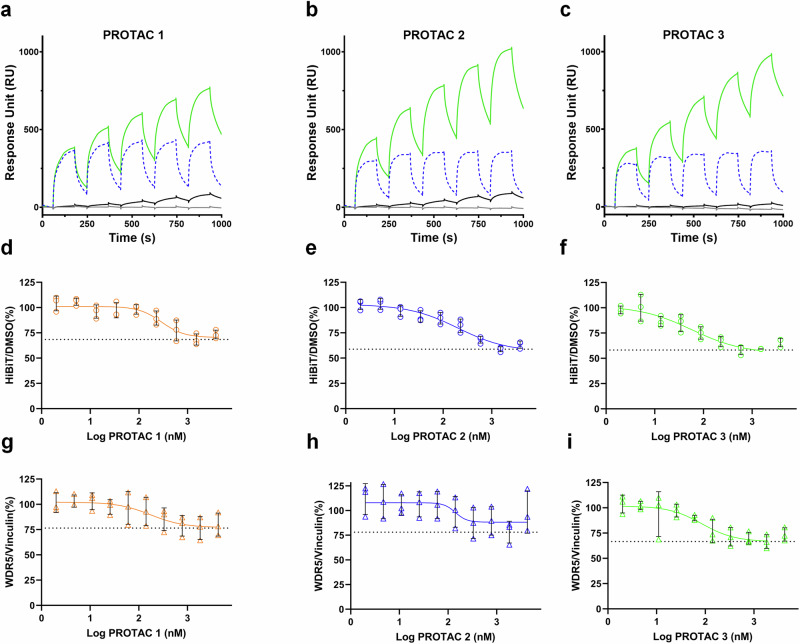
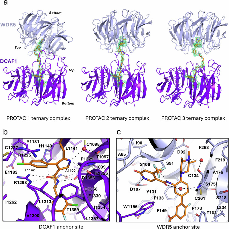
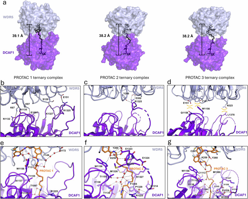
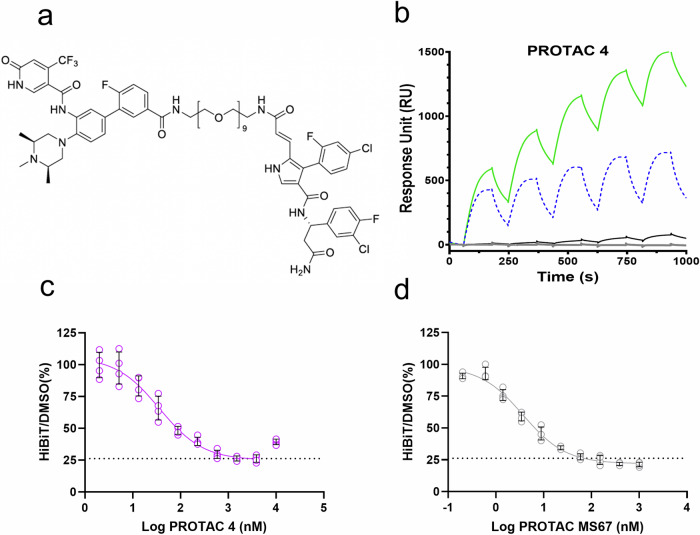
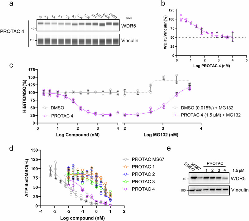
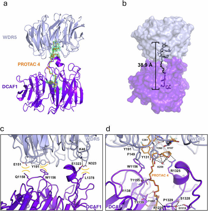
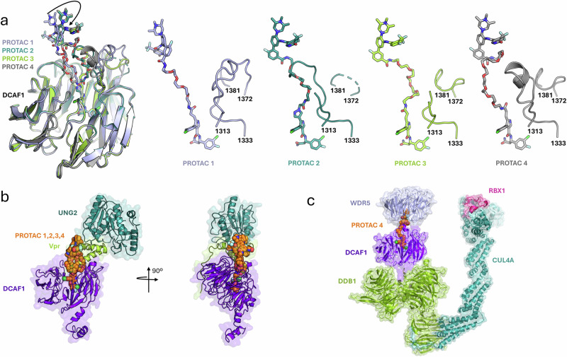
Proteolysis-targeting chimeras (PROTACs) have been explored for the degradation of drug targets for more than two decades. However, only a handful of E3 ligase substrate receptors have been efficiently used. Downregulation and mutation of these receptors would reduce the effectiveness of such PROTACs. We recently developed potent ligands for DCAF1, a substrate receptor of EDVP and CUL4 E3 ligases. Here, we focus on DCAF1 toward the development of PROTACs for WDR5, a drug target in various cancers. We report four DCAF1-based PROTACs with endogenous and exogenous WDR5 degradation effects and high-resolution crystal structures of the ternary complexes of DCAF1-PROTAC-WDR5. The structures reveal detailed insights into the interaction of DCAF1 with various WDR5-PROTACs, indicating a significant role of DCAF1 loops in providing needed surface plasticity, and reflecting the mechanism by which DCAF1 functions as a substrate receptor for E3 ligases with diverse sets of substrates.


Conflict of interest statement

Competing interests: The authors declare no competing interests.

Figures

Fig. 1
Fig. 1. Design and chemical synthesis of DCAF1-based PROTACs.
a Crystal structure of DCAF1 in complex with OICR-8268 (PDB 8F8E). PROTAC design extends from the pyrazole group of OICR-8268 close to the top side of DCAF1 into the solvent space. b The pyrazole group of the DCAF1 anchor was changed to pyrrole to facilitate PROTAC synthesis without loss of activity. c The WDR5 anchor (left) was connected to the DCAF1 anchor through various potential linkers (right).
Fig. 2
Fig. 2. Chemical structures of PROTAC 1 (OICR-40333), PROTAC 2 (OICR-40407), and PROTAC 3 (OICR-40792).
Binding affinity of each PROTAC for WDR5 and DCAF1 was evaluated by SPR, and the related KD values are indicated on each structure as mean ± SD. SPR experiments were performed in triplicate. Source data are provided as a Source Data file.
Fig. 3
Fig. 3. Ternary complex formation and WDR5 degradation effect of PROTAC 1-3.
ac DCAF1-PROTAC-WDR5 ternary complex formation of (a) PROTAC 1, (b) PROTAC 2, and (c) PROTAC 3 were evaluated by SPR. Biotinylated DCAF1 was immobilized on a high-affinity streptavidin chip. A blank injection was used as a baseline (gray lines). Each PROTAC alone was tested for binding to DCAF1 (black lines). WDR5 at 0.25 µM was injected independently over the flow cells with the immobilized DCAF1 as a reference (blue dash lines). Solutions with ratio of 1 (PROTAC):10 (WDR5; fixed final concentration of 0.25 µM) were injected for ternary complex formation (green line). Although some background nonspecific binding for WDR5 was observed, in the presence of all three PROTACs a significant increase in RU (response unit) was observed indicating DCAF1-PROTAC-WDR5 ternary complex formation. The effect of PROTAC 1–3 treatment of MV4-11 cells on (df) exogenous and (gi) endogenous WDR5 degradation was assessed. The amount of WDR5 was quantified using the exogenous (HiBiT) and endogenous (ProteinSimple’s Jess) WDR5 degradation assays as described in Methods. Results are shown as average ± SD of biological replicates (n = 3). Source data are provided as a Source Data file.
Fig. 4
Fig. 4. Crystal structures of ternary complexes of DCAF1 and WDR5 with PROTACs.
a Cartoon representation of ternary complexes showing the top-to-top orientation of the WDR domains of DCAF1 (purple) and WDR5 (light blue). PROTAC 1, 2, and 3 are depicted in orange sticks. A 2Fo-Fc electron density map contoured at 1σ surrounds each PROTAC. b, c The DCAF1 and WDR5 anchors of PROTAC 1-3 are stabilized in their respective binding sites by various electrostatic and hydrogen bonding interactions, in addition to surrounding hydrophobic and van der Waals interactions, with protein side chains (sticks). Some hydrogen bonds between PROTAC and DCAF1/WDR5 residues are mediated by water molecules (red spheres). Hydrogen/electrostatic bonds are shown as broken black lines.
Fig. 5
Fig. 5. Structural details of the DCAF1-WDR5-PROTAC interactions and their significance to PROTAC activity.
a PROTAC 1-3 bring DCAF1 and WDR5 together to form a ternary complex. The longer PROTAC 2 and 3 are compressed to a similar effective length as PROTAC 1 in the ternary structures. Surface representations also indicate diminishing contacts between WDR domains with increasing PROTAC linker length. DCAF1 and WDR5 are colored as in Fig. 4. bd Interactions between the top side of the WDR domains of DCAF1 and WDR5 in ternary complex with PROTAC change depending on linker length. Hydrogen bonding and electrostatic interactions are shown as broken black lines. Hydrophobic/van der Waals interactions are shown as brown concave lines. In the presence of PROTAC 1 or 2, the DCAF1-WDR5 interaction is mediated by various hydrogen bonds (b, c), which disappear in the interface as the linker becomes longer. Weak hydrophobic/van der Waals interactions sustain the DCAF1-WDR5 interface in the presence of the longest PROTAC 3 (d). eg PROTAC linkers of different lengths are accommodated inside the space formed by DCAF1 and WDR5 by compacting. e PROTAC 1 (orange sticks) assumes a fully extended configuration inside the pocket. A flexible loop, formed by DCAF1 residues 1313-1333, is retracted away from the PROTAC linker, resulting in an open cavity filled with water molecules. Only water molecules participating in hydrogen bonding with nearby solvent and/or protein residues are shown. f The longer PROTAC 2 linker is kinked inside the DCAF1-WDR5 interface. Notably, the flexible loop formed by DCAF1 residues 1313–1333 assumes a different configuration that cradles the curved portion of the linker. In addition, a rotamer shift for DCAF1’s W1156 occurs to complement linker shape. g The compacted linker of PROTAC 3, longer than those of PROTAC 1 and 2, engenders a new interaction pattern with protein residues. The longer linker is further compressed inside the binding space, resulting in a more curved configuration. The nearby loop of DCAF1 (residues 1313–1333, with residues 1315–1326 disordered and not modeled) is displaced from its original location in the PROTAC 1 and 2 ternary complexes.
Fig. 6
Fig. 6. PROTAC 4 (OICR41114) degrades WDR5 better than PROTAC 1-3.
a PROTAC 4 chemical structure. b Confirmation of DCAF1-PROTAC 4-WDR5 ternary complex formation by SPR. Experiments were performed the same as for PROTAC 1-3 in Fig. 3. Biotinylated DCAF1 was immobilized onto a sensor SA chip. A blank injection was used as a baseline (gray line). PROTAC 4 (black line) and WDR5 (dashed blue line) were injected independently before combining both in a 1:10 ratio of PROTAC with 0.25 µM WDR5 (green line) for ternary complex formation. In the presence of both WDR5 and PROTAC 4, the binding response significantly increased. c PROTAC 4 degraded exogenous WDR5 in cells. The amount of WDR5 was quantified using the exogenous (HiBiT) WDR5 degradation assay as described in Methods. d PROTAC MS67 was used as a control and degraded HiBiT-WDR5 with an EC50 and Dmax values of 3.8 ± 1.0 nM and 78.7 ± 1.8%, respectively. Values are included as average ± SD of biological replicates (n = 4). Source data are provided as a Source Data file.
Fig. 7
Fig. 7. Degradation of the endogenous WDR5.
The amount of WDR5 was quantified using the endogenous WDR5 degradation assays (ProteinSimple’s Jess) as described in Methods. a A representative lane view image generated by Compass software, which is used to analyze simple western results. In MV4-11, PROTAC 4 degraded endogenous WDR5 in a dose-dependent manner after 24 h of treatment. Vinculin was used as a loading control. The concentration of PROTAC 4 is indicated above each lane. b PROTAC 4 degraded endogenous WDR5 with an EC50 and Dmax values of 40 ± 24 nM and 49 ± 1.9%, respectively. Values are included as average ± SD of biological replicates (n = 3). c PROTAC 4 degraded HiBiT-tagged WDR5 in a proteasome-dependent manner. MV4-11 cells expressing HiBiT tagged WDR5 were treated with PROTAC 4 for 5 h. PROTAC 4 decreased exogenous WDR5 in a dose dependent manner (purple circles). In the same plate but different wells, cells were co-treated with a fixed concentration of PROTAC 4 (1.5 μM) and increasing concentrations of MG132 (purple dashed line) or with DMSO and MG132 (gray dashed line). MG132 fully rescued the levels of degraded HiBiT WDR5 after 5 h of PROTAC 4 and MG132 treatment. Values are included as average ± SD of biological replicates (n = 3). d ATPlite assay results showing the cytotoxic effects of DCAF1-WDR5 PROTACs on MV4-11 cells after 5 days of treatment. Cells were treated with varying concentrations of MS67 (control), PROTAC 1, PROTAC 2, PROTAC 3, and PROTAC 4. Cell viability was assessed using the ATPlite assay, and results were normalized to DMSO-treated controls. Values are included as average ± SD of biological replicates (n ≥ 4). e Representative western blot image of 3 biological replicates. Western blot analysis was performed to assess the protein levels of WDR5 after 24 h of treatment of MV4-11 cells with 1.5 μM of PROTACs. MS67 and PROTAC 4 significantly decreased the levels of WDR5. Source data are provided as a Source Data file.
Fig. 8
Fig. 8. Crystal structure of the PROTAC 4 ternary complex with DCAF1 and WDR5.
a Cartoon representation of the crystal structure showing PROTAC 4 (orange sticks), DCAF1 (purple) and WDR5 (blue). A 2Fo-Fc electron density map contoured at 1.0σ surrounds PROTAC 4. Unlike PROTAC 1-3, PROTAC 4 forms a ternary complex with WDR5 and DCAF1 in which the latter’s long loop (residues 1313–1333) forms a loop-helix-loop structure. b PROTAC 4 (black sticks) is compressed to an effective length of 38.9 Å, similar with those observed for PROTAC 1-3 in their ternary complexes. Surface representation indicates contacts between protein components enabled by PROTAC 4. c Electrostatic/hydrogen bonding (broken black line) and hydrophobic/van der Waals interactions (brown concave lines) occur between the top side of the WDR domains of DCAF1 and WDR5 in the presence of PROTAC 4. d The PROTAC 4 linker is accommodated inside the space formed by DCAF1 and WDR5 by coiling and compacting. A portion of the PROTAC 4 linker close to the WDR5 anchor forms a coiled structure stabilized by an ordered water molecule (red sphere) located below the center of the coil, and interaction with the loop-helix-loop structure formed by residues 1313–1333. Specifically, R1325 of the loop-helix-loop structure of DCAF1 interacts with the coiled structure using hydrogen bonds. The coiled structure fills the space bordered by WDR5 residues D107, Y131, F149, Y191, and Y260. The coiled structure is further stabilized by water-mediated interactions with WDR5 residues and the WDR5 anchor of PROTAC 4 itself. The rest of the PROTAC 4 linker is curved and is also supported by the helix-loop-helix structure of DCAF1. Near the DCAF1 anchor, several water-mediated interactions with DCAF1 residues stabilize the linker conformation. As is also seen in the PROTAC 2 and PROTAC 3 ternary complexes, residue R1225 helps stabilize the linker and the base of the loop-helix-loop structure through direct and water-mediated hydrogen bonding interactions.
Fig. 9
Fig. 9. DCAF1 uses flexible loops for substrate recognition.
a Superposition of DCAF1 component of ternary complexes shows rotation of the WDR anchor in the PROTAC 2, 3, and 4 complexes relative to that of PROTAC 1. In response to PROTAC linker length and curvature, DCAF1’s specificity loop (residues 1313-1333) rearranges to accommodate it, as shown in the series of structures on the right. A nearby loop (residues 1372-1381) in blade 7 also changes conformations. Here, each PROTAC-DCAF1 pair is colored the same: PROTAC 1-DCAF1 is light blue, PROTAC 2-DCAF1 is teal, PROTAC 3-DCAF1 is yellow green, PROTAC 4-DCAF1 is gray. b Superposition of PROTAC ternary complex structures with that of DCAF1-Vpr-UNG2 complex (PDB id 5JK7, DDB1 is not shown for clarity) shows that PROTAC 1, 2, 3, and 4 (spheres) together partially occupy the Vpr (yellow green cartoon) volume between DCAF1 (purple) and UNG2 (teal). PROTACs with longer linkers may fill the extra volume and lead to more stable ternary complexes and exhibit greater efficiency. c Superposition of the PROTAC 4 ternary complex structure with that of DDB1-DCAF1-CUL4A-RBX1 complex orientates WDR5 for ubiquitination by the E3 ligase. DCAF1 and WDR5 are colored as in Fig. 8a, DDB1 is moss green, CUL4A is teal, RBX1 is magenta. PROTAC 4 is shown as spheres. The extra surface for DCAF1 (purple) inserted into the DDB1 adaptor corresponds to the N-terminus that is removed from the crystallized sequence of DCAF1.

References

    1. Scheer, S. et al. A chemical biology toolbox to study protein methyltransferases and epigenetic signaling. Nat. Commun.10, 19 (2019). - PMC - PubMed
    1. Wu, Q. et al. A chemical toolbox for the study of bromodomains and epigenetic signaling. Nat. Commun.10, 1915 (2019). - PMC - PubMed
    1. Cui, Q., Huang, C., Liu, J. Y. & Zhang, J. T. Small molecule inhibitors targeting the “Undruggable” survivin: the past, present, and future from a medicinal chemist’s perspective. J. Med. Chem.66, 16515–16545 (2023). - PMC - PubMed
    1. Poso, A. The future of medicinal chemistry, PROTAC, and undruggable drug targets. J. Med. Chem.64, 10680–10681 (2021). - PMC - PubMed
    1. Sakamoto, K. M. et al. Protacs: chimeric molecules that target proteins to the Skp1-Cullin-F box complex for ubiquitination and degradation. Proc. Natl Acad. Sci. USA98, 8554–8559 (2001). - PMC - PubMed

Publication types

MeSH terms

Substances

LinkOut - more resources