. 2025 Feb;39(2):438-450.
doi: 10.1038/s41375-024-02479-1. Epub 2024 Nov 23.

Dimethyl fumarate and extracorporeal photopheresis combination-therapy synergize in inducing specific cell death and long-term remission in cutaneous T cell lymphoma

Affiliations

Dimethyl fumarate and extracorporeal photopheresis combination-therapy synergize in inducing specific cell death and long-term remission in cutaneous T cell lymphoma

Özge Ç Şener et al. Leukemia. 2025 Feb.

Abstract

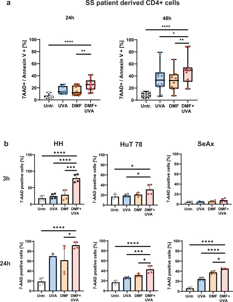
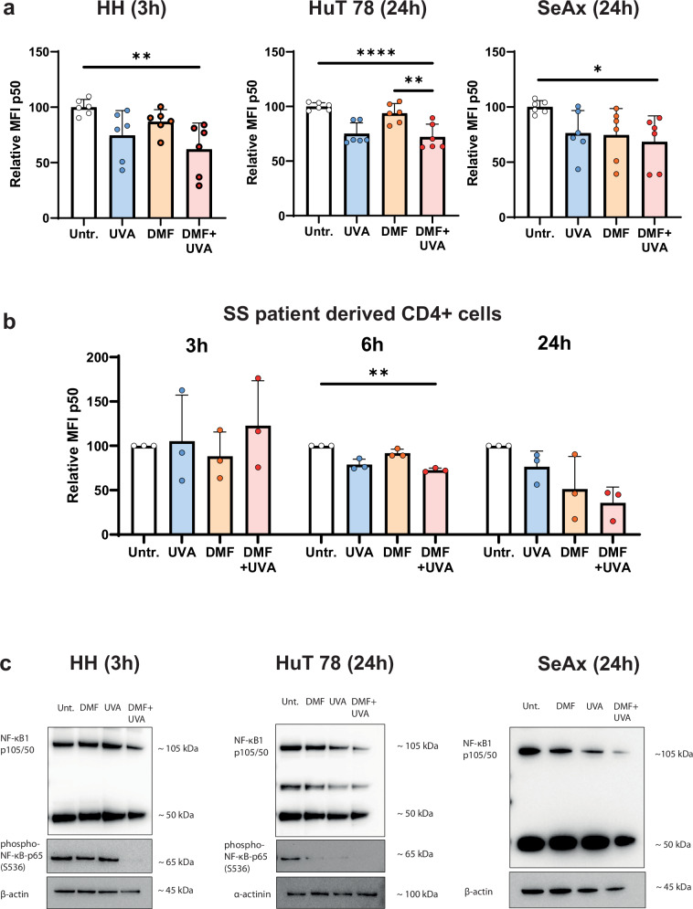
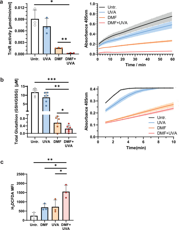
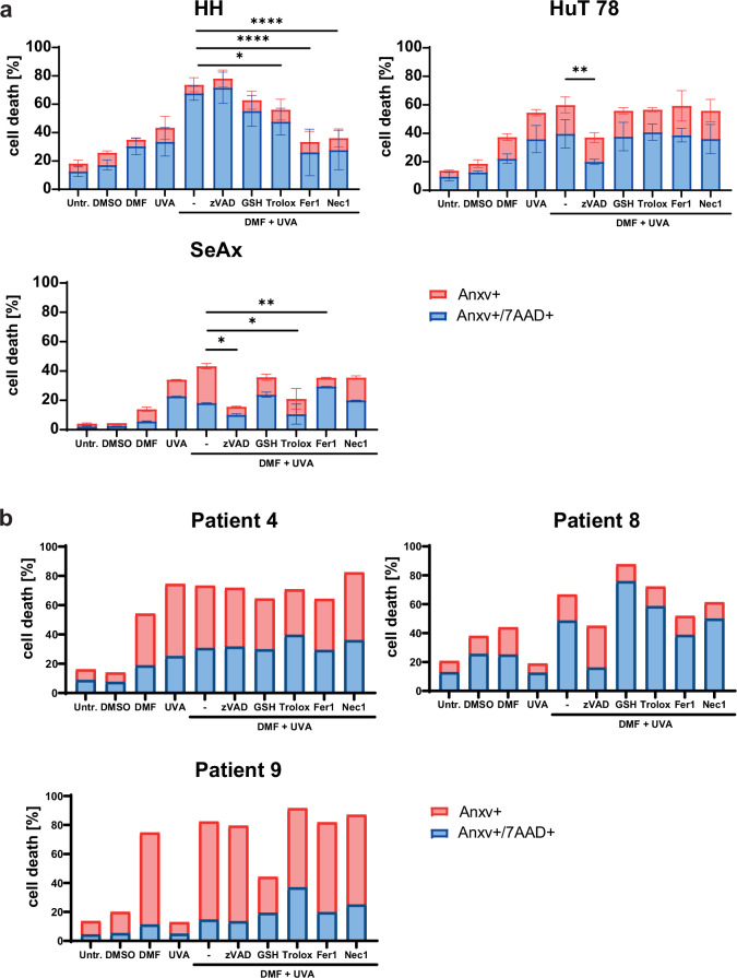
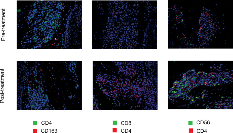
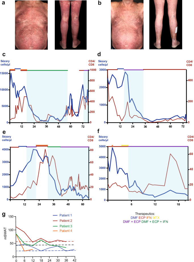
Primary cutaneous T cell lymphomas (CTCL) are characterized by high relapse rates to initially highly effective therapies. Combination therapies have proven beneficial, particularly if they incorporate extracorporeal photopheresis (ECP). The NF-κB inhibitor dimethyl fumarate (DMF) has proven a new, effective drug in CTCL in a clinical phase II study. In vitro experiments with patient-derived SS cells and the CTCL cell lines HH, HuT 78, and SeAx revealed a synergistic effect of DMF and ECP on cell death induction in CTCL cells. Furthermore, an additional increase in the capacity to inhibit NF-κB in CTCL was detected for the combination treatment compared to DMF monotherapy. The same synergistic effects could be measured for ROS production via decreased Thioredoxin reductase activity and glutathione levels. Consequently, a cell death inhibitor screen indicated that the DMF/ECP combination treatment induces a variety of cell death mechanisms in CTCL. As a first step into clinical translation, 4 patients were already treated with the DMF/ECP combination therapy with an overall response rate of 100% and a time to next treatment in skin and blood of up to 57 months. Therefore, our study introduces the combination treatment of DMF and ECP as a highly effective and long-lasting CTCL therapy.


Conflict of interest statement

Competing interests: SM received honoraria and travel funding by Kyowa Kirin. KG received consulting fees from Biogen and supports BioMed X as an academic mentor. JSU is on the advisory board or has received honoraria and travel support from Amgen, Bristol Myers Squibb, GSK, Immunocore, LeoPharma, Merck Sharp and Dohme, Novartis, Pierre Fabre, Roche, Sanofi outside the submitted work. JPN received travel and congress participation funding by TEVA and Novartis as well as consulting fees by TEVA, Almirall, Biogen, Novartis, Kyowa Kirin, Innate Pharma, Takeda and Actelion, UCB Pharma and Recordati. ÖÇŞ, LT, PLB, JDA, TH, DT, and SG have no conflict of interest to declare.

Figures

Fig. 1
Fig. 1. DMF-8-MOP/UVA combination treatment induces increased cell death in SS patient-derived CD4+ T cells and CTCL cell lines in vitro.
a CD4+ T cells were isolated from the peripheral blood of SS patients (n = 6, the patients’ cells were measured at n = 2 different timepoints resulting in overall 12 data points) and treated with DMF- and 8-MOP/UVA-monotreatment, and the DMF-8-MOP/UVA-combination treatment, and compared to untreated controls. Cell death was measured by 7AAD/Annexin V staining with FACS after 24 h and 48 h. b HH, HuT 78 and SeAx cells were treated with DMF- and 8-MOP/UVA-monotreatment, and the DMF-8-MOP/UVA-combination treatment, and compared to untreated controls. Cell death was measured by 7AAD staining with FACS after 3 h and 24 h (n = 4). The level of significance is indicated by asterisks (**** ≤0.0001; *** ≤0.001; ** ≤0.01; * ≤0.05). Error bars show the standard deviation.
Fig. 2
Fig. 2. DMF-8-MOP/UVA combination treatment significantly decreases NF-κB expression in CTCL cell lines and SS patient cells compared to monotreatment.
HH, HuT 78 and SeAx cells as well as patient-derived CD4+ T cells were treated with DMF- and 8-MOP/UVA-monotreatment, and the DMF-8-MOP/UVA-combination treatment, and compared to untreated controls. a A p105/p50 staining was performed and the relative MFI was measured by FACS after 3 h for HH cells, and after 24 h for HuT 78 and SeAx cells. b The MFI of p105/p50 was measured in patient CD4+ T cells after 3 h, 6 h and 24 h. Error bars show the standard deviation (n = 6 for cell lines and n = 3 patients). The level of significance is indicated by asterisks (**** ≤0.0001; *** ≤0.001; ** ≤0.01; * ≤0.05). c NF-κB1 p105/50 and phospho-NF-κB-p65 (S536) were detected in the cell lines by immunoblot (representative immunoblot, n = 3).
Fig. 3
Fig. 3. DMF-8-MOP/UVA combination treatment induces ROS production via decreased Thioredoxin reductase activity and diminished free glutathione levels.
HH cells were treated with DMF- and 8-MOP/UVA-monotreatment, and the DMF-8-MOP/UVA-combination treatment, and compared to untreated controls. a Thioredoxin reductase activity was calculated via the increase in absorbance at a wavelength of 405 nm over the course of 60 min according to manufacturer’s instructions (n = 3). b The concentration of total glutathione (GSSG/GSH) was calculated via the increase in absorbance at 405 nm wavelength over the course of 10 min according to manufacturer’s protocol (n = 6). c The MFI of intracellular H2DCFDA was measured by FACS as a surrogate parameter for intracellular reactive oxygen species (ROS) levels (n = 3). The level of significance is indicated by asterisks (**** ≤0.0001; *** ≤0.001; ** ≤0.01; * ≤0.05). Error bars show the standard deviation.
Fig. 4
Fig. 4. DMF-8-MOP/UVA-combination treatment induces cell death in CTCL cell lines and SS patient cells via different mechanisms and pathways.
a HH, HuT 78 and SeAx cells as well as (b) CD4+ T cells from three patients were treated with DMF- and 8-MOP/UVA-monotreatment, and the DMF-8-MOP/UVA-combination treatment. Additionally, the combination treatment was performed together with the cell death inhibitors zVAD (50 µM), Ferrostatin 1 (1 µM), Necrostatin 1 (50 µM), and the anti-oxidants Trolox (1 mM) and glutathione (GSH; 0.5 mM), and compared to untreated controls. Cell death was measured by Annexin V/7-AAD staining (n = 4 for cell lines). The level of significance is indicated by asterisks (**** ≤0.0001; *** ≤0.001; ** ≤0.01; * ≤0.05). Error bars show the standard deviation.
Fig. 5
Fig. 5. Immunofluorescent stainings of patient 1, patient 2, and patient 3 before and under the DMF/ECP combination treatment.
Representative pictures of immunofluorescent stainings of patient 1, patient 2, and patient 3 before and under the DMF/ECP combination treatment stained for CD4, CD8, CD163, CD56, and with DAPI, the latter to counterstain nuclei.
Fig. 6
Fig. 6. DMF and ECP combination therapy shows excellent clinical responses in skin and blood in SS patients.
Clinical photographs from patient 2 at week 0 (a) and week 52 (b) of the combination treatment of dimethyl fumarate (DMF) and extracorporeal photopheresis (ECP). Course of the Sézary cells (CD3+CD4+CD7- cell population; in blue) and the CD4/CD8 ratio (red) from patient 1 (c), patient 2 (d), patient 3 (e) and patient 4 (f) over time (in months). The patients’ treatments since the introduction of ECP in the therapeutic regimen are marked with colored lines (DMF = blue, ECP = red, IFN = orange, MTX = yellow, combination therapy with DMF and ECP = purple, combination therapy with DMF, ECP, and IFN = green). The period of combination therapy with DMF and ECP is shaded in light blue. g mSWAT scores during the combination therapy of DMF and ECP of patient 1 (blue), patient 2 (red), patient 3 (green), and patient 4 (orange) over time (in months).

References

    1. Alaggio R, Amador C, Anagnostopoulos I, Attygalle AD, Araujo IBO, Berti E, et al. The 5th edition of the World Health Organization Classification of Haematolymphoid Tumours: Lymphoid Neoplasms. Leukemia. 2022;36:1720–48. - PMC - PubMed
    1. Willemze R, Cerroni L, Kempf W, Berti E, Facchetti F, Swerdlow SH, et al. The 2018 update of the WHO-EORTC classification for primary cutaneous lymphomas. Blood. 2019;133:1703–14. - PMC - PubMed
    1. Latzka J, Assaf C, Bagot M, Cozzio A, Dummer R, Guenova E, et al. EORTC consensus recommendations for the treatment of mycosis fungoides/Sezary syndrome - Update 2023. Eur J Cancer. 2023;195:113343. - PubMed
    1. Willemze R, Hodak E, Zinzani PL, Specht L, Ladetto M, Committee EG. Primary cutaneous lymphomas: ESMO Clinical Practice Guidelines for diagnosis, treatment and follow-up. Ann Oncol. 2018;29:iv30–iv40. - PubMed
    1. Knobler R, Arenberger P, Arun A, Assaf C, Bagot M, Berlin G, et al. European dermatology forum: Updated guidelines on the use of extracorporeal photopheresis 2020 - Part 2. J Eur Acad Dermatol Venereol. 2021;35:27–49. - PMC - PubMed