. 2024 Dec 24;43(12):114942.
doi: 10.1016/j.celrep.2024.114942. Epub 2024 Nov 23.

High-affinity agonists reveal recognition motifs for the MRGPRD GPCR

Affiliations

High-affinity agonists reveal recognition motifs for the MRGPRD GPCR

Chunyu Wang et al. Cell Rep. .

Abstract

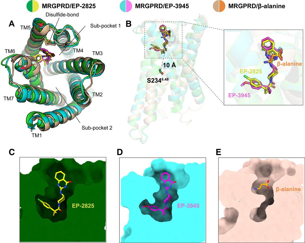
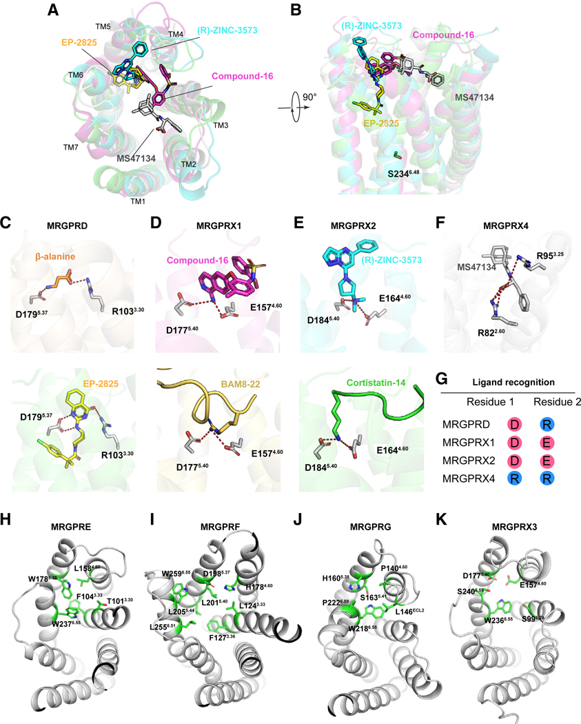
The human MRGPRD protein is a member of the Mas-related G protein-coupled receptors (MRGPRs) that is involved in the sensing of pain, itch, and other inflammatory stimuli. As with other MRGPRs, MRGPRD is a relatively understudied receptor with few known agonists. The most potent small-molecule agonist of MRGPRD reported so far is β-alanine, with an affinity in the micromole range, which largely restricts its functional study. Here, we report two MRGPRD agonists, EP-2825 and EP-3945, that are approximately 100-fold more potent than β-alanine and determine the structures of MRGPRD-Gq in complex with EP-2825 and EP-3945, respectively. The structures reveal distinct agonist binding modes of MRGPRD and large conformational plasticity of the orthosteric pocket. Collectively, the discovery of high-affinity MRGPRD agonists and their distinct binding modes will facilitate the functional study and the structure-based design of ligands targeting this understudied receptor.

Keywords: CP: Molecular biology; GPCR; MRGPRD; agonist recognition; drug discovery; structure.


Conflict of interest statement

Declaration of interests A pending patent application has been filed by Escient Pharmaceuticals that includes compounds EP-2825 and EP-3945. B.L.R. is on the scientific advisory board of Escient Pharmaceuticals.

Figures

Figure 1.
Figure 1.. Structure of agonist-bound MRGPRD-Gq complexes
(A) The chemical structures of β-alanine, GABA, EP-2825, and EP-3945. (B) EP-2825 and EP-3945 activate MRGPRD much more potently than β-alanine and GABA. Data are presented as mean ± SEM of four biological repeats. Emax, maximum effect. (C and D) Cryo-EM map of the EP-2825-bound MRGPRD-Gq complex (C) and EP-3945-bound MRGPRD-Gq complex (D). See also Figures S1–S3 and Tables S1.
Figure 2.
Figure 2.. The overall structure of agonist-bound MRGPRD
(A) Structural comparison of MRGPRD structures in EP-2825-, EP-3945-, and β-alanine-bound (PDB: 7Y12) states. (B) EP-2825 and EP-3945 bound to MRGPRD at a position that is 10 Å away from S2346.48. (C–E) Cross-section images of the EP-2825 (C), EP-3945 (D), and β-alanine (E) binding pocket of MRGPRD. See also Figures S4 and S5.
Figure 3.
Figure 3.. The agonist binding mode of EP-2825
(A–C) Key interactions between MRGPRD and the top part (A), middle part (B), and bottom part (C) of EP-2825. Polar interactions are shown as red dashed lines. (D–F) The effects of alanine substitutions of the key residues interacting with the top part (D), middle part (E), and bottom part (F) of ligand on EP-2825-stimulated MRGPRD activation. Data are presented as mean ± SEM of four biological repeats. WT, wild-type. See also Figures S6, S7, and S9 and Table S3.
Figure 4.
Figure 4.. Conformational plasticity of the orthosteric pocket
(A and B) Structural comparison of EP-2825-bound MRGPRD with β-alanine-bound MRGPRD (PDB: 7Y12), highlighting the conformational changes of residues W2416.55 and F2426.56 (A) and the residues F1765.34 and Y2456.59 (B). The red arrows indicate the relative movements of MRGPRD from the β-alanine-bound state to the EP-2825-bound state. (C and D) Structural comparison of EP-2825-bound MRGPRD with apo MRGPRD (PDB: 7Y15), highlighting the conformational changes of residues W2416.55 and F2426.56 (C) and the residues F1765.34 and Y2456.59 (D). The red arrows indicate the relative movements of MRGPRD from the apo state to the EP-2825-bound state. See also Figures S6 and S7.
Figure 5.
Figure 5.. Structural comparison of the ligand recognition mode of MRGPRD with other MRGPR-family receptors
(A and B) Structural superpositions of the EP-2825-bound MRGPRD structure with compound-16-bound MRGPRX1 (PDB: 8DWH), (R)-ZINC-3573-bound MRGPRX2 (PDB: 7S8N), and MS47134-bound MRGPRX4 (7S8P), highlighting the different agonist binding modes in MRGPRs. The MRGPR structures are shown in top view (A) and side view (B). (C–F) The key charge interactions between agonist and receptor observed in MRGPRD (C), MRGPRX1 (D), MRGPRX2 (E), and MRGPRX4 (F). Polar interactions are highlighted as red dashed lines. Residue D1795.37 of MRGPRD and residue D5.40 of MRGPRX1 and MRGPRX2 are structurally conserved despite their different Ballesteros-Weinstein numbers. The β-alanine-bound MRGPRD (PDB: 7Y12), EP-2825-bound MRGPRD, compound-16-bound MRGPRX1 (PDB: 8DWH), BAM8–22-bound MRGPRX1 (PDB: 8DWG), (R)-ZINC3573-bound MRGPRX2 (PDB: 7S8N), Cortistatin-14-bound MRGPRX2 (PDB: 7S8L), and MS47134-bound MRGPRX4 (PDB:7S8P) structures are used for analysis. (G) The key charged residues that are critical for MRGPRD, MRGPRX1, MRGPRX2, and MRGPRX4 agonist recognition, highlighting that MRGPRs recognize agonists through differently charged residues. Negatively charged residues are colored red, whereas positively charged residues are colored blue. (H–K) The potential orthosteric pocket of the AlphaFold-predicted models of MRGPRE (H), MRGPRF (I), MRGPRG (J), and MRGPRX3 (K). See also Figures S5 and S8.

References

    1. Zylka MJ, Rice FL, and Anderson DJ (2005). Topographically distinct epidermal nociceptive circuits revealed by axonal tracers targeted to Mrgprd. Neuron 45, 17–25. 10.1016/j.neuron.2004.12.015. - DOI - PubMed
    1. Vrontou S, Wong AM, Rau KK, Koerber HR, and Anderson DJ (2013). Genetic identification of C fibres that detect massage-like stroking of hairy skin in vivo. Nature 493, 669–673. 10.1038/nature11810. - DOI - PMC - PubMed
    1. Wang H, and Zylka MJ (2009). Mrgprd-expressing polymodal nociceptive neurons innervate most known classes of substantia gelatinosa neurons. J. Neurosci. 29, 13202–13209. 10.1523/JNEUROSCI.3248-09.2009. - DOI - PMC - PubMed
    1. Rau KK, McIlwrath SL, Wang H, Lawson JJ, Jankowski MP, Zylka MJ, Anderson DJ, and Koerber HR (2009). Mrgprd enhances excitability in specific populations of cutaneous murine polymodal nociceptors. J. Neurosci 29, 8612–8619. 10.1523/JNEUROSCI.1057-09.2009. - DOI - PMC - PubMed
    1. Xu M, Zhang Z, and Lan L. (2022). Identification of MrgprD expression in mouse enteric neurons. Cell Tissue Res. 388, 479–484. 10.1007/s00441-022-03608-x. - DOI - PubMed

Publication types

Substances

LinkOut - more resources