. 2024 Nov 25;21(1):302.
doi: 10.1186/s12985-024-02575-9.

Tropism of adeno-associated virus serotypes in mouse lungs via intratracheal instillation

Affiliations

Tropism of adeno-associated virus serotypes in mouse lungs via intratracheal instillation

Haoyu Wu et al. Virol J. .

Abstract

Background: Gene therapy holds great potential for treating various acquired and inherited pulmonary diseases. Adeno-associated viral (AAV) vectors have been thought to be primary candidates for gene delivery in patients with pulmonary diseases. However, the tropism of AAVs in the lungs remains largely unknown.

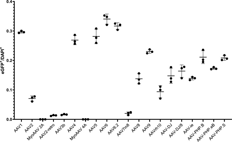
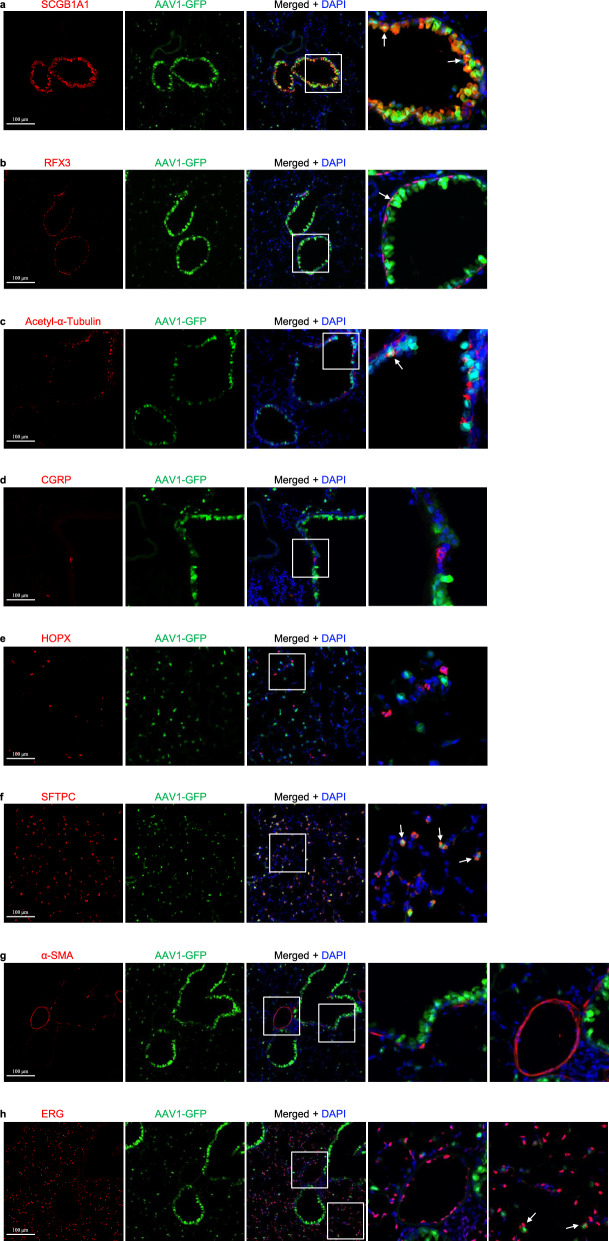
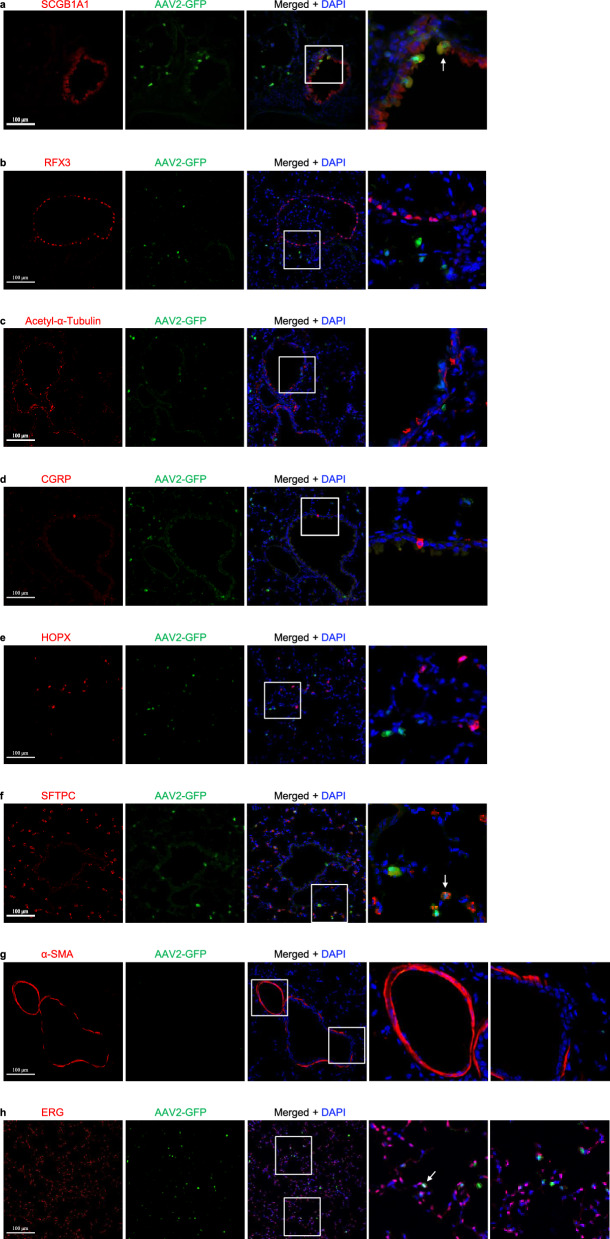
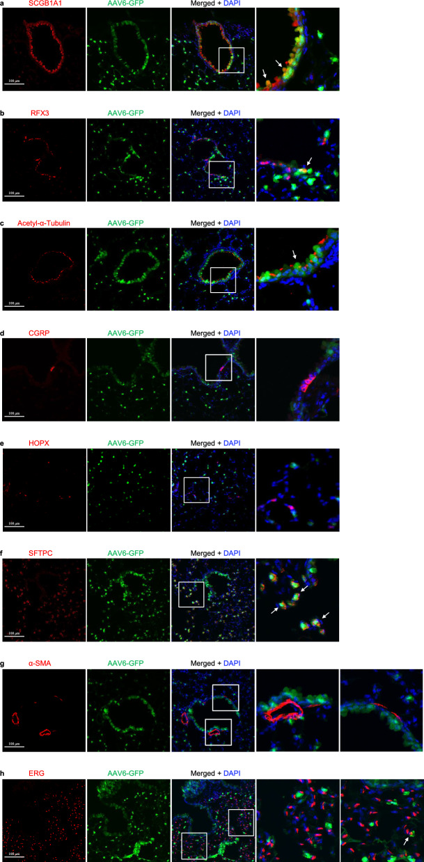
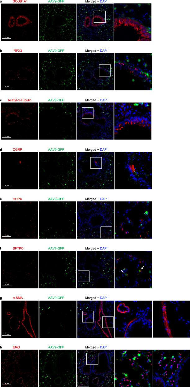
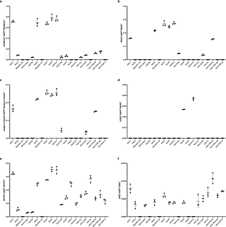
Results: Here, we investigate the tropism of twenty serotypes of AAVs by examining AAV-packed vector expression of the enhanced green fluorescent protein (eGFP) in mice. AAV1, AAV4, AAV5, AAV6, AAV6.2, AAV-PHP.B, and AAV-PHP.S exhibit high transduction rates in the airway epithelium. AAV1, AAV4, AAV5, AAV6, and AAV6.2 highly infect club cells. AAV1, AAV4, AAV5, AAV6, AAV6.2, and AAV-PHP.B efficiently infect ciliated cells. AAV8 and AAVrh10 can infect a few alveolar type I cells. AAV1, AAV5, AAV6, AAV6.2, AAV9, and AAVie can infect alveolar type II cells. AAV1, AAV5, AAVie, AAV-PHP.B, AAV-PHP.eB, and AAV-PHP.S can infect a few endothelial cells. However, none of these AAVs can efficiently infect neuroendocrine or smooth muscle cells.

Conclusions: Our findings provide comprehensive information about the tropism of AAVs in pulmonary epithelium in mice, which might be helpful in developing efficient AAV-mediated gene therapy strategies for pulmonary disease treatment.

Keywords: Adeno-associated virus; Alveolar epithelial cells; Ciliated cells; Club cells; Tropism.


Conflict of interest statement

Declarations. Ethics approval and consent to participate: Not applicable. Consent for publication: All authors agree to this publication. Competing interests: The authors declare no competing interests.

Figures

Fig. 1
Fig. 1
Analysis of infection efficiency of AAVs in the mouse lungs. The ratio of eGFP+ cells to DAPI+ cells. 2746, 695, 0, 115, 26, 2435, 0, 2588, 3118, 2919, 205, 1386, 2366, 1016, 1525, 1673, 1352, 2200, 1681, 2078 eGFP+ cells were analyzed for AAV1, AAV2, MyoAAV 2A, AAV2-retro, AAV3b, AAV4, MyoAAV 4A, AAV5, AAV6, AAV6.2, AAV7m8, AAV8, AAV9, AAVrh10, AAV-DJ, AAV-DJ/8, AAVie, AAV-PHP.B, AAV-PHP.eB, AAV-PHP.S, and 9263, 9804, 9701, 8762, 10,703, 9052, 9592, 9193, 9145, 9217, 10,157, 10,084, 10,272, 10,672, 10,330, 10,242, 9735, 10,423, 9715, 9958 DAPI+ cells, respectively. n = 3
Fig. 2
Fig. 2
AAV1-eGFP expression in the lungs infected by AAV1 in mice. a Immunostaining for eGFP (green), SCGB1A1 (red) and DAPI staining (blue) of 2-month-old mouse lungs infected with AAV1-eGFP. b Immunostaining for eGFP (green), RFX3 (red), and DAPI staining (blue) of 2-month-old mouse lungs infected with AAV1-eGFP. c Immunostaining for eGFP (green), Acetylated-α-tubulin (red), and DAPI staining (blue) of 2-month-old mouse lungs infected with AAV1-eGFP. d Immunostaining for eGFP (green), CGRP (red), and DAPI staining (blue) of 2-month-old mouse lungs infected with AAV1-eGFP. e Immunostaining for eGFP (green), HOPX (red), and DAPI staining (blue) of 2-month-old mouse lungs infected with AAV1-eGFP. f Immunostaining for eGFP (green), SFTPC (red), and DAPI staining (blue) of 2-month-old mouse lungs infected with AAV1-eGFP. g Immunostaining for eGFP (green), α-SMA (red), and DAPI staining (blue) of 2-month-old mouse lungs infected with AAV1-eGFP. h Immunostaining for eGFP (green), ERG (red), and DAPI staining (blue) of 2-month-old mouse lungs infected with AAV1-eGFP. Scale bars: 100 μm
Fig. 3
Fig. 3
AAV2-eGFP expression in the lungs infected by AAV2 in mice. a Immunostaining for eGFP (green), SCGB1A1 (red) and DAPI staining (blue) of 2-month-old mouse lungs infected with AAV2-eGFP. b Immunostaining for eGFP (green), RFX3 (red), and DAPI staining (blue) of 2-month-old mouse lungs infected with AAV2-eGFP. c Immunostaining for eGFP (green), Acetylated-α-tubulin (red), and DAPI staining (blue) of 2-month-old mouse lungs infected with AAV2-eGFP. d Immunostaining for eGFP (green), CGRP (red), and DAPI staining (blue) of 2-month-old mouse lungs infected with AAV2-eGFP. e Immunostaining for eGFP (green), HOPX (red), and DAPI staining (blue) of 2-month-old mouse lungs infected with AAV2-eGFP. f Immunostaining for eGFP (green), SFTPC (red), and DAPI staining (blue) of 2-month-old mouse lungs infected with AAV2-eGFP. g Immunostaining for eGFP (green), α-SMA (red), and DAPI staining (blue) of 2-month-old mouse lungs infected with AAV2-eGFP. h Immunostaining for eGFP (green), ERG (red), and DAPI staining (blue) of 2-month-old mouse lungs infected with AAV2-eGFP. Scale bars: 100 μm
Fig. 4
Fig. 4
AAV3b-eGFP expression in the lungs infected by AAV3b in mice. a Immunostaining for eGFP (green), SCGB1A1 (red), and DAPI staining (blue) of 2-month-old mouse lungs infected with AAV3b-eGFP. b Immunostaining for eGFP (green), RFX3 (red), and DAPI staining (blue) of 2-month-old mouse lungs infected with AAV3b-eGFP. c Immunostaining for eGFP (green), Acetylated-α-tubulin (red), and DAPI staining (blue) of 2-month-old mouse lungs infected with AAV3b-eGFP. d Immunostaining for eGFP (green), CGRP (red), and DAPI staining (blue) of 2-month-old mouse lungs infected with AAV3b-eGFP. e Immunostaining for eGFP (green), HOPX (red), and DAPI staining (blue) of 2-month-old mouse lungs infected with AAV3b-eGFP. f Immunostaining for eGFP (green), SFTPC (red), and DAPI staining (blue) of 2-month-old mouse lungs infected with AAV3b-eGFP. g Immunostaining for eGFP (green), α-SMA (red), and DAPI staining (blue) of 2-month-old mouse lungs infected with AAV3b-eGFP. h Immunostaining for eGFP (green), ERG (red), and DAPI staining (blue) of 2-month-old mouse lungs infected with AAV3b-eGFP. Scale bars: 100 μm
Fig. 5
Fig. 5
AAV6-eGFP expression in the lungs infected by AAV6 in mice. a Immunostaining for eGFP (green), SCGB1A1 (red), and DAPI staining (blue) of 2-month-old mouse lungs infected with AAV6-eGFP. b Immunostaining for eGFP (green), RFX3 (red), and DAPI staining (blue) of 2-month-old mouse lungs infected with AAV6-eGFP. c Immunostaining for eGFP (green), acetylated-α-tubulin (red), and DAPI staining (blue) of 2-month-old mouse lungs infected with AAV6-eGFP. d Immunostaining for eGFP (green), CGRP (red), and DAPI staining (blue) of 2-month-old mouse lungs infected with AAV6-eGFP. e Immunostaining for eGFP (green), HOPX (red), and DAPI staining (blue) of 2-month-old mouse lungs infected with AAV6-eGFP. f Immunostaining for eGFP (green), SFTPC (red), and DAPI staining (blue) of 2-month-old mouse lungs infected with AAV6-eGFP. g Immunostaining for eGFP (green), α-SMA (red), and DAPI staining (blue) of 2-month-old mouse lungs infected with AAV1-eGFP. h Immunostaining for eGFP (green), ERG (red), and DAPI staining (blue) of 2-month-old mouse lungs infected with AAV6-eGFP. Scale bars: 100 μm
Fig. 6
Fig. 6
AAV8-eGFP expression in the lungs infected by AAV8 in mice. a Immunostaining for eGFP (green), SCGB1A1 (red), and DAPI staining (blue) of 2-month-old mouse lungs infected with AAV8-eGFP. b Immunostaining for eGFP (green), RFX3 (red), and DAPI staining (blue) of 2-month-old mouse lungs infected with AAV8-eGFP. c Immunostaining for eGFP (green), Acetylated-α-tubulin (red), and DAPI staining (blue) of 2-month-old mouse lungs infected with AAV8-eGFP. d Immunostaining for eGFP (green), CGRP (red), and DAPI staining (blue) of 2-month-old mouse lungs infected with AAV8-eGFP. e Immunostaining for eGFP (green), HOPX (red), and DAPI staining (blue) of 2-month-old mouse lungs infected with AAV8-eGFP. f Immunostaining for eGFP (green), SFTPC (red), and DAPI staining (blue) of 2-month-old mouse lungs infected with AAV8-eGFP. g Immunostaining for eGFP (green), α-SMA (red), and DAPI staining (blue) of 2-month-old mouse lungs infected with AAV8-eGFP. h Immunostaining for eGFP (green), ERG (red), and DAPI staining (blue) of 2-month-old mouse lungs infected with AAV8-eGFP. Scale bars: 100 μm
Fig. 7
Fig. 7
AAV9-eGFP expression in the lungs infected by AAV9 in mice. a Immunostaining for eGFP (green), SCGB1A1 (red), and DAPI staining (blue) of 2-month-old mouse lungs infected with AAV9-eGFP. b Immunostaining for eGFP (green), RFX3 (red), and DAPI staining (blue) of 2-month-old mouse lungs infected with AAV9-eGFP. c Immunostaining for eGFP (green), Acetylated-α-tubulin (red), and DAPI staining (blue) of 2-month-old mouse lungs infected with AAV9-eGFP. d Immunostaining for eGFP (green), CGRP (red), and DAPI staining (blue) of 2-month-old mouse lungs infected with AAV9-eGFP. e Immunostaining for eGFP (green), HOPX (red), and DAPI staining (blue) of 2-month-old mouse lungs infected with AAV9-eGFP. f Immunostaining for eGFP (green), SFTPC (red), and DAPI staining (blue) of 2-month-old mouse lungs infected with AAV9-eGFP. g Immunostaining for eGFP (green), α-SMA (red), and DAPI staining (blue) of 2-month-old mouse lungs infected with AAV9-eGFP. h Immunostaining for eGFP (green), ERG (red), and DAPI staining (blue) of 2-month-old mouse lungs infected with AAV9-eGFP. Scale bars: 100 μm
Fig. 8
Fig. 8
Quantification of pulmonary epithelial cells that are infected by AAVs. a Ratio of SCGB1A1+ cells that are eGFP+. 1754, 260, 0, 0, 94, 1308, 0, 1282, 1240, 1470, 74, 126, 0, 0, 126, 254, 0, 276, 366, 0 for eGFP+SCGB1A1+cells were analyzed for AAV1, AAV2, MyoAAV 2A, AAV2-retro, AAV3b, AAV4, MyoAAV 4A, AAV5, AAV6, AAV6.2, AAV7m8, AAV8, AAV9, AAVrh10, AAV-DJ, AAV-DJ/8, AAVie, AAV-PHP.B, AAV-PHP.eB, AAV-PHP.S, and 2456, 3116, 2478, 2856, 2094, 1904, 2226, 1904, 1586, 1932, 1724, 1940, 1204, 1372, 2824, 2996, 1778, 2300, 2460, 1736 for SCGB1A1+ cells, respectively. n = 3. b Ratio of RFX3+ cells that are eGFP+. 612, 0, 0, 0, 0, 496, 0, 802, 448, 634, 178, 0, 0, 0, 0, 176, 0, 712, 0, 0 for RFX3+eGFP+ cells were analyzed for AAV1, AAV2, MyoAAV 2A, AAV2-retro, AAV3b, AAV4, MyoAAV 4A, AAV5, AAV6, AAV6.2, AAV7m8, AAV8, AAV9, AAVrh10, AAV-DJ, AAV-DJ/8, AAVie, AAV-PHP.B, AAV-PHP.eB, AAV-PHP.S, and 3838, 3808, 5224, 5976, 6526, 2280, 4572, 2978, 1818, 2328, 3848, 2734, 2602, 2800, 4156, 5136, 3778, 4634, 4940, 4376 for RFX3+ cells, respectively. n = 3. c Ratio of Acetyl-α-Tubulin+ cells that are eGFP+.778, 0, 0, 0, 0, 642, 0, 822, 562, 666, 174, 0, 0, 0, 0, 178, 0, 616, 0, 0 for Acetyl-α-Tubulin+eGFP+ cells were analyzed for AAV1, AAV2, MyoAAV 2A, AAV2-retro, AAV3b, AAV4, MyoAAV 4A, AAV5, AAV6, AAV6.2, AAV7m8, AAV8, AAV9, AAVrh10, AAV-DJ, AAV-DJ/8, AAVie, AAV-PHP.B, AAV-PHP.eB, AAV-PHP.S, and 4704, 4590, 6018, 6834, 8024, 2930, 5508, 3204, 2320, 2634, 3974, 3672, 5916, 5032, 5270, 5780, 4216, 4090, 4046, 5678 for Acetyl-α-Tubulin+ cells, respectively. n = 3. d Ratio of HOPX+ cells that are eGFP+. 0, 0, 0, 0, 0, 0, 0, 0, 0, 0, 0, 78, 0, 132, 0, 0, 0, 0, 0, 0 for HOPX+eGFP+ cells were analyzed for AAV1, AAV2, MyoAAV 2A, AAV2-retro, AAV3b, AAV4, MyoAAV 4A, AAV5, AAV6, AAV6.2, AAV7m8, AAV8, AAV9, AAVrh10, AAV-DJ, AAV-DJ/8, AAVie, AAV-PHP.B, AAV-PHP.eB, AAV-PHP.S, and 1470, 3780, 2478, 3136, 3234, 2828, 2226, 4074, 2996, 3052, 3780, 2898, 2772, 3606, 3262, 2142, 1778, 1148, 1890, 1302 for HOPX+ cells, respectively. n = 3. e Ratio of SFTPC+ cells that are eGFP+. 1020, 408, 0, 252, 444, 766, 0, 2242, 2534, 2330, 1668, 1512, 1054, 1040, 950, 1144, 2142, 662, 860, 490 for SFTPC+eGFP+ cells were analyzed for AAV1, AAV2, MyoAAV 2A, AAV2-retro, AAV3b, AAV4, MyoAAV 4A, AAV5, AAV6, AAV6.2, AAV7m8, AAV8, AAV9, AAVrh10, AAV-DJ, AAV-DJ/8, AAVie, AAV-PHP.B, AAV-PHP.eB, AAV-PHP.S, and 1586, 3782, 2478, 4250, 6468, 1568, 2226, 4070, 3582, 3348, 9194, 5282, 5366, 5340, 3070, 3260, 3728, 2436, 2600, 2120 for SFTPC+ cells, respectively. n = 3. f Ratio of ERG+ cells that are eGFP+. 66, 58, 0, 72, 0, 52, 0, 96, 98, 76, 0, 78, 0, 0, 76, 76, 114, 122, 64, 70 for ERG+eGFP+ cells were analyzed for AAV1, AAV2, MyoAAV 2A, AAV2-retro, AAV3b, AAV4, MyoAAV 4A, AAV5, AAV6, AAV6.2, AAV7m8, AAV8, AAV9, AAVrh10, AAV-DJ, AAV-DJ/8, AAVie, AAV-PHP.B, AAV-PHP.eB, AAV-PHP.S, and 8364, 19,142, 10,392, 22,746, 13,582, 12,748, 9490, 16,240, 26,052, 19,534, 11,782, 20,028, 8488, 8262, 16,880, 14,944, 15,482, 11,352, 10,870, 9828 for ERG+ cells, respectively. n = 3

References

    1. Aneja MK, Geiger JP, Himmel A, Rudolph C. Targeted gene delivery to the lung. Expert Opin Drug Deliv. 2009;6:567–83. - PubMed
    1. Ikeda Y, Sun Z, Ru X, Vandenberghe LH, Humphreys BD. Efficient gene transfer to kidney mesenchymal cells using a synthetic adeno-associated viral vector. J Am Soc Nephrol. 2018;29:2287–97. - PMC - PubMed
    1. Chen H, Durinck S, Patel H, Foreman O, Mesh K, Eastham J, Caothien R, Newman RJ, Roose-Girma M, Darmanis S, Warming S, Lattanzi A, Liang Y, Haley B. Population-wide gene disruption in the murine lung epithelium via AAV-mediated delivery of CRISPR-Cas9 components. Mol Ther Methods Clin Dev. 2022;27:431–49. - PMC - PubMed
    1. Schulz M, Levy DI, Petropoulos CJ, Bashirians G, Winburn I, Mahn M, Somanathan S, Cheng SH, Byrne BJ. Binding and neutralizing anti-AAV antibodies: detection and implications for rAAV-mediated gene therapy. Mol Ther. 2023;31:616–30. - PMC - PubMed
    1. Challis RC, Ravindra Kumar S, Chan KY, Challis C, Beadle K, Jang MJ, Kim HM, Rajendran PS, Tompkins JD, Shivkumar K, Deverman BE, Gradinaru V. Systemic AAV vectors for widespread and targeted gene delivery in rodents. Nat Protoc. 2019;14:379–414. - PubMed

Publication types

Substances

LinkOut - more resources