

Perturbation of mammary epithelial cell apicobasal polarity by RHBDF1-facilitated nuclear translocation of PKCζ
- PMID: 39582014
- PMCID: PMC11587606
- DOI: 10.1186/s40659-024-00566-2
Perturbation of mammary epithelial cell apicobasal polarity by RHBDF1-facilitated nuclear translocation of PKCζ
Abstract
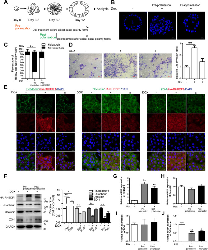
Background: The establishment of apicobasal polarity in epithelial cells is of critical importance in morphogenesis of mammary gland and other secretive gland tissues. The demise of the polarity is a critical step in early stages of tumorigenesis such as in breast ductal carcinoma in situ. The underlying molecular mechanism thus warrants in-depth investigations.
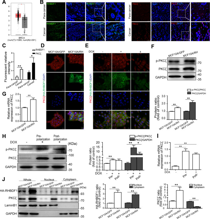
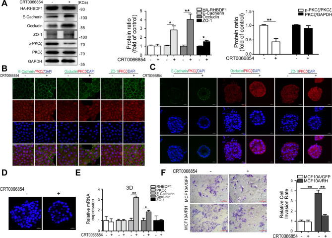
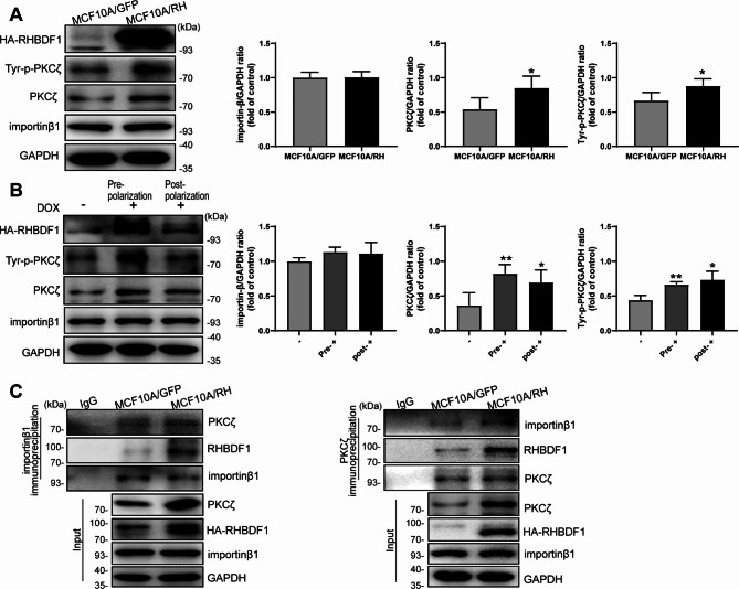
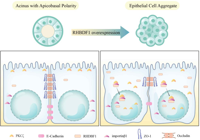
Results: Protein kinase C isoform ζ (PKCζ), which is highly expressed in breast cancer cells, accumulates in the nuclei of human mammary epithelial cells overexpressing human rhomboid family-1 (RHBDF1), an endoplasmic reticulum membrane protein. Nuclear translocation of PKCζ results in the failure of the formation of the cytosolic apicobasal polarity complex Par, of which PKCζ is an essential component. Additionally, enhanced nuclear translocation of PKCζ is accompanied by an inhibition of the expression of cell tight junction and adherens junction proteins and an increase of cell mobility. Mechanistically, RHBDF1 is able to interact with importin β1 and PKCζ and promote PKCζ phosphorylation. Consistently, treatment of RHBDF1-overexpressing cells with an inhibitor of PKCζ phosphorylation leads to restoration of apicobasal polarity and cell-cell junctions, as well as suppressed cell mobility.
Conclusions: RHBDF1-facilitated nuclear translocation of PKCζ is critically responsible for the dismantlement of epithelial cell apicobasal polarity, and thus may serve as a target in the development of therapeutic approaches against early stages of breast cancer.
Keywords: Adherens junction; Apicobasal polarity; Cell invasion; PKCζ; RHBDF1; Tight junction.
© 2024. The Author(s).
Conflict of interest statement
Declarations. Ethics approval and consent to participate: Breast tissue samples were collected from the full information database of breast cancer patients in the Breast Cancer Pathology and Research Laboratory of Cancer Hospital of Tianjin Medical University (Tianjin, China), including para-cancerous tissue and breast cancer tissue diagnosed as invasive breast cancer by two senior pathologists of six patients. The research procedure of this work is in line with the ethical standards of Tianjin Medical Oncology Institute and Hospital Ethics Committee. This study was approved by the Evaluation Committee of Cancer Hospital of Tianjin Medical University and all breast cancer patients were fully informed and signed the informed consent form. Conflict of interest: The authors declare no conflict of interest. Consent to participate and consent to publish: All authors consent to the publication of the data.
Figures
Similar articles
-
Perturbation of epithelial apicobasal polarity by rhomboid family-1 gene overexpression.FASEB J. 2018 Oct;32(10):5577-5586. doi: 10.1096/fj.201800016R. Epub 2018 May 4. FASEB J. 2018. PMID: 29727209
-
PKCzeta regulates cell polarisation and proliferation restriction during mammary acinus formation.J Cell Sci. 2010 Oct 1;123(Pt 19):3316-28. doi: 10.1242/jcs.065243. J Cell Sci. 2010. PMID: 20844151
-
PKCζ Promotes Breast Cancer Invasion by Regulating Expression of E-cadherin and Zonula Occludens-1 (ZO-1) via NFκB-p65.Sci Rep. 2015 Jul 28;5:12520. doi: 10.1038/srep12520. Sci Rep. 2015. PMID: 26218882 Free PMC article.
-
Tissue polarity-dependent control of mammary epithelial homeostasis and cancer development: an epigenetic perspective.J Mammary Gland Biol Neoplasia. 2010 Mar;15(1):49-63. doi: 10.1007/s10911-010-9168-y. Epub 2010 Jan 27. J Mammary Gland Biol Neoplasia. 2010. PMID: 20101444 Free PMC article. Review.
-
Cell polarity in motion: redefining mammary tissue organization through EMT and cell polarity transitions.J Mammary Gland Biol Neoplasia. 2010 Jun;15(2):149-68. doi: 10.1007/s10911-010-9180-2. Epub 2010 May 12. J Mammary Gland Biol Neoplasia. 2010. PMID: 20461450 Review.
References
-
- Freeman M. Rhomboid proteases and their biological functions. Annu Rev Genet. 2008;42:191–210. - PubMed
-
- Zhou Z, Liu F, Zhang ZS, Shu F, Zheng Y, Fu L, Li LY. Human rhomboid family-1 suppresses oxygen-independent degradation of hypoxia-inducible factor-1α in breast cancer. Cancer Res. 2014;74(10):2719–30. - PubMed
MeSH terms
Substances
Grants and funding
LinkOut - more resources
Full Text Sources
