. 2024 Nov 23;16(4):e70040.
doi: 10.1002/dad2.70040. eCollection 2024 Oct-Dec.

Elevated tau in the piriform cortex in Alzheimer's but not Parkinson's disease using PET-MR

Affiliations

Elevated tau in the piriform cortex in Alzheimer's but not Parkinson's disease using PET-MR

Hossein Moein Taghavi et al. Alzheimers Dement (Amst). .

Abstract

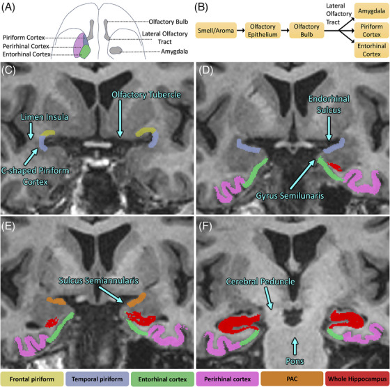
Introduction: Olfactory dysfunction can be an early sign of Alzheimer's disease (AD). We used tau positron emission tomography-magnetic resonance (PET-MR) to analyze a key region of the olfactory circuit, the piriform cortex, in comparison to the adjacent medial temporal lobe.

Methods: Using co-registered magnetic resonance imaging (MRI) and 18F-PI-2620 tau PET-MR scans in 94 older adults, we computed tau uptake in the piriform-periamygdaloid cortex, amygdala, entorhinal-perirhinal cortices, and hippocampus.

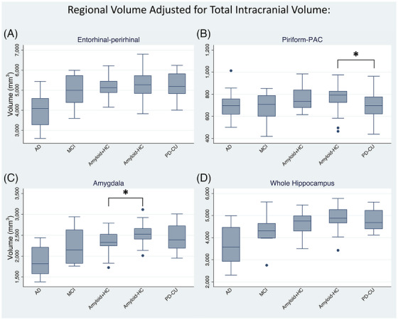
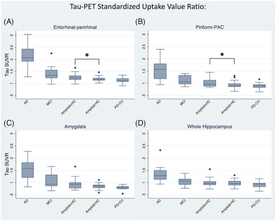
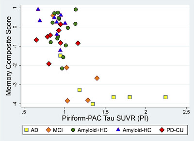
Results: We found an ordinal cross-sectional increase in piriform cortex tau uptake with increasing disease severity (amyloid-negative controls, amyloid-positive controls, mild cognitive impairment [MCI] and AD), comparable to entorhinal-perirhinal cortex. Amyloid-positive controls showed significantly greater tau uptake than amyloid-negative controls. Negative correlations were present between memory performance and piriform uptake. Piriform uptake was not elevated in cognitively unimpaired Parkinson's disease.

Discussion: Cross-sectionally, there is an early increase in tau uptake in the piriform cortex in AD but not in Parkinson's disease.

Highlights: Positron emission tomography-magnetic resonance (PET-MR) analysis of the piriform cortex sheds light on its role as a potential early region affected by neurodegenerative disorders underlying olfactory dysfunction.Uptake of tau tracer was elevated in the piriform cortex in Alzheimer's disease (AD) and mild cognitive impairment (MCI) but not in Parkinson's disease (PD).Memory performance was worse with greater piriform uptake.

Keywords: Alzheimer's; MRI; PD; PET; PET‐MR; olfaction; piriform cortex; tau.


Conflict of interest statement

Dr Kathleen Poston has been funded by grants to conduct research from the Michael J Fox Foundation for Parkinson's Research, the Knight Initiative for Brain Resilience, the Wu Tsai Neurosciences Institute, the Lewy Body Dementia Association, the Alzheimer's Drug Discovery Foundation, the Sue Berghoff LBD Research Fellowship, and the National Institutes of Health (NIH). She is on the Scientific Advisory Board for Curasen where she receives consulting fees and stock options. She is on the Scientific Advisory Board for Amprion, where she receives stock options. She is a consultant for Novartis, Biohaven, and Neuron23, where she receives consulting fees. Dr Michael Zeineh receives research funding from GE Healthcare. All other authors have no disclosures relevant to this manuscript. Author disclosures are available in the Supporting Information.

Figures

FIGURE 1
FIGURE 1
Piriform segmentation procedure overlaid on a T1‐weighted three‐dimensional (3D) inversion‐recovery fast spoiled gradient echo (IR‐FSPGR) image. (A) Inferior‐perspective brain illustration depicting the location of the piriform cortex with respect to other regions of the olfactory circuit. (B) Schematic view of the olfactory circuit connections. (C) The first most anterior slice starts where the white matter of the limen insula is completely fused. Frontal piriform (PirF) and temporal piriform (PirT) are segmented, forming halves of a “C.” PirF is segmented ~50% of the approximate distance to the olfactory tubercle, whereas PirT is segmented ~30% to the apex of the gyrus semilunaris. (D) PirF was not segmented. PirT was extended to 100% of the distance to the sulcus semiannularis. (E) PirT was replaced with periamygdaloid cortex (PAC), which was segmented the entire length of the gyrus semilunaris to the sulcus semiannularis. (F) Slice ~8: The appearance of the hippocampal fissure along the inferior aspect of the undulations of the hippocampal head marked the end of periamygdaloid segmentation. Note that the amygdala is segmented separately.
FIGURE 2
FIGURE 2
Regional volume corrected for total intracranial volume in Alzheimer's disease (AD), mild cognitive impairment (MCI), amyloid‐positive/‐negative healthy controls (amyloid+HC/amyloid–HC), and cognitively unimpaired Parkinson's disease (PD‐CU). (A) Entorhinal‐perirhinal, (B) Piriform‐PAC (periamygdaloid cortex), (C) amygdala, and (D) whole hippocampus volumes. All show cross‐sectionally increased volumes with decreasing disease status along the AD spectrum. * = Normal (cognitively unimpaired) PD‐CU showed decreased piriform‐PAC volume compared to amyloid–HC (p = 0.041), and amyloid+HC showed decreased amygdala volume compared to amyloid–HC (p = 0.006).
FIGURE 3
FIGURE 3
18F‐PI‐2620 tau uptake referenced to the inferior cerebellum. (A) Entorhinal‐perirhinal volume, (B) piriform‐PAC (periamygdaloid cortex), (C) amygdala, and (D) whole hippocampus. All show cross‐sectionally increased tau with increasing disease status. * = Amyloid+HC showed increased tau uptake compared to amyloid–HC in entorhinal‐perirhinal (p = 0.025) and piriform‐PAC (p = 0.042).
FIGURE 4
FIGURE 4
Alzheimer's Disease Research Center (ADRC) behavioral correlations with piriform‐PAC (periamygdaloid cortex) tau uptake. The memory composite score displayed a negative monotonic relationship relative to tau uptake in all four regions across participants, with piriform‐PAC tau shown here, and other regions in Figure S4.

References

    1. Braak H, Alafuzoff I, Arzberger T, Kretzschmar H, Tredici K. Staging of Alzheimer disease‐associated neurofibrillary pathology using paraffin sections and immunocytochemistry. Acta Neuropathol. 2006;112(4):389‐404. doi:10.1007/S00401-006-0127-Z/FIGURES/5 - DOI - PMC - PubMed
    1. Gallardo G, Holtzman DM. Amyloid‐β and Tau at the Crossroads of Alzheimer's Disease. Adv Exp Med Biol. 2019;1184:187‐203. doi:10.1007/978-981-32-9358-8_16/FIGURES/1 - DOI - PubMed
    1. Soria Lopez JA, González HM, Léger GC. Chapter 13 ‐ Alzheimer's disease. In: Dekosky ST, Asthana S, eds. Handbook of Clinical Neurology. Geriatric Neurology. Elsevier; 2019:231‐255. doi:10.1016/B978-0-12-804766-8.00013-3 - DOI - PubMed
    1. Doty RL. Handbook of Olfaction and Gustation. 3rd ed. 2015:1‐1264. doi:10.1002/9781118971758. Published online June. - DOI
    1. Tian Q, Bilgel M, Moghekar AR, Ferrucci L, Resnick SM. Olfaction, Cognitive Impairment, and PET Biomarkers in Community‐Dwelling Older Adults. J Alzheimers Dis : JAD. 2022;86(3):1275‐1285. doi:10.3233/JAD-210636 - DOI - PubMed

LinkOut - more resources