Review
. 2024 Oct 31;10(21):e39996.
doi: 10.1016/j.heliyon.2024.e39996. eCollection 2024 Nov 15.

Mechanistic insights and therapeutic potential of astilbin and apigenin in diabetic cardiomyopathy

Affiliations
Review

Mechanistic insights and therapeutic potential of astilbin and apigenin in diabetic cardiomyopathy

Sachin Dhiman et al. Heliyon. .

Abstract

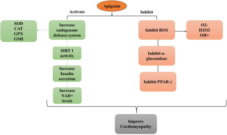
Diabetic cardiomyopathy (DCM) represents a critical complication of Diabetes mellitus (DM), characterized by structural and functional changes in the myocardium independent of coronary artery disease or hypertension. Emerging evidence highlights the significant roles of phytochemicals, particularly astilbin and apigenin, in modulating key molecular pathways implicated in DCM. This review synthesizes current mechanistic insights and therapeutic potential of these compounds, focusing on their interactions with AMP-activated protein kinase (AMPK), peroxisome proliferator-activated receptors (PPARs), O-linked N-acetylglucosamine (O-GlcNAc), sodium-glucose co-transporter 2 (SGLT2), protein kinase C (PKC), nuclear factor kappa B (NF-κB), mitogen-activated protein kinase (MAPK), and c-Jun N-terminal kinase (JNK) pathways. Astilbin and apigenin have demonstrated the ability to improve cardiac function, mitigate oxidative stress, and reduce inflammatory responses in diabetic conditions. By activating AMPK and PPARs, these flavonoids enhance glucose uptake and fatty acid oxidation, contributing to improved metabolic homeostasis. Their inhibition of O-GlcNAcylation, SGLT2 activity, and PKC signaling further attenuates hyperglycemia-induced cellular damage. Additionally, suppression of NF-κB, MAPK, and JNK pathways by astilbin and apigenin results in reduced pro-inflammatory cytokine production and apoptotic cell death. Collectively, these interactions position astilbin and apigenin as promising therapeutic agents for ameliorating DCM, offering novel avenues for treatment strategies aimed at modulating multiple pathogenic pathways.

Keywords: Apigenin; Astilbin; Cardiomyopathy; Diabetes.


Conflict of interest statement

The authors declare that they have no known competing financial interests or personal relationships that could have appeared to influence the work reported in this paper.

Figures

Fig. 1
Fig. 1
The following figure depicts the molecular pathways involved in diabetic cardiomyopathy (DCM) under the conditions of diabetes. Diabetes is marked by increased levels of hyperglycemia, high free fatty acids and impaired insulin signaling. These metabolic alterations precipitate an imbalance between fatty acid and glucose oxidation within the mitochondria that enhances fatty acid oxidation as well as reduces glucose oxidation.
Fig. 2
Fig. 2
This figure depicts the signalling pathways that describe the defective metabolism in diabetic cardiomyopathy. Diabetes increases the levels of fatty acids, hyperglycemia, and hyperinsulinemia due to which the balance of the oxidation of fatty acids versus oxidation of glucose within the mitochondria gets disturbed. Pathways involving AMPK and SIRT1 that regulate mitochondrial biogenesis and energy homeostasis are shown on the left. Impaired function of AMPK reduces PGC-1α recruitment, contributing to decreased mitochondrial biogenesis and impaired energy production in the diabetic cardiomyopathy.
Fig. 3
Fig. 3
This figure indicates the results of PPAR-γ activation in diabetic cardiomyopathy: it acts in the context of metabolic regulation and inflammation. This increases the uptake of cholesterol and lipid catabolism as well to support mitochondrial function in energy production and reduction of lipid accumulation in the diabetic heart. Another effect of PPAR-γ is its ability to decrease the formation of reactive oxygen species (ROS) and factors pro-inflammatory, increase insulin sensitivity, thus decrease oxidative stress and inflammation, both of which are highly involved in the pathogenesis of DCM. Essentially, activation of PPAR-γ conveys protective effects in the sense that it augments the metabolic function and decreases inflammation of DCM.
Fig. 4
Fig. 4
This figure depicts the molecular pathways that affect functional changes in diabetic cardiomyopathy. Elevated O-GlcNAcylation activates CaMKII; activated CaMKII then disrupts the function of phospholamban and SERCA, impairs calcium handling, reduces contractility and relaxation in myocardium. Myocardial impairment is further worsened by ROS and altered potassium channel function. Collectively, these changes make significant contribution to the heart failure progression in the diabetic heart.
Fig. 5
Fig. 5
This figure captures the cardiovascular effects of SGLT2i, indicating how they affect pathways in the heart and in endothelial cells. SGLT2i activate pathways of AKT and AMP-activated protein kinase (AMPK), and through this, leads to the activation of endothelial nitric oxide synthase (eNOS) to produce nitric oxide (NO). This is very important for vascular functions. Additionally, SGLT2i increases AMPK activation, promoting the expression of proteins PGC-1α and ULK-1 that contribute to mitochondrial biogenesis and autophagy, respectively. Activating Sestrin 2 with SGLT2i activates additional downstream targets with antioxidant and anti-inflammatory effects that support cardiovascular benefits.
Fig. 6
Fig. 6
The figure illustrates the process that hyperglycemia triggers to endothelial dysfunction, but more specifically earmarks PKC activation as the mediator of selective insulin resistance. Its effects have been detected to lower downstream signalling molecules such as PI3K, MAPK, NO, VEGF, ET-1, and CTGF. Hyperglycemia activates PKC, and this dramatically depresses both PI3K and NO in a way that decreases VEGF. This results in a parallel increase in activity of MAPK and an increase in the levels of ET-1. Both of these pathways can result in endothelial dysfunction and contribute to the expression of CTGF, thereby explaining the molecular interactions in complications of diabetes involving vascular health.
Fig. 7
Fig. 7
This figure represents a pathway model that explains the mechanism by which elevated blood glucose levels leads to diabetic cardiomyopathy through a cascade of biochemical events. Elevated glucose levels favor increased levels of advanced glycation end-products (AGEs) along with their interaction with receptors (RAGE), generation of reactive oxygen species (ROS), and elevations in levels of inflammatory cytokines (IL-1/TNFα). These activations activate nuclear factor kappa-light-chain-enhancer of activated B cells, NF-κB, leading to the increased expression of mitogen-activated protein kinase, protein kinase B, and peroxisome proliferator-activated receptors. The enhanced signaling molecules cause inflammation, fibrosis, and hypertrophy, thus resulting in the creation of diabetic cardiomyopathy.
Fig. 8
Fig. 8
Structure of apigenin.
Fig. 9
Fig. 9
Pharmacological properties of Apigenin.
Fig. 10
Fig. 10
The diagram depicts cardioprotective effects of Apigenin with a variety of biochemical pathways. Through activation of SOD, CAT, GPX, and GSH to the body's endogenous defense mechanism, Apigenin triggers subsequent increase in SIRT 1 activity that, in turn further stimulates insulin secretion and enhances NAD + levels. The other end is the inhibition of the formation of ROS molecules that include O2-, H2O2, and OH+, in addition to a downregulating activity that affects the activities of α-glucosidase and peroxisome proliferator-activated receptor gamma (PPAR-γ). These accumulate to result in improved outcomes for cardiomyopathy.
Fig. 11
Fig. 11
Structure of astilbin.
Fig. 12
Fig. 12
A mechanistic pathway of inflammatory stimulation and its relationship with diabetic cardiomyopathy can be described on a diagram focusing on Astilbin as an inhibitor. Inflammatory stimulators including phospholipase A2 and phospholipase C are engaged into arachidonic acid release, which ends in prostaglandins synthesis through COX, in association with leukotriene synthesis due to LOX, causing anaphylaxis, vasodilation, and chemotaxis. These processes activate polymorphonuclear cells, mononuclear cells, and lymphocytes and release inflammatory mediators such as SOD, iNOS, ROS, and hydrolytic enzymes to deal with injury. Activation of the key transcription factors Nrf2, JNK, and NF-κB and proinflammatory cytokines also contribute to inflammation and tissue damage. Astilbin acts on several steps in this cascade, such as phospholipase A2, COX, and the function of immunocompetent cells like CD4+ and CD8+, and it reduced the emission of proinflammatory cytokines and inflammation thus, alleviating diabetic cardiomyopathy.

References

    1. Tabish S.A. Is diabetes becoming the biggest epidemic of the twenty-first century? Int. J. Health Sci. 2007;1(2):V. - PMC - PubMed
    1. Low Wang C.C., et al. Clinical update: cardiovascular disease in diabetes mellitus: atherosclerotic cardiovascular disease and heart failure in type 2 diabetes mellitus–mechanisms, management, and clinical considerations. Circulation. 2016;133(24):2459–2502. - PMC - PubMed
    1. Whelton P.K., Williams B. The 2018 European society of cardiology/European society of hypertension and 2017 American college of cardiology/American heart association blood pressure guidelines: more similar than different. JAMA. 2018;320(17):1749–1750. - PubMed
    1. Davies M.J., et al. Management of hyperglycaemia in type 2 diabetes, 2018. A consensus report by the American diabetes association (ada) and the European association for the study of diabetes (easd) Diabetologia. 2018;61:2461–2498. - PubMed
    1. Wang M, et al. Endothelial dysfunction and diabetic cardiomyopathy. Front. Endocrinol. 2022;13:851941. - PMC - PubMed

LinkOut - more resources