Review
. 2024 Sep 24;11(4):309-317.
doi: 10.1080/23328940.2024.2401674. eCollection 2024.

Prostaglandin E2 production in the brainstem parabrachial nucleus facilitates the febrile response

Affiliations
Review

Prostaglandin E2 production in the brainstem parabrachial nucleus facilitates the febrile response

Anders Blomqvist. Temperature (Austin). .

Abstract

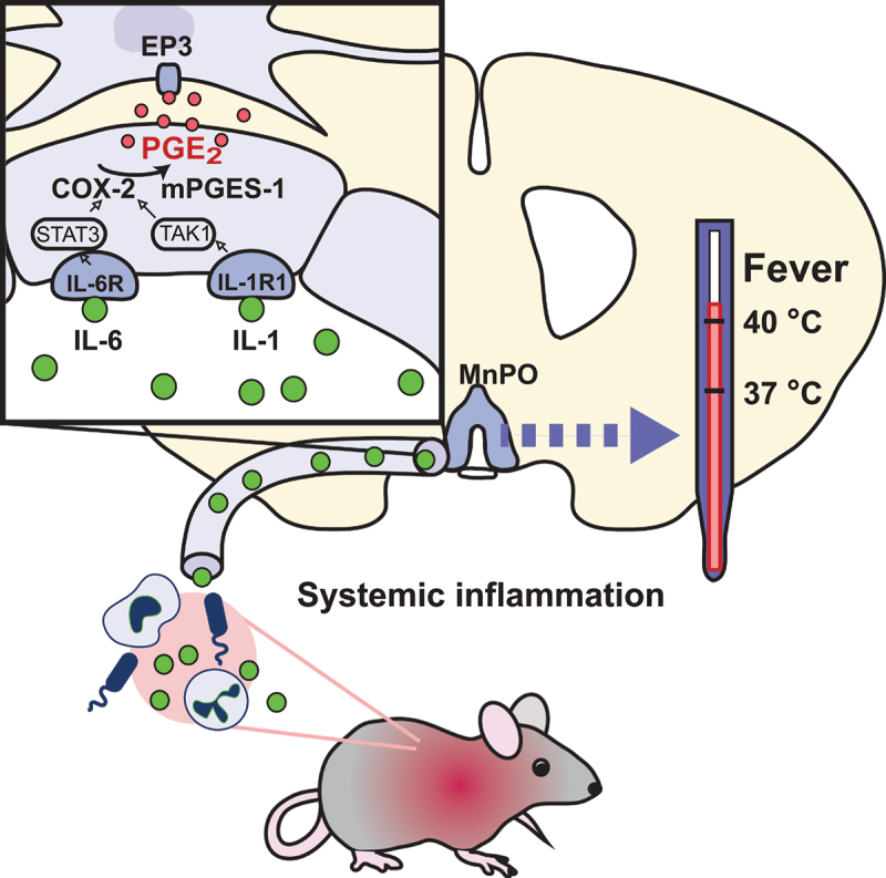
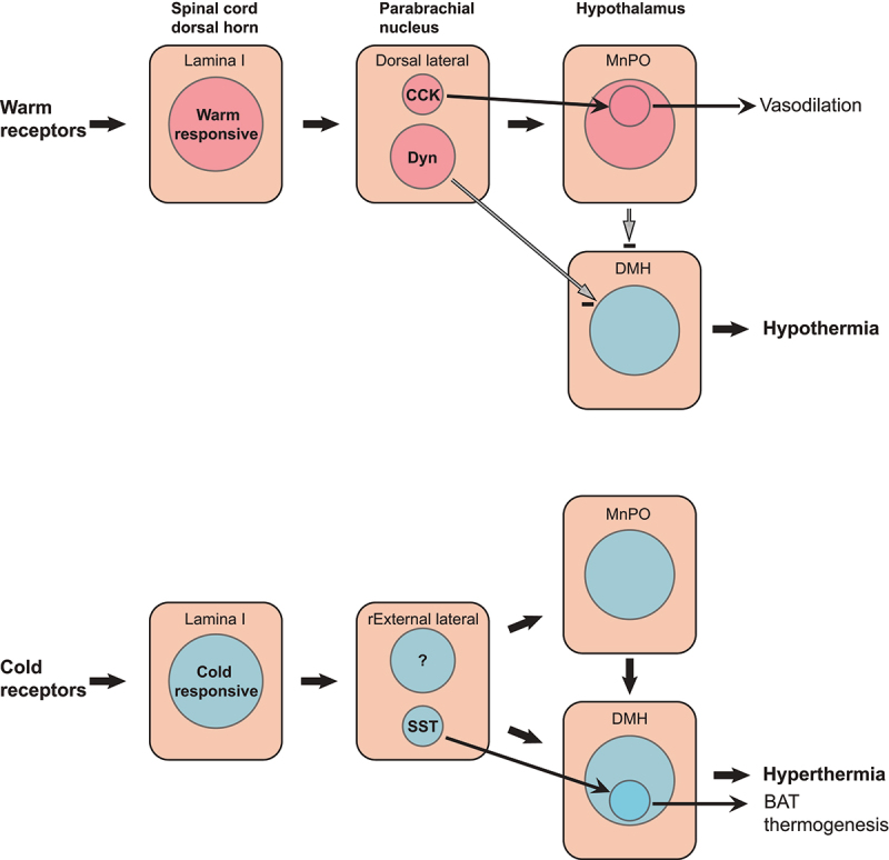
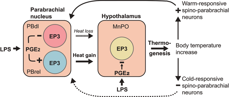
Our body temperature is normally kept within a narrow range of 1°C. For example, if our body temperature rises, such as in a hot environment or due to strenuous exercise, our thermoregulatory system will trigger a powerful heat defense response with vasodilation, sweating, and lowered metabolism. During fever, which often involves body temperatures of up to 41°C, this heat defense mechanism is apparently inhibited; otherwise, the rising body temperature would be immediately combated, and fever would not be allowed to develop. New evidence suggests how and where this inhibition takes place. In two consecutive studies from Cheng et al. and Xu et al., it has been shown that prostaglandin E2, which generates fever by acting on thermosensory neurons in the preoptic hypothalamus, also acts on neurons in the brainstem parabrachial nucleus, which receive temperature information from temperature-activated spinal cord neurons and relay this information to the thermoregulatory center in the hypothalamus to either induce cold or heat defenses. By acting on the same type of prostaglandin E2 receptor that is critical for fever generation in the preoptic hypothalamus, the EP3 receptor, prostaglandin E2 inhibits the signaling of the heat-responsive parabrachial neurons, while stimulating the cold-responsive neurons. These novel findings thus show that prostaglandin E2, by binding to the same receptor subtype in the parabrachial nucleus as in the preoptic hypothalamus, adjusts the sensitivity of the thermosensory system in a coordinated manner to allow the development of febrile body temperatures.

Keywords: EP3 receptors; Fever; PGE2; median preoptic nucleus; thermosensory pathways.


Conflict of interest statement

No potential conflict of interest was reported by the author(s).

Figures

Figure 1.
Figure 1.
Mechanisms of inflammation-induced fever. During systemic inflammation, cytokines, such as IL-1 and IL-6, are released into the circulation. The binding of these cytokines to their receptors (IL-1R1, IL-6R) on brain endothelial cells induces via the NFκB (TAK1) and STAT3 pathways the expression of COX-2 and mPGES-1, resulting in the production of PGE2 that is released into the brain parenchyma where it by binding to EP3 receptors in the median preoptic nucleus (MnPO) evokes thermogenesis. Adapted from [14].
Figure 2.
Figure 2.
Top: Radio-labeled in situ hybridization showing EP3 receptor expression in the parabrachial nucleus of the rat. a–d are frontal sections ordered from rostral to caudal. cl, central lateral nucleus; dl, dorsal lateral nucleus; el, external lateral nucleus; il, internal lateral nucleus; KF, Kölliker–fuse nucleus; m, medial parabrachial nucleus; scp, superior cerebellar peduncle; sl, superior lateral nucleus; vl, ventral lateral nucleus. Adapted from [41]. Bottom: The cytoarchitecture of the different parabrachial subnuclei is shown in thionin-stained sections (taken from [42]) that are approximately from the same rostrocaudal levels as those showing the EP3 receptor expression. Sections are separated by 140 μm, with section b being at the level of separation of the inferior colliculus from the pons. Scale bar = 100 μm but note that magnification in the top dark-field micrographs is about two times higher.
Figure 3.
Figure 3.
Co-expression of digoxigenin labeled preprodynorpin (ppDYN) mRNA (a) and radiolabeled EP3 receptor mRNA (a1) in the lateral parabrachial nucleus of the rat. The dynorphin neurons in the dorsal lateral subnucleus (dl, a2) display extensive double labeling with EP3 (wide arrowheads). Another ppDYN expressing population, located in the inner part of the external lateral subnucleus (el, a3), is single labeled. cl, central lateral subnucleus; scp, superior cerebral peduncle. Adapted from [53].
Figure 4.
Figure 4.
The thermoregulatory spino-parabrachial-hypothalamic pathways. Warm-responsive spinal dorsal horn neurons activate median preoptic (MnPO)-projecting dynorphinergic (Dyn) and cholecytokininergic (CCK) neurons in the dorsal lateral parabrachial subnucleus to evoke a cold defense response, with the CCK neurons specifically being involved in vasodilation. The dynorphinergic neurons also directly project to the dorsomedial hypothalamus (DMH), where they inhibit thermogenesis-promoting neurons. Cold-responsive dorsal horn neurons activate MnPO and DMH projecting neurons in the rostral-to-external (rExternal) lateral subnucleus, which evoke hyperthermia, with neurons expressing somatostatin (SST) being specifically involved in brown adipose tissue (BAT) thermogenesis.
Figure 5.
Figure 5.
Concerted action of the thermogenic and afferent thermoregulatory systems in creating the febrile response. An inflammatory stimulus, such as lipopolysaccharide (LPS) results in the formation of PGE2 in the brain. The binding of PGE2 to EP3 receptors on neurons in the median preoptic nucleus (MnPO) inhibits these neurons, resulting in release of thermogenesis and subsequent rise in body temperature (fever). The increased body temperature will, while inhibiting cold-responsive thermoreceptive neurons, activate warm-responsive spino-parabrachial neurons that target heat defense eliciting neurons in dorsal lateral subnucleus of the parabrachial nucleus (PBdl). Their activation would hence counteract the PGE2 induced thermogenesis. However, the PGE2 produced in the parabrachial nucleus will through binding to EP3 receptors on the PBdl neurons inhibit these neurons, while at the same time activating the neurons in the rostral-to-external lateral subnucleus (PBrel) that are involved in the cold defense, hence creating a concerted action with the PGE2 released in the MnPO. Solid and dotted lines indicate activated and inhibited pathways, respectively.

Similar articles

References

    1. Evans SS, Repasky EA, Fisher DT.. Fever and the thermal regulation of immunity: the immune system feels the heat. Nat Rev Immmunol. 2015;15(6):335–349. doi: 10.1038/nri3843 - DOI - PMC - PubMed
    1. Earn DJ, Andrews PW, Bolker BM. Population-level effects of suppressing fever. Proc Biol Sci. 2014;281(1778):20132570. doi: 10.1098/rspb.2013.2570 - DOI - PMC - PubMed
    1. Schulman CI, Namias N, Doherty J, et al. The effect of antipyretic therapy upon outcomes in critically ill patients: a randomized, prospective study. Surg Infect. 2005;6(4):369–375. doi: 10.1089/sur.2005.6.369 - DOI - PubMed
    1. Kurosawa S, Kobune F, Okuyama K, et al. Effects of antipyretics in rinderpest virus infection in rabbits. J Infect Dis. 1987;155(5):991–997. doi: 10.1093/infdis/155.5.991 - DOI - PubMed
    1. Engblom D, Saha S, Engstrom L, et al. Microsomal prostaglandin E synthase-1 is the central switch during immune-induced pyresis. Nat Neurosci. 2003;6(11):1137–1138. doi: 10.1038/nn1137 - DOI - PubMed