. 2024 Dec;13(1):2432372.
doi: 10.1080/22221751.2024.2432372. Epub 2024 Nov 28.

Development and characterization of high-efficiency cell-adapted live attenuated vaccine candidate against African swine fever

Affiliations

Development and characterization of high-efficiency cell-adapted live attenuated vaccine candidate against African swine fever

Min Ho Kim et al. Emerg Microbes Infect. 2024 Dec.

Abstract

African swine fever (ASF), a contagious and lethal haemorrhagic disease of domestic pigs and wild boars, poses a significant threat to the global pig industry. Although experimental vaccine candidates derived from naturally attenuated, genetically engineered, or cell culture-adapted ASF virus have been tested, no commercial vaccine is accepted globally. We developed a safe and effective cell-adapted live attenuated vaccine candidate (ASFV-MEC-01) by serial passage of a field isolate in CA-CAS-01-A cells. ASFV-MEC-01, isolated via repeated plaque purification using next-generation sequencing analysis, was obtained at passage 18 and showed significant attenuation in 4- and 6-week-old pigs. ASFV-MEC-01 conferred 100% protection against challenge with lethal parental ASFV, which correlated with high ASFV-specific humoral and cellular immune responses. Additionally, ASFV-MEC-01 was not detected in blood until 28 days post-inoculation. Global transcriptome analysis showed that ASFV-MEC-01 lacking 12 genes triggered stronger innate antiviral responses than the parental virus, as exemplified by high levels of mRNA encoding interferon regulatory and inflammatory genes in PAM cells. Ectopic expression of most deleted genes increased replication of DNA viruses by suppressing production of interferons and pro-inflammatory cytokines. Among the genes deleted from ASFV-MEC-01, MGF100-1R interacted specifically with the scaffold dimerization domain of TBK1, thereby preventing TBK1 dimerization and impairing TBK1-mediated type I IFN and NF-κB signalling. These results suggest that attenuation of ASFV-MEC-01 may be mediated by induction of stronger type I IFN and NF-κB signalling within the host innate immune system. Thus, ASFV-MEC-01 could be a safe and effective live attenuated ASFV vaccine candidate with commercial potential.

Keywords: African swine fever, CA-CAS-01-A cell, live-attenuated virus, ASFV-MEC-01, vaccine, Type I IFN, MGF100-1R.


Conflict of interest statement

No potential conflict of interest was reported by the author(s).

Figures
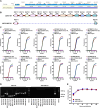
Figure 1.
Figure 1.
Characterization of the CA-CAS-01-A cell-adapted ASFV strain. (A) Next-generation sequencing of the CA-CAS-01-A cell-adapted ASFV strain (ASFV-MEC-01). Blue peaks denote coverage when mapped to the Georgia 2007 strain. (B) Schematic depiction of the genetic deletion in the left variable region of ASFV-MEC-01. The dashed line indicates the deleted genome region. (C) Assessment of the genetic deletions by qPCR. Fluorescence amplification curves of ASFV-MEC-01 displayed positive signals for viral p72, but not for the deleted genes. (D) Assessment of genetic deletions by PCR. Viral DNA obtained from parental ASFV or ASFV-MEC-01 were subjected to PCR amplification. PCR products were visualized by agarose gel electrophoresis. (E) In vitro growth characteristics of the ASFV-MEC-01 compared with those of parental ASFV. Primary PAMs were infected with each virus at an MOI = 0.01, and viral replication was measured indirectly at 2, 24, 48, 72, 96, and 120 h post-infection by detecting expression of the p72 gene by qPCR.
Figure 2.
Figure 2.
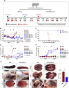
Safety and efficacy of ASFV-MEC-01 as a live attenuated virus vaccine in domestic pigs: Experiment 1. (A) A schematic representation of the strategy used to evaluate the safety and efficacy of ASFV-MEC-01. The test group (n = 3 pigs) was vaccinated intramuscularly (IM) with 105 HAD50 ASFV-MEC-01. The control group (n = 3) was mock-vaccinated with the same volume of PBS. A booster vaccination was given at 14 days post-vaccination (DPV). At 28 DPV, all pigs were challenged IM with 102 HAD50 parental ASFV. Rectal temperatures were measured and blood samples were collected on the days depicted in the schematic diagram. Survival was monitored for 21 DPC. At the point of death or experiment termination, all pigs were subjected to necropsy. (B) Rectal temperature, as measured during the period of vaccination and virulent challenge. A temperature >40°C (dashed line) was defined as fever. (C) Survival of ASFV-MEC-01-vaccinated or mock-vaccinated pigs. (D) Systemic viremia in blood during the vaccination and virulent challenge periods were measured indirectly by detecting the expression of the p72 gene by qPCR. (E) Representative images show macroscopic changes in the spleen, lymph node, lung, kidney, liver, and heart of one mock-vaccinated and one ASFV-MEC-01 pig at the point of death or experimental termination; the overall macroscopic lesion score for mock-vaccinated and ASFV-MEC-01-vaccinated pigs is also shown. (F) ASFV p32-, p62-, and p72-specific antibody titres were measured in an indirect ELISA. Data are presented as the mean ± SE. *p < 0.05 (student's t-test; panel E). Data in panels B, D, and F are presented as individual values for each animal in each of the groups.
Figure 3.
Figure 3.
Safety and efficacy of ASFV-MEC-01 as a live attenuated virus vaccine in domestic pigs: Experiment 2. (A) Schematic representation showing the strategy used to evaluate the safety and efficacy of ASFV-MEC-01. The test group (n = 4) was vaccinated intramuscularly (IM) with 105 HAD50 ASFV-MEC-01. The control group (n = 2) received the same volume of PBS. A booster was given 21 DPV. All pigs were challenged IM with 102 HAD50 parental ASFV at 35 DPV. Rectal temperatures were measured and blood samples were collected on the days depicted in the schematic diagram. Survival was monitored for 14 days post-challenge. (B) Rectal temperature during the period of vaccination and virulent challenge. A temperature >40°C (dashed line) was defined as fever. (C) Survival of ASFV-MEC-01-vaccinated or mock-vaccinated pigs. (D) Systemic viremia in the blood during the period of vaccination and virulent challenge was measured indirectly by detecting the expression of the p72 gene by qPCR. (E) ASFV p32-, p62-, and p72-specific antibody titres were measured in an indirect ELISA. (F) ASFV-specific IFN-γ responses by PBMCs from vaccinated pigs were measured in an IFN-γ ELISA (PHA: phytohemaglutinin). Data are presented as the mean ± SE. Statistical analyses were performed using two-way ANOVA with Dunnett's multiple comparisons test. ***p < 0.001 (panel F). The data presented in panels B, D, and E are individual values for each animal in each group.
Figure 4.
Figure 4.
ASFV-MEC-01-infected primary PAMs show an antiviral innate immune signature. (A) Volcano map of differentially expressed genes (DEGs) in primary PAMs infected with parental ASFV or ASFV-MEC-01 (red dots = significantly upregulated DEGs; grey dots = no significant change; change green dots = significantly downregulated DEGs. (B) Scatter plots showing changes in expression of genes in primary PAMs infected with parental ASFV or ASFV-MEC-01 (red dots = upregulated; black dots = no change; green dots = downregulated; line = upregulated antiviral innate immune genes). (C) Clustering heatmap depicting changes in the transcript levels of IFN regulatory and inflammatory cytokines in parental ASFV- or ASFV-MEC-01-infected primary PAMs relative to those in non-infected primary PAMs or ASFV-MEC-01-infected primary PAMs relative to parental ASFV infected primary PAMs (cutoff: log2-fold change > 1.5). The colour scale shows the z-score, which represents the expression of mRNA encoding each gene: blue = low; red = high. (D) qRT-PCR analysis of interferon, interferon regulatory gene, and inflammatory cytokine mRNA expression at 12 and 24 h post-infection of primary PAMs with parental ASFV or ASFV-MEC-01. (E) Primary PAMs were infected for 18 h with ASFV-WT or ASFV-MEC-01 at an MOI of 0.1, 0.5 or 1. Cell supernatant was collected and subjected to an ELISA assay. Data are presented as the mean ± S.D. *p < 0.05 and **p < 0.01; unpaired Student's t-test (panel D).
Figure 5.
Figure 5.
Genes deleted from ASFV-MEC-01 induce virus replication and negatively regulate the innate immune response of 3D4/21 cells. (A) 3D4/21 cells were transfected for 12 h with the indicated genes or an empty vector, followed by infection with ADV-GFP at an MOI of 1. After 2 h, the medium was replaced with a complete cell culture medium containing 10% FBS. GFP expression was measured at 24 hpi. (B) GFP absorbance was measured in a Gloma multi-detection luminometer (Promega). (C) Titration of the virus in the cell supernatant and in cells was performed in a standard plaque assay, and the results were expressed as plaque-forming units (PFU). ELISA to measure the concentration of (D) IFN-β, (E) IL-6, (F) IFN-α, and (G) IL-12 in the cell supernatant. Data are presented as the mean ± S.D. *p < 0.05 and **p < 0.01; unpaired Student's t-test.
Figure 6.
Figure 6.
MGF100-1R, MGF110-8L, MGF110-10L-14L, and MGF300-1L negatively regulate antiviral immune responses in 3D4/21 cells. (A-D) Fluorescence microscopy (left) of 3D4/21 cells transiently overexpressing an empty vector plasmid or a plasmid encoding Flag-tagged (A) MGF100-1R, (B) MGF110-8L, (C) MGF110-10L-14L, and (D) MGF300-1L to assess green fluorescence absorbance (middle) and virus replication (right) at 12 and 24 hpi post-infection with HSV-GFP (MOI = 1). (E-H) ELISA of IFN-β (left) and IL-6 (right) secretion in the supernatant in HSV-GFP stimulated Flag-tagged (E) MGF100-1R, (F) MGF110-8L, (G) MGF110-10L-14L, and (H) MGF300-1L overexpressed cells compared with the empty vector expressed samples stimulated with HSV-GFP. Data are representative of at least two independent experiments, each with similar results, and are expressed as the mean ± S.D. of two biological replicates. *p < 0.05, **p < 0.01 and ***p < 0.001 (unpaired Student's t-test).
Figure 7.
Figure 7.
MGF100-1R inhibits cGAS-STING pathway signalling and transcription of antiviral genes. (A and B) MGF100-1R-expressing 3D4/21 or PK15 cells and control cells were infected with ADV-GFP (MOI = 1) and harvested at the indicated times. The phosphorylated versus intact forms of TBK1, IRF3, IKBα, p65, and STAT1 were detected by immunoblotting. β-actin was used as the internal control to ensure equal amounts of protein in the samples. All immunoblot data are representative of at least two independent experiments, each with similar results. (C and D) MGF100-1R-expressing 3D4/21 or PK15 cells were infected with ADV-GFP (MOI = 1) and harvested at the indicated times. Expression of interferon regulatory genes and inflammatory genes were analysed by RT-qPCR. Data are representative of at least two independent experiments, each with similar results, and are expressed as the mean ± S.D. of two biological replicates. *p < 0.05, **p < 0.01 and ***p < 0.001 (unpaired Student's t-test).
Figure 8.
Figure 8.
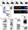
MGF100-1R interacts with TBK1 and inhibits dimerization. (A) IFN-β luciferase activity in HEK293 T cells expressing the IFN-β promoter (Firefly) and TK-Renilla (internal control), stimulated with Poly (dA:dT), 2′,3′ cGAMP, STING, TBK1, or IRF3-5D and increasing doses (50, 100, 200, and 400 ng) of MGF100-1R for 24 h. (B) HEK293 T cells were co-transfected with an empty vector (Flag) or Flag-MGF100-1R together with GST-TBK1 plasmids. The cell lysates were subjected to immunoprecipitation (IP) with an anti-Flag antibody, followed by immunoblotting (IB) with the indicated antibodies. (C) HEK293 T cells were transfected with an empty vector (Flag) or Flag-MGF100-1R. The cell lysates were then subjected to IP with an anti-Flag antibody, followed by IB with the indicated antibodies. (D) Immunofluorescence analysis (confocal microscopy) shows colocalization of Strep-tagged MGF100-1R (red) and Flag-tagged TBK1 (green) in PK15 cells. Nuclei were stained with DAPI (blue). (E) Immunofluorescence analysis (confocal microscopy) shows colocalization of Flag-tagged MGF100-1R (red) and endogenous TBK1 (green) in PK15 cells. Nuclei were stained with DAPI (blue). Scale bar 16.5 μm. (F and G) Schematic representation of the GST-tagged domain constructs of TBK1, the kinase domain (KD), the ubiquitin-like domain (ULD), coil-coiled domain 1/scafold dimerization domain (CCD1/SDD), and coil-coiled domain 2 (CCD2). (H) HEK293 T cells were cotransfected with an empty vector (GST) or plasmids containing GST-tagged TBK1 constructs together with a Flag-MGF100-1R plasmid. The cell lysates were then subjected to GST bead pulldown (GST-PD) and IB with the indicated antibodies. (I) HEK293 T cells were cotransfected with a control vector (GST) or with GST-TBK1 or V5-TBK1, along with increasing doses of a Flag-tagged MGF100-1R plasmid. (J) HEK293 T cells transfected with a V5-tagged empty vector or TBK1 along with increasing doses of Flag-tagged MGF100-1R and HA-tagged ubiquitin plasmids. Cell lysates were subjected to IP with anti-V5 antibodies, followed by IB with the indicated antibodies. (K) HEK293 T cells transfected with a V5-tagged empty vector or TBK1 plasmids together with an increasing dose of the Flag-tagged MGF100-1R plasmid. Cell lysates were subjected to IB with the indicated antibodies to detect phosphorylated (p-)TBK1. Data are representative of two independent experiments, each with similar results, and are expressed as the mean ± S.D. of two biological replicates. *p < 0.05, and **p < 0.01 (unpaired Student's t-test).

References

    1. Dixon LK, Chapman DA, Netherton CL, et al. . African swine fever virus replication and genomics. Virus Res. 2013;173(1):3–14. doi:10.1016/j.virusres.2012.10.020. - DOI - PubMed
    1. Qu H, Ge S, Zhang Y, et al. . A systematic review of genotypes and serogroups of African swine fever virus. Virus Genes. 2022;58(2):77–87. doi:10.1007/s11262-021-01879-0. - DOI - PMC - PubMed
    1. Lee HS, Bui VN, Dao DT, et al. . Pathogenicity of an African swine fever virus strain isolated in Vietnam and alternative diagnostic specimens for early detection of viral infection. Porcine Health Manage. 2021;7:1–11. doi:10.1186/s40813-020-00179-7. - DOI - PMC - PubMed
    1. Ranganatha S, Rathnamma D, Isloor S, et al. . African swine fever: analysing its epidemiology, pathogenesis and control strategies: A review. Indian J Anim Res; 1(10.
    1. Chandana M, Nair SS, Chaturvedi V, et al. . Recent progress and major gaps in the vaccine development for African swine fever. Braz J Microbiol. 2024;55(1):997–1010. doi:10.1007/s42770-024-01264-7. - DOI - PMC - PubMed

MeSH terms

LinkOut - more resources