. 2024 Nov 30;38(22):e70185.
doi: 10.1096/fj.202401664RR.

Overexpression of enhanced yellow fluorescent protein fused with Channelrhodopsin-2 causes contractile dysfunction in skeletal muscle

Affiliations

Overexpression of enhanced yellow fluorescent protein fused with Channelrhodopsin-2 causes contractile dysfunction in skeletal muscle

Syeda N Lamia et al. FASEB J. .

Abstract

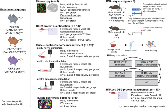
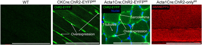
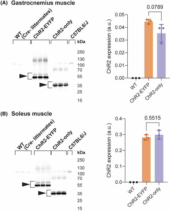
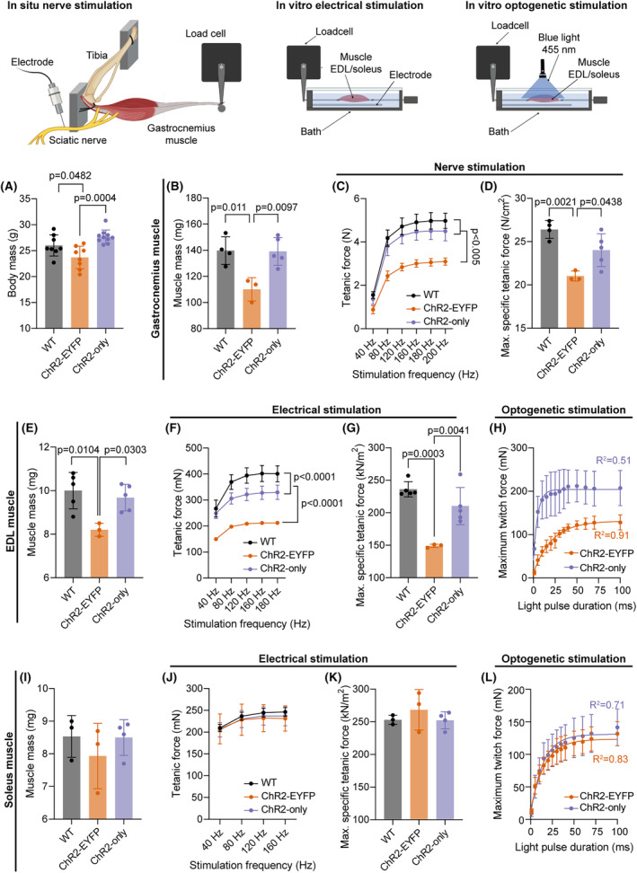
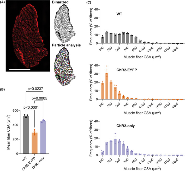
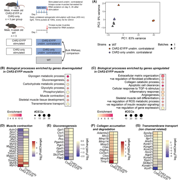
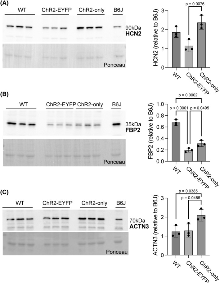
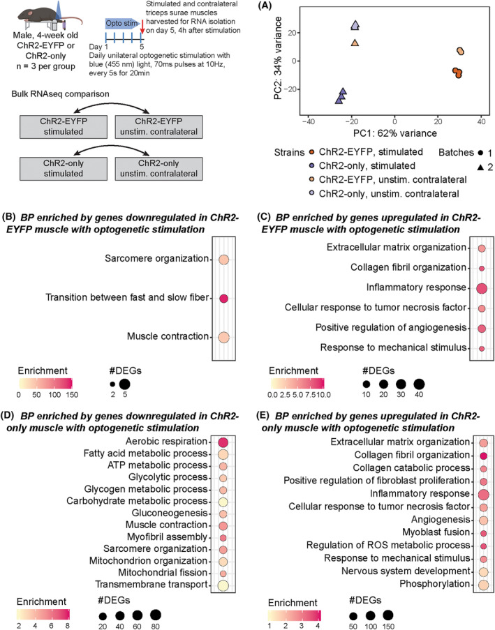
Skeletal muscle activation using optogenetics has emerged as a promising technique for inducing noninvasive muscle contraction and assessing muscle function both in vivo and in vitro. Transgenic mice overexpressing the optogenetic fusion protein, Channelrhodopsin 2-EYFP (ChR2-EYFP) in skeletal muscle are widely used; however, overexpression of fluorescent proteins can negatively impact the functionality of activable tissues. In this study, we characterized the contractile properties of ChR2-EYFP skeletal muscle and introduced the ChR2-only mouse model that expresses light-responsive ChR2 without the fluorescent EYFP in their skeletal muscles. We found a significant reduction in the contractile ability of ChR2-EYFP muscles compared with ChR2-only and WT mice, observed under both electrical and optogenetic stimulation paradigms. Bulk RNAseq identified the downregulation of genes associated with transmembrane transport and metabolism in ChR2-EYFP muscle, while the ChR2-only muscle did not demonstrate any notable deviations from WT muscle. The RNAseq results were further corroborated by a reduced protein-level expression of ion channel-related HCN2 in ChR2-EYFP muscles and gluconeogenesis-modulating FBP2 in both ChR2-EYFP and ChR2-only muscles. Overall, this study reveals an intrinsic skeletal dysfunction in the widely used ChR2-EYFP mice model and underscores the importance of considering alternative optogenetic models, such as the ChR2-only, for future research in skeletal muscle optogenetics.

Keywords: Channelrhodopsin‐2; function; optogenetics; skeletal muscle; structure.


Conflict of interest statement

The authors have stated explicitly that there are no conflicts of interest in connection with this article.

Figures

FIGURE 1
FIGURE 1
Study design: schematic of experiments with mouse strains and number of mice utilized. Mice with muscle‐specific expression of optogenetic ChR2‐EYFP fusion or ChR2‐only protein (referred to as ChR2‐EYFP or ChR2‐only strains, respectively) and their wild‐type (WT) littermates were used in various experiments. Young adult (2–3‐month‐old) mice were used in microscopy, muscle contractility with both optogenetic and gold standard nerve or electrical stimulation, and fiber cross‐sectional area measurement. Muscles from young (5‐week‐old) mice were harvested for protein measurements. For RNA sequencing, right triceps surae muscles of young (4‐week‐old) ChR2‐EYFP and ChR2‐only mice were stimulated with blue light (455 nm) to induce optogenetic muscle contraction. After 5 daily bouts of 20 min stimulation, stimulated and age‐matched WT mice were euthanized to harvest the triceps surae for sequencing. The superscripts ‘u’ or ‘v’ indicate same mice were used across different experiments. The number of mice used in each experiment are presented as WT, ChR2‐EYFP, and ChR2‐only, respectively.
FIGURE 2
FIGURE 2
Abnormal ChR2‐EYFP clustering was observed in EYFP‐positive optogenetic mouse skeletal muscle. Confocal imaging demonstrated ChR2‐EYFP overexpression in the longitudinal plane of EDL muscle (CKCre;ChR2‐EYFPfl/fl muscle) and the transverse plane of gastrocnemius muscle (Acta1Cre;ChR2‐EYFPfl/fl muscle). Normal muscle membranes were visible in Acta1Cre;ChR2‐onlyfl/fl mice when stained with Di8ANEPPS. Scale bar: 100 μm.
FIGURE 3
FIGURE 3
ChR2‐EYFP gastrocnemius muscle had moderate, but not significant, elevation of ChR2 protein compared with ChR2‐only gastrocnemius muscle, and no difference was observed in the soleus muscle. ChR2 western blot and quantification of (A) gastrocnemius and (B) soleus muscle. n = 3 mice per group. Data were compared using one‐way analysis of variance (ANOVA) with Tukey's correction for multiple comparisons. Error bars denote means ± SD. WT group included n = 1 ChR2‐EYFPfl/fl and n = 2 ChR2‐onlyfl/fl mice (all cre‐negative).
FIGURE 4
FIGURE 4
The presence of EYFP with ChR2 compromised the electrical and optogenetic contractile properties of fast‐twitch skeletal muscle compared with ChR2 only and WT muscle. (A) Body mass of all mice used in the experiment (N = 26; n ≥ 8 per genotype; WT group included n = 6 ChR2‐EYFPfl/fl and n = 2 ChR2‐onlyfl/fl mice, all cre‐negative). (B, E, I) Gastrocnemius and EDL, but not soleus muscle masses, were significantly different in ChR2‐EYFP mice compared with WT and ChR2‐only mice. (C, F, J) Tetanic forces of the gastrocnemius and EDL, but not soleus muscles, were significantly reduced in ChR2‐EYFP mice compared with WT and ChR2‐only at incremental electrical stimulation frequencies. (D, G, K) Maximum specific tetanic forces of the gastrocnemius, EDL, and soleus muscles, respectively; and (H, L) Light induced twitch forces of EDL and soleus muscle, respectively, measured at light pulse durations ranging from 1 to 100 ms. For (A), (B), (D), (E), (G), (I), and (K), data were compared using one‐way ANOVA with Tukey's correction for multiple comparisons. For (C), (F), and (J), data were compared using repeated measures two‐way ANOVA with Tukey's correction. For (H) and (L), data points from the average maximum twitch within strains were fitted into one‐phase decay model. Error bars denote means ± SD. EDL, extensor digitorum longus. Each dot represents a biological replicate (individual mouse).
FIGURE 5
FIGURE 5
The presence of EYFP in ChR2‐expressing EDL muscle led to smaller fiber size. (A) WGA‐stained EDL muscle section. Images were binarized before particle analysis. (B) Mean muscle fiber area; and (C) fiber size frequency distribution. WGA, wheat germ agglutinin; CSA, cross‐sectional area. Scale bar = 500 μm. n = 3 per group; WT group included n = 1 ChR2‐EYFPfl/fl and n = 2 ChR2‐onlyfl/fl mice (all cre‐negative). Error bars denote mean ± SEM. For (B), data were compared using one‐way ANOVA with Tukey's multiple correction.
FIGURE 6
FIGURE 6
RNA sequencing revealed differential gene expression between WT, unstimulated ChR2‐EYFP, and unstimulated ChR2‐only triceps surae (i.e., gastrocnemius and soleus) muscles. (A) Principal component analysis of gene expression shows clustering of WT (all cre‐negative ChR2‐onlyfl/fl), ChR2‐EYFP, and ChR2‐only muscles. Shapes indicate different sequencing batches. Biological processes enriched in ChR2‐EYFP skeletal muscle compared with WT indicated (B) downregulation of metabolism, muscle contraction, and transmembrane transport‐related genes and (C) upregulation of inflammatory response and ECM related genes. (D–G) Gene expressions associated with enriched muscle contraction, gluconeogenesis, collagen catabolism biological processes, and transmembrane transport, respectively. n = 3 per group.
FIGURE 7
FIGURE 7
Presence of EYFP led to reduced HCN2 and FBP2 protein levels in gastrocnemius skeletal muscle. Significantly reduced protein‐level expressions of (A) HCN2 in the ChR2‐EYFP muscle and (B) FBP2 in both ChR2‐EYFP and ChR2‐only muscles were observed, compared with ChR2‐only and WT muscles, respectively. (C) ACTN3 protein expression was significantly higher in ChR2‐only muscle compared with WT and ChR2‐EYFP muscles. Error bars denote means ± SD. WT group included n = 1 ChR2‐EYFPfl/fl and n = 2 ChR2‐onlyfl/fl mice (all cre‐negative).
FIGURE 8
FIGURE 8
Repeated bouts of optogenetic stimulation led to a more robust transcriptional response in ChR2‐only muscle compared with ChR2‐EYFP muscle. (A) Principal component analysis of gene expression. Shapes indicate different sequencing batches. Biological processes obtained from genes (B) downregulated and (C) upregulated in ChR2‐EYFP stimulated muscle and (D) downregulated and (E) upregulated in ChR2‐only stimulated muscle compared with unstimulated contralateral muscle. n = 3 per group.

References

    1. Gundelach LA, Hüser MA, Beutner D, Ruther P, Bruegmann T. Towards the clinical translation of optogenetic skeletal muscle stimulation. Pflugers Arch. 2020;472:527‐545. - PMC - PubMed
    1. Peckham PH, Knutson JS. Functional electrical stimulation for neuromuscular applications. Annu Rev Biomed Eng. 2005;7:327‐360. - PubMed
    1. Thomas CK, Zaidner EY, Calancie B, Broton JG, Bigland‐Ritchie BR. Muscle weakness, paralysis, and atrophy after human cervical spinal cord injury. Exp Neurol. 1997;148:414‐423. - PubMed
    1. Magown P, Shettar B, Zhang Y, Rafuse VF. Direct optical activation of skeletal muscle fibres efficiently controls muscle contraction and attenuates denervation atrophy. Nat Commun. 2015;6:8506. - PMC - PubMed
    1. Bruegmann T, van Bremen T, Vogt CC, Send T, Fleischmann BK, Sasse P. Optogenetic control of contractile function in skeletal muscle. Nat Commun. 2015;6:7153. - PMC - PubMed

LinkOut - more resources