. 2024 Dec 1;4(12):3165-3179.
doi: 10.1158/2767-9764.CRC-24-0382.

Nuclear Focal Adhesion Kinase Protects against Cisplatin Stress in Ovarian Carcinoma

Affiliations

Nuclear Focal Adhesion Kinase Protects against Cisplatin Stress in Ovarian Carcinoma

Yichi Zhang et al. Cancer Res Commun. .

Abstract

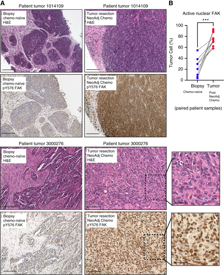
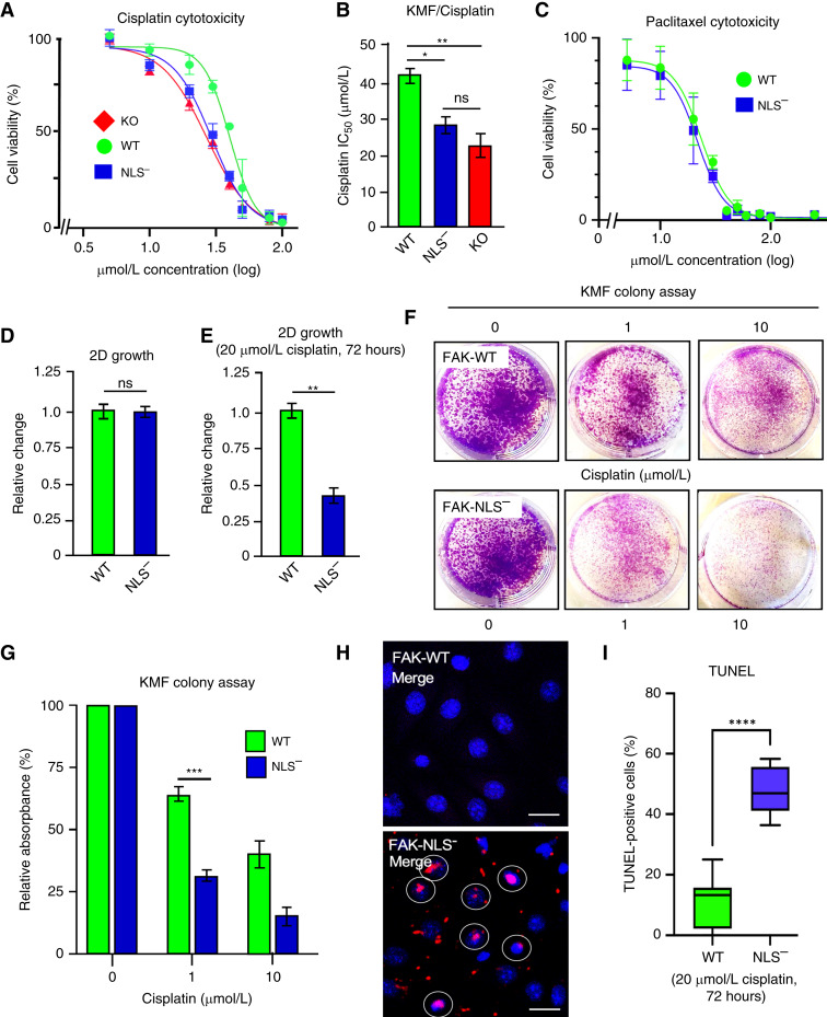
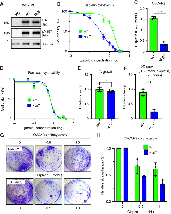
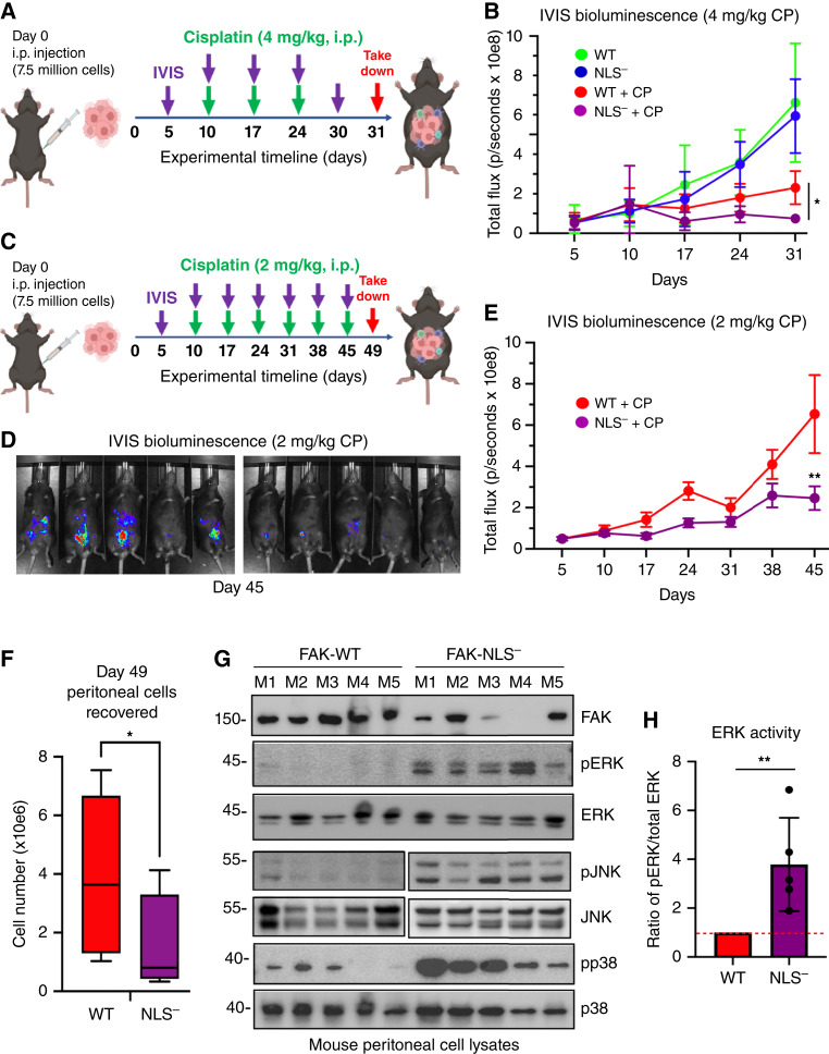
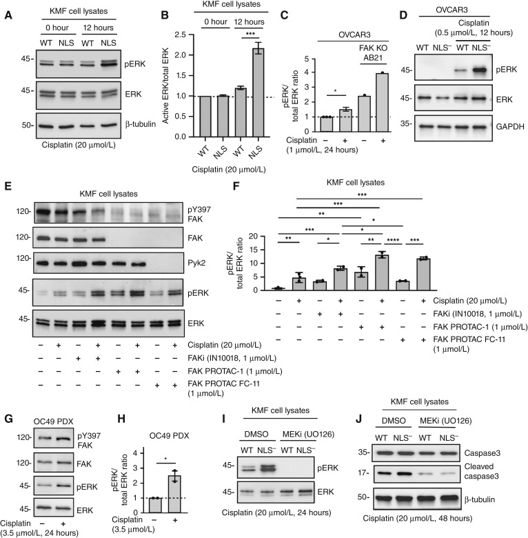
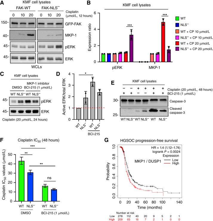
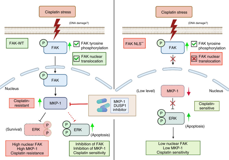
Abstract: Tumor chemotherapy resistance arises frequently and limits high-grade serous ovarian cancer (HGSOC) patient survival. Focal adhesion kinase (FAK) is an intracellular protein–tyrosine kinase encoded by PTK2, a gene that is often gained in HGSOC. Canonically, FAK functions at the cell periphery. However, FAK also transits to the nucleus to modulate gene expression. We find that FAK is tyrosine-phosphorylated and nuclear-localized in tumors of patients with HGSOC surviving neoadjuvant platinum–paclitaxel chemotherapy and that FAK nuclear accumulation occurs upon subcytotoxic cisplatin exposure to ovarian tumor cells in vitro. FAK nuclear localization sequence (NLS) mutational inactivation resulted in tumor cell sensitization to cisplatin in vitro and in vivo relative to wild-type FAK-reconstituted ovarian tumor cells. Cisplatin cytotoxicity was associated with elevated ERK MAPK activation in FAK NLS− cells, cisplatin-stimulated ERK activation was also enhanced upon loss of FAK activity or expression, and cisplatin-stimulated cell death was prevented by an inhibitor of ERK signaling. MAPK phosphastase-1 (MKP1) negatively regulates ERK signaling, and cisplatin-induced MKP1 levels were significantly elevated in wild-type FAK compared with FAK NLS− ovarian tumor cells. Notably, small-molecule MKP1 inhibition enhanced both cisplatin-stimulated ERK phosphorylation and ovarian tumor cell death. Together, our results show that FAK expression, activity, and nuclear localization limit cisplatin cytotoxicity in part by regulating MKP1 levels and preventing noncanonical ERK/MAPK activation.

Significance: FAK inhibitors are in combinatorial clinical testing with agents that prevent Ras-Raf-MAPK pathway activation in various cancers. This study suggests that nuclear FAK limits ERK/MAPK activation in supporting HGSOC cell survival to cisplatin stress. Overall, it is likely that targets of FAK-mediated survival signaling may be tumor type- and context-dependent.


Conflict of interest statement

D.C. Connolly reports grants from the NCI during the conduct of the study. M.T. McHale reports grants from the UCSD during the conduct of the study. D.G. Stupack reports personal fees from Amplia Therapeutics outside the submitted work. No disclosures were reported by the other authors.

Figures

Figure 1
Figure 1
Active FAK staining increases in the nucleus of HGSOC patient tumors after neoadjuvant chemotherapy. A, IHC staining of paraffin-embedded serial initial tumor biopsy sections (patient 1014109 and patient 3000276) and corresponding tumor resection after neoadjuvant chemotherapy treatment. Samples were stained with H&E and phosphospecific antibodies to the FAK activation loop Y576 (pY576) within the kinase domain. Scale is 100 μm (inset scale is 25 μm). B, Image quantification of percent of tumor cell area exhibiting FAK pY576 staining. Paired tumor samples (n = 8) from initial biopsy (blue circles) and from surgical tumor debulking after neoadjuvant chemotherapy (red circles). Dotted lines connect paired patient tumor samples collected prior to and after neoadjuvant chemotherapy (***, P < 0.001). Chemo, chemotherapy; H&E, hematoxylin and eosin; NeoAdj, neoadjuvant.
Figure 2
Figure 2
Nuclear FAK accumulation occurs upon cisplatin treatment of human and murine ovarian tumor cells. A, Nuclear and cytoplasmic cell fractionation of human OVCAR4 cells treated with DMSO (control) or cisplatin (1 μmol/L) for 12 hours followed by immunoblotting for FAK pY397, total FAK, β-tubulin (cytoplasmic marker), and histone H3 (nuclear marker). B, Schematic of GFP fusion to FAK denoting N-terminal band 4.1, ezrin, radixin, moesin homology (FERM), kinase, and focal adhesion targeting (FAT) domains. FAK FERM arginine R177 and R178 to alanine mutational changes prevent FAK nuclear localization. C, Stable reexpression of GFP-FAK-WT (WT) and GFP-FAK-NLS (NLS) in KMF FAK KO murine ovarian tumor cells as analyzed by FAK, pY397 FAK, and β-actin immunoblotting. D, KMF FAK-WT and KMF FAK-NLS cells treated with DMSO (control) or cisplatin (20 μmol/L) for 1 hour and immunoblotting for FAK pY397 and total FAK. E, Confocal immunofluorescence imaging was used to visualize GFP-FAK and DAPI stain in FAK-WT and FAK-NLS cells treated with DMSO (control) or cisplatin (20 μmol/L) for 12 hours. Shown are representative images of GFP-FAK (images shown at midline of Z-stack), nucleus, and merged images. Scale bar, 10 μm. F, Nuclear and cytoplasmic fractionation of FAK-WT and FAK-NLS cells treated with cisplatin (20 μmol/L) for 0, 12, or 24 hours followed by immunoblotting for FAK, β-tubulin, and histone H3. G, Representative images of GFP-FAK-WT and FAK-NLS expressing (images shown at midline of Z-stack) and DAPI-stained cells treated with leptomycin B (50 nmol/L) for 6 hours. Scale bar, 10 μm. Cyto, cytoplasmic.
Figure 3
Figure 3
FAK nuclear localization enhances murine ovarian tumor cell survival to cisplatin. A, FAK-KO, FAK-WT, and FAK-NLS KMF cells were evaluated for cisplatin cytotoxicity after 48 hours in culture. Shown is percent cell viability vs. cisplatin concentration (μmol/L, log10), and points are means of triplicate samples ± SD (n = 3 independent experiments). B, Determination of cisplatin IC50 values as performed in A (*, P < 0.05; **, P < 0.01). C, FAK-WT and FAK-NLS KMF cells were evaluated for paclitaxel cytotoxicity (μmol/L, log10) after 48 hours in culture. D, Analysis of KMF-FAK-WT and KMF-FAK-NLS cells for growth in culture and (E) in the presence of 20 μmol/L cisplatin for 72 hours. Values are means ± SD from two independent experiments with triplicate points and FAK-WT values set to 1 (**, P < 0.01). F, Representative images of crystal violet–stained KMF FAK-WT and FAK-NLS cell colonies formed in the presence of 0, 1, or 10 μmol/L cisplatin after 10 days. G, Quantitation of FAK-WT (green bars) and FAK-NLS (blue bars) colony formation. Values are means ± SD from three independent experiments with triplicate points (***, P < 0.001). H, Representative images of TUNEL staining to detect changes in FAK-WT or FAK-NLS cell DNA fragmentation after cisplatin (20 μmol/L, 48 hours) addition. TUNEL-positive nuclei (identified by DAPI costain) are marked (white circle) in the merged images. Scale bar, 10 μm. I, Quantification of FAK-WT (green bars) and FAK-NLS (blue bars) TUNEL staining. Box and whisker plots show the mean ± SD from three independent experiments (****, P < 0.0001). ns, not significant.
Figure 4
Figure 4
FAK KO and reexpression show that nuclear FAK promotes cisplatin but not paclitaxel resistance in human OVCAR3 cells. A, Immunoblots of cultured FAK KO OVCAR3 (clone AB21) and GFP-FAK-WT or FAK-NLS reconstituted AB21 cells for HA-tag (at FAK C-terminal), FAK pY397, and β-tubulin as a loading control. B, FAK-WT, and FAK-NLS OVCAR3 cells were evaluated for cisplatin cytotoxicity after 48 hours in culture. Shown is percent cell viability vs. cisplatin concentration (μmol/L, log10), and points are means of triplicate samples ± SD (n = 3 independent experiments). C, Determination of IC50 values to cisplatin as performed in B (***, P < 0.001). D, FAK-WT and FAK-NLS OVCAR3 cells were evaluated for paclitaxel cytotoxicity (μmol/L, log10) after 48 hours in culture. E, Analysis of OVCAR3 FAK-WT and FAK-NLS cells for growth in culture and (F) in the presence of 0.5 μmol/L cisplatin for 72 hours. Values are means ± SD from two independent experiments with triplicate points and FAK-WT values set to 1 (***, P < 0.001; ns not significant). G, Representative images of crystal violet–stained OVCAR3 FAK-WT and FAK-NLS cell colonies formed in the presence of 0, 0.5, or 1.0 μmol/L cisplatin after 10 days. H, Quantitation of FAK-WT (green bars) and FAK-NLS (blue bars) colony formation. Values are means ± SD from three independent experiments with triplicate points (*, P < 0.05).
Figure 5
Figure 5
FAK nuclear localization supports KMF tumor cisplatin resistance in vivo with effects on downstream signaling. A, Experimental schematic: 7.5 million luciferase-expressing KMF FAK-WT or FAK-NLS cells were intraperitoneally injected into C57Bl6 mice (day 0), and IVIS luciferase imaging at day 5 was used to randomize experimental groups (n = 8 each). Mice were administered saline or cisplatin (CP, 4 mg/kg) and imaged on the indicated days. B, Graphical presentation of IVIS tumor burden from day 5 to day 31 as expressed as total flux in photons per second. Experimental groups are FAK-WT saline (green), FAK-WT plus CP (red), FAK-NLS saline (blue), and FAK-NLS + CP (violet). Values are means ± SD (n = 8, *, P < 0.05). C, Experimental schematic (similar as described in A) with decreased and more frequent cisplatin (2 mg/kg) administration over a longer experimental period. D, Representative IVIS images of CP-treated FAK-WT and FAK-NLS tumor-bearing mice on day 45. E, Graphical presentation of IVIS tumor burden from day 5 to day 45. Experimental groups are FAK-WT + CP (red) and FAK-NLS + CP (violet). Values are means ± SD (n = 8, **, P < 0.01). F, Enumeration (trypan blue–negative) of peritoneal cells recovered after euthanasia. Box and whisker plots show the mean (±SD, *, P < 0.05) for FAK-WT (red) and FAK-NLS (violet) tumor-bearing mice. G, Protein cell lysates were made from peritoneal collected cells from individual FAK-WT and FAK-NLS tumor-bearing mice (n = 5 each, M1 to M5) and immunoblotted for FAK, β-tubulin, active ERK (pERK), total ERK, active p38 (pp38), and total p38 MAPK. Immunoblotting for active JNK (pJNK) and total JNK were from the same samples but independent gels. H, Image quantitation of pERK to total ERK ratio from samples in G. Values are means ± SD (n = 5 experimental points, **, P < 0.01). FAK-WT values set to 1 (dotted line).
Figure 6
Figure 6
Prevention of FAK expression, activity, or nuclear localization results in elevated cisplatin-stimulated ERK activation. A, KMF FAK-WT and FAK-NLS cells were treated for 0 or 12 hours with cisplatin (20 μmol/L), and cell lysates were immunoblotted for active ERK (pERK), total ERK, and β-tubulin. B, Image quantitation of pERK to total ERK ratio from two independent experiments from A. Values are means ± SD with control FAK-WT values set to 1 (***, P < 0.001). C, Image quantitation of pERK to total ERK ratio from parental OVCAR3 and FAK-KO AB21 cells treated with cisplatin (1 μmol/L) for 24 hours. Values are means ± SD from two independent experiments with control OVCAR3 values set to 1 (*, P < 0.05). D, Representative pERK, total ERK, and GAPDH loading control immunoblotting of OVCAR3 FAK-WT and FAK-NLS lysates ± cisplatin (0.5 μmol/L, 12 hours). E, KMF parental cells were preincubated (48 hours) with FAKi (IN10018, 1 μmol/L), FAK-specific PROTAC (FAK PROTAC-1, 1 μmol/L), and FAK-Pyk2 targeting PROTAC (FAK PROTAC FC-11, 1 μmol/L) followed by with cisplatin addition (20 μmol/L, 24 hours), as indicated. Cell lysates were immunoblotted for pY397 FAK, FAK, and Pyk2 and for active (pERK) and total ERK. F, Image quantitation of pERK to total ERK ratio from three independent experiments described in E. Values are means ± SD with control set to 1 (*, P < 0.05; **, P < 0.01; ***, P < 0.001; ****, P < 0.0001). G, Representative immunoblotting of OC49 ovarian PDX cell lysates for pY397 FAK, total FAK, pERK, and total ERK ± cisplatin (3.5 μmol/L, 24 hours). H, Image quantitation of pERK to total ERK ratio from two independent experiments described in G. Values are means ± SD with control set to 1 (*, P < 0.05). I, KMF FAK-WT and FAK-NLS cells were treated 20 μmol/L cisplatin (24 hours) with (DMSO) control or MEK1 inhibitor (U0125, 10 μmol/L) addition. Cell lysates were immunoblotted for active and total ERK. J, KMF FAK-WT and FAK-NLS cells were treated 20 μmol/L cisplatin (48 hours) as above with MEK1 inhibitor and cell lysates blotted for caspase-3, cleaved caspase-3, and β-tubulin. MEKi, MEK inhibitor.
Figure 7
Figure 7
Nuclear FAK regulation of MKP1 expression is associated with cisplatin resistance. A, KMF FAK-WT and FAK-NLS cells were treated with increased concentrations of cisplatin (0, 10, or 20 μmol/L) for 12 hours and protein lysates were immunoblotted for GFP-FAK, MKP1, pERK, and total ERK. B, Image quantitation of pERK to total ERK ratio or MKP1 levels (FAK-WT set to 0) from two independent experiments described in A. Values are means ± SD (***, P < 0.001). C, KMF FAK-WT and FAK-NLS cells were treated 20 μmol/L cisplatin (24 hours) under control (DMSO) or with MKP1 inhibitor (BCI-215, 1 μmol/L) addition. Protein lysates were immunoblotted for pERK and total ERK. D, Image quantitation of pERK to total ERK ratio from two independent experiments described in C. Values are means ± SD with FAK-WT control set to 1 (*, P < 0.05). E, KMF FAK-WT and FAK-NLS cells were treated 20 μmol/L cisplatin (48 hours) as above with MKP1 inhibitor (BCI-215, 1 μmol/L) addition, and cell lysates were immunoblotted for caspase-3 and cleaved caspase-3. F, Determination of IC50 values to cisplatin KMF FAK-WT (green bars) and FAK-NLS (blue bars) cells in the presence of MKP1 inhibitor (BCI-215, 1 μmol/L) or DMSO control as determined by alamarBlue cell viability assay. Values are means ± SD from three independent experiments (*, P < 0.05; **, P < 0.01; ***, P < 0.001). G, Kaplan–Meier curves from 949 HGSOC patient samples that received adjuvant carbo- or cisplatin-based chemotherapy. Plots show probability of relapse-free survival in months with tumors high (red) or low (black) for MKP1 mRNA (HR = 1.4, P = 0.0024). ns, not significant; WCL, whole-cell protein lysate.
Figure 8
Figure 8
Model of cisplatin-stimulated nuclear FAK protection against ERK-associated cell death in ovarian cancer. Left, Patient tumors surviving neoadjuvant chemotherapy exhibit a high level of active FAK staining in the cell nucleus. This is a tumor cell–intrinsic response, as subcytotoxic cisplatin treatment was associated with FAK nuclear accumulation in human and mouse ovarian tumor cells. Cisplatin stress also increased MKP1 tyrosine phosphatase expression that limits phosphorylation and activity of ERK. Inhibition of MKP1 results in elevated cisplatin-stimulated ERK activity associated with cell death. FAK inhibition also results in increased ERK activity, and FAK inhibition potentiated cisplatin-induced cell death (28). Right, Mutational inactivation of FAK nuclear targeting (FAK-NLS) does not prevent basal or cisplatin-stimulated FAK-NLS phosphorylation at Y397. FAK-NLS does not accumulate in the nucleus upon cisplatin addition to cells. FAK-NLS cells exhibit sensitivity to cisplatin cytotoxicity, reduced MKP1 expression, and elevated ERK activation. Our results support the notion that nuclear FAK supports MKP1 expression and that combinatorial small-molecule targeting of FAK kinase and MKP1 phosphatase activities may function as an effective therapy combination with cisplatin for HGSOC. Created in BioRender (RRID: SCR_018361). Schlaepfer, D. (2024) BioRender.com/n24i207.

Similar articles

References

    1. Siegel RL, Giaquinto AN, Jemal A. Cancer statistics, 2024. CA Cancer J Clin 2024;74:12–49. - PubMed
    1. Kim S, Han Y, Kim SI, Kim H-S, Kim SJ, Song YS. Tumor evolution and chemoresistance in ovarian cancer. NPJ Precis Oncol 2018;2:20. - PMC - PubMed
    1. Agarwal R, Kaye SB. Ovarian cancer: strategies for overcoming resistance to chemotherapy. Nat Rev Cancer 2003;3:502–16. - PubMed
    1. Bowtell DD, Böhm S, Ahmed AA, Aspuria P-J, Bast RC Jr, Beral V, et al. . Rethinking ovarian cancer II: reducing mortality from high-grade serous ovarian cancer. Nat Rev Cancer 2015;15:668–79. - PMC - PubMed
    1. Sulzmaier FJ, Jean C, Schlaepfer DD. FAK in cancer: mechanistic findings and clinical applications. Nat Rev Cancer 2014;14:598–610. - PMC - PubMed

Publication types

MeSH terms