. 2025 Jan;399(1):97-117.
doi: 10.1007/s00441-024-03930-6. Epub 2024 Nov 25.

Enhanced cell survival in prepubertal testicular tissue cryopreserved with membrane lipids and antioxidants rich cryopreservation medium

Affiliations

Enhanced cell survival in prepubertal testicular tissue cryopreserved with membrane lipids and antioxidants rich cryopreservation medium

Reyon Dcunha et al. Cell Tissue Res. 2025 Jan.

Abstract

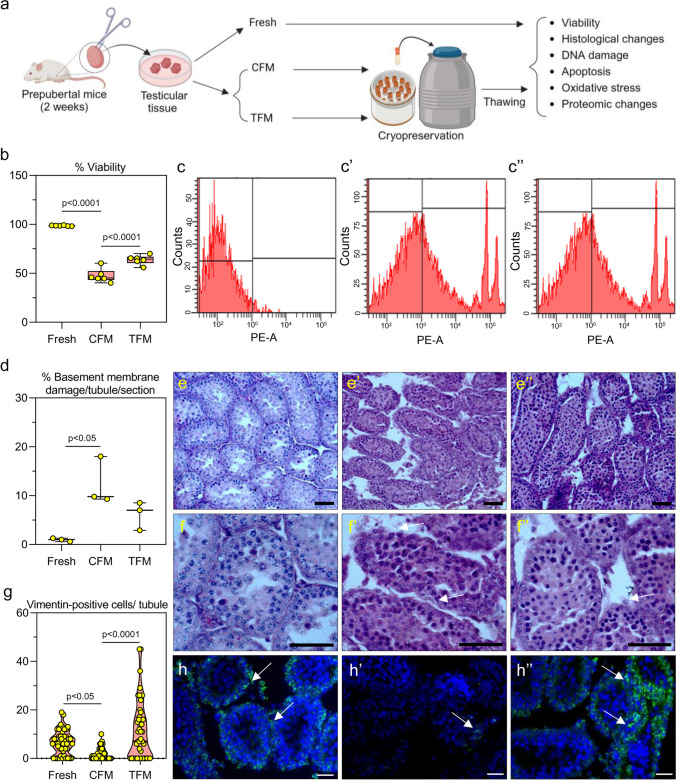
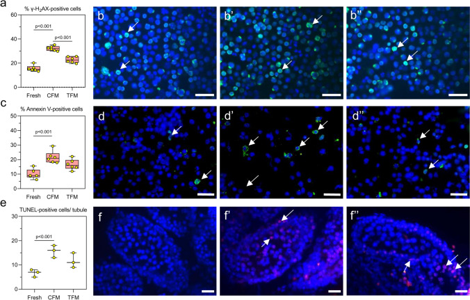
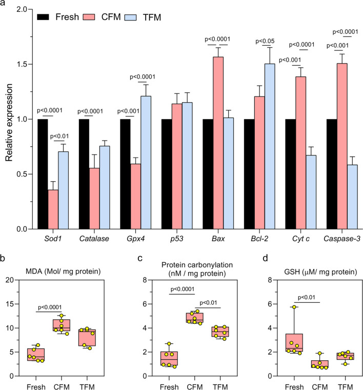
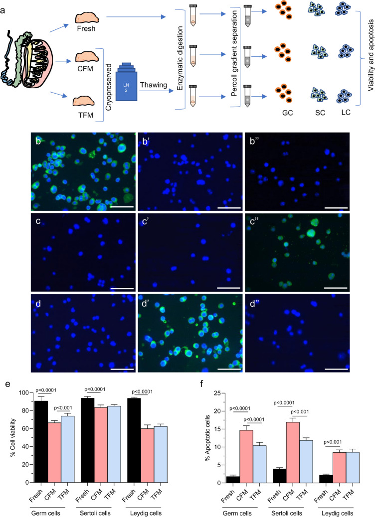
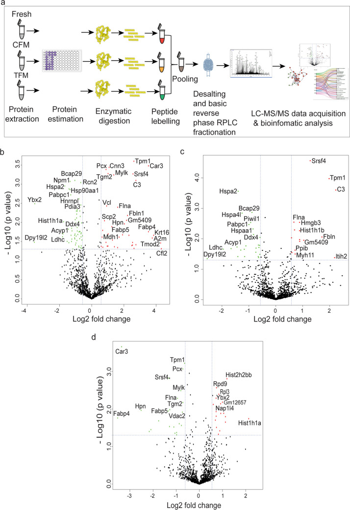
The present study explores the advantages of enriching the freezing medium with membrane lipids and antioxidants in improving the outcome of prepubertal testicular tissue cryopreservation. For the study, testicular tissue from Swiss albino mice of prepubertal age group (2 weeks) was cryopreserved by slow freezing method either in control freezing medium (CFM; containing DMSO and FBS in DMEM/F12) or test freezing medium (TFM; containing soy lecithin, phosphatidylserine, phosphatidylethanolamine, cholesterol, vitamin C, sodium selenite, DMSO and FBS in DMEM/F12 medium) and stored in liquid nitrogen for at least one week. The tissues were thawed and enzymatically digested to assess viability, DNA damage, and oxidative stress in the testicular cells. The results indicate that TFM significantly mitigated freeze-thaw-induced cell death, DNA damage, and lipid peroxidation compared to tissue cryopreserved in CFM. Further, a decrease in Cyt C, Caspase-3, and an increase in Gpx4 mRNA transcripts were observed in tissues frozen with TFM. Spermatogonial germ cells (SGCs) collected from tissues frozen with TFM exhibited higher cell survival and superior DNA integrity compared to those frozen in CFM. Proteomic analysis revealed that SGCs experienced a lower degree of freeze-thaw-induced damage when cryopreserved in TFM, as evident from an increase in the level of proteins involved in mitigating the heat stress response, transcriptional and translational machinery. These results emphasize the beneficial role of membrane lipids and antioxidants in enhancing the cryosurvival of prepubertal testicular tissue offering a significant stride towards improving the clinical outcome of prepubertal testicular tissue cryopreservation.

Keywords: Fertility preservation; Membrane integrity; Oncofertility; Proteomics; Spermatogonial germ cells.


Conflict of interest statement

Declarations. Ethical Approval: The Institutional Animal Ethical Committee of Kasturba Medical College, Manipal, India approved the current study (IAEC/KMC/134/2019). The experiments performed were in accordance with the guidelines advocated by the institutional and national Committee for the Purpose of Control and Supervision of Experiments on Animals (CPCSEA), New Delhi, India, and in accordance with ARRIVE guidelines. Conflicts of interests: The authors have no relevant financial or non-financial interests to disclose.

Figures

Fig. 1
Fig. 1
a Experimental design of mice prepubertal testicular tissue. The images were created with Biorender.com; b Effect of TFM on cell viability assessed by flow cytometry assay in prepubertal testicular tissue subjected to cryopreservation by slow freezing method (N = 6); Representative histogram for flow cytometry assessment of cells that are c unstained cells, from c’ CFM and c’’ TFM. The data is represented as Mean ± SEM; d Basement membrane damage assessed in prepubertal mice testicular tissue cryopreserved with CFM and TFM (N = 3); e–f’’ Histopathological changes in prepubertal mice testicular tissue cryopreserved with CFM (e’ and f’ at 100 × and 400 × magnification, respectively) and TFM (e’’ and f’’ at 100 × and 400 × magnification, respectively). Arrows indicate basement membrane damage. Scale bar represents 100 μm; g Expression of vimentin in prepubertal testicular tissue after freeze–thaw process (N = 3). The data is represented as Mean ± SEM; Representative images of IHC showing vimentin expression in prepubertal testicular tissue cryopreserved with h’ CFM and h’’ TFM. White arrowheads indicate cells positive for vimentin. The scale bar represents 20 μm
Fig. 2
Fig. 2
Assessment of DNA integrity, and apoptosis in prepubertal mice testicular tissue cryopreserved with CFM and TFM. a Assessment of DNA damage by γ-H2AX expression in prepubertal testicular tissue (N = 6); Representative images showing γ-H2AX positive cells (400x) in b Fresh, b’ CFM and b’’ TFM group. White arrowheads indicate γ-H2AX positive cells; the Scale bar represents 20 μm. c Annexin V expression (N = 6). The data is represented as Mean ± SEM; Representative images showing testicular cells positive for Annexin V from d Fresh, d’ CFM and d’’ TFM group; e Effect of TFM on DNA damage assessed by TUNEL assay (N = 3); The data is represented as Mean ± SEM; Representative images showing TUNEL-positive cells in f Fresh tissue and prepubertal mice testicular tissue subjected to freeze–thaw process using f’ CFM and f’’ TFM. White arrowheads indicate TUNEL-positive cells; Scale bar represents 20 μm
Fig. 3
Fig. 3
a Gene expression pattern for Gpx4, Sod1, Cat, p53, Bcl-2, Bax, Cytc, and Casp3 analyzed by qRT-PCR in prepubertal mice testicular tissue cryopreserved with CFM and TFM (N = 6). The data is represented as Mean ± SEM; Assessment of b Malondialdehyde; c Protein carbonyl content; d Glutathione level in prepubertal mice testicular tissue cryopreserved with CFM and TFM (N = 6). The data is represented as Mean ± SEM
Fig. 4
Fig. 4
Effect of freeze–thaw process on the survival of germ, Sertoli, and Leydig cells of prepubertal mice testicular tissue. a Experimental outline for isolation of various testicular cells; Representative images to depict characterization of germ cells by studying VASA expression- b Germ cells, b’ Leydig cells, and b’’ Sertoli cells; Representative images to depict characterization of Sertoli cells by studying SOX9 expression- c Germ cells, c’ Leydig cells, and c’’ Sertoli cells; Representative images to depict characterization of germ cells by studying β-HSD expression- d Germ cells, d’ Leydig cells, and d’’ Sertoli cells; e Assessment of viability by trypan blue dye exclusion method (N = 6); f Apoptosis in isolated germ, Sertoli, and Leydig cell fraction isolated from prepubertal mice testicular tissue assessed by Annexin-V staining (N = 6). The data is represented as Mean ± SEM
Fig. 5
Fig. 5
Proteomic alterations in spermatogonial germ cells (SGCs) isolated from prepubertal mice testicular tissue cryopreserved with CFM and TFM. a Experimental outline; b Volcano plot depicting significantly up- and downregulated proteins in spermatogonial germ cells from tissue cryopreserved in CFM compared to fresh tissue; c Volcano plot depicting significantly up- and downregulated proteins in spermatogonial germ cells from tissue cryopreserved in TFM compared to fresh tissue; d Volcano plot depicting significantly up- and downregulated proteins in spermatogonial germ cells from tissue cryopreserved in CFM compared to TFM; Red color indicates significantly upregulated and green color indicates significantly downregulated proteins
Fig. 6
Fig. 6
The altered protein expression in spermatogonial germ cells isolated from prepubertal mice testicular tissue cryopreserved with CFM and TFM. a Venn diagram showing proteins that are common and uniquely expressed in the three experimental conditions; b Differential expression of proteins from spermatogonial germ cells isolated from fresh, CFM, and TFM c Differential expression of proteins from spermatogonial germ cells isolated from CFM, and TFM; d Interactome analysis of the common proteins expressed in spermatogonial germ cells from fresh, CFM, and TFM; Node color indicates the protein expression and edge color indicates the various biological processes they are involved in
Fig. 7
Fig. 7
Gene ontology analysis of the significantly up- and downregulated proteins in spermatogonial germ cells from tissue cryopreserved in a CFM compared to fresh tissue; b CFM compared to TFM
Fig. 8
Fig. 8
Pathway analysis of the differentially expressed proteins in spermatogonial germ cells isolated from prepubertal mice tissue cryopreserved with CFM and TFM

References

    1. Allen CM, Lopes F, Mitchell RT, Spears N (2018) How does chemotherapy treatment damage the prepubertal testis? Reproduction 156:R209–R233. 10.1530/REP-18-0221 - PMC - PubMed
    1. Andrae CS, Oliveira ECS, Ferraz MAMM, Nagashima JB (2021) Cryopreservation of grey wolf (Canis lupus) testicular tissue. Cryobiology 100:173–179. 10.1016/j.cryobiol.2021.01.010 - PubMed
    1. Avarbock MR, Brinster CJ, Brinster RL (1996) Reconstitution of spermatogenesis from frozen spermatogonial stem cells. Nat Med 2:693–696. 10.1038/nm0696-693 - PMC - PubMed
    1. Baert Y, Onofre J, Van Saen D, Goossens E (2018) Cryopreservation of Human Testicular Tissue by Isopropyl-Controlled Slow Freezing. In: Alves MG, Oliveira PF (eds) Sertoli Cells: Methods and Protocols. Springer, New York, New York, NY, pp 287–294 - PubMed
    1. Beckman JK, Coniglio JG (1979) A comparative study of the lipid composition of isolated rat sertoli and germinal cells. Lipids 14:262–267. 10.1007/BF02533912 - PubMed