Review
. 2024 Nov 25;5(1):61.
doi: 10.1186/s43556-024-00226-7.

Human cytomegalovirus: pathogenesis, prevention, and treatment

Affiliations
Review

Human cytomegalovirus: pathogenesis, prevention, and treatment

Zifang Shang et al. Mol Biomed. .

Abstract

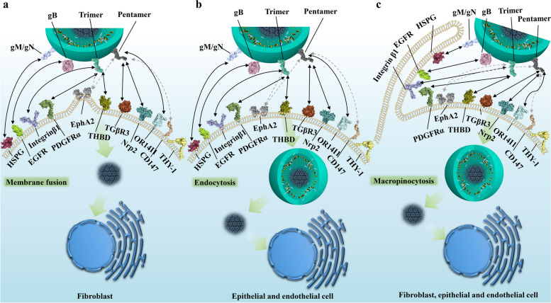
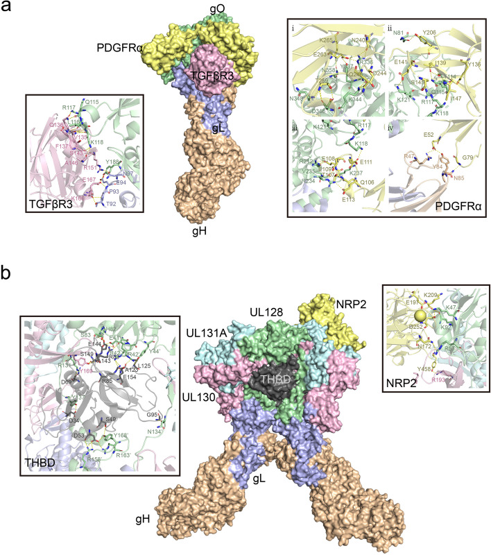
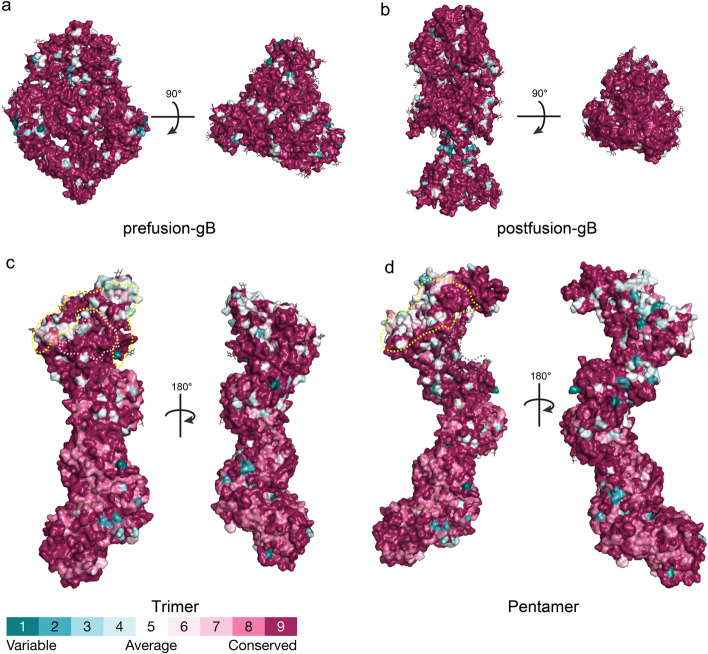
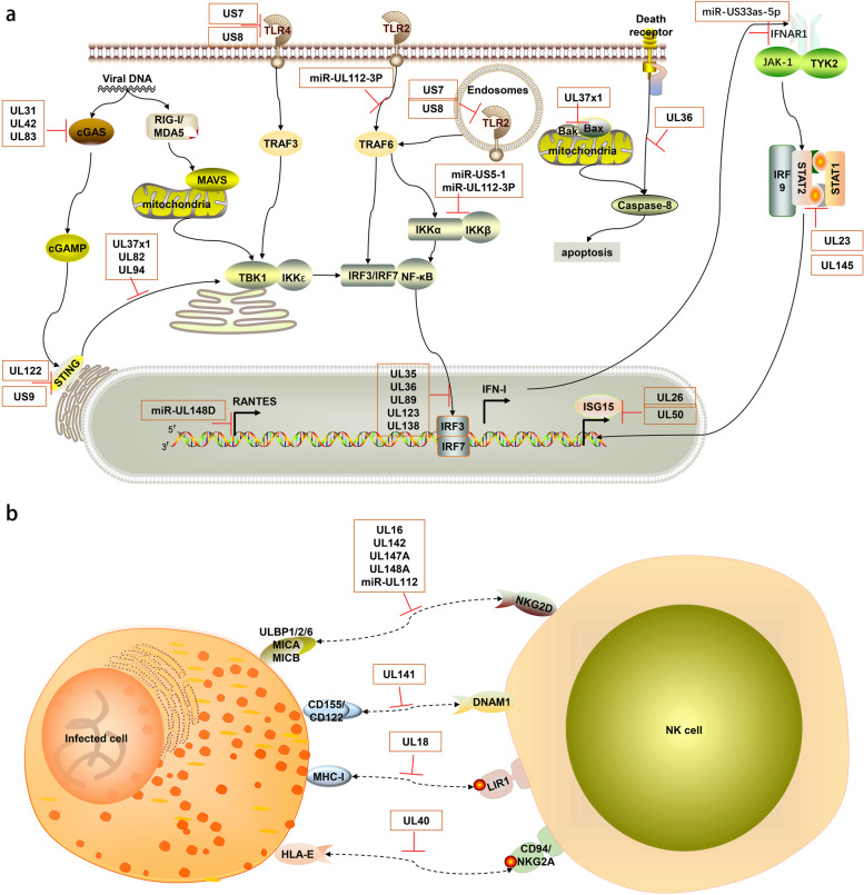
Human cytomegalovirus (HCMV) infection remains a significant global health challenge, particularly for immunocompromised individuals and newborns. This comprehensive review synthesizes current knowledge on HCMV pathogenesis, prevention, and treatment strategies. We examine the molecular mechanisms of HCMV entry, focusing on the structure and function of key envelope glycoproteins (gB, gH/gL/gO, gH/gL/pUL128-131) and their interactions with cellular receptors such as PDGFRα, NRP2, and THBD. The review explores HCMV's sophisticated immune evasion strategies, including interference with pattern recognition receptor signaling, modulation of antigen presentation, and regulation of NK and T cell responses. We highlight recent advancements in developing neutralizing antibodies, various vaccine strategies (live-attenuated, subunit, vector-based, DNA, and mRNA), antiviral compounds (both virus-targeted and host-targeted), and emerging cellular therapies such as TCR-T cell approaches. By integrating insights from structural biology, immunology, and clinical research, we identify critical knowledge gaps and propose future research directions. This analysis aims to stimulate cross-disciplinary collaborations and accelerate the development of more effective prevention and treatment strategies for HCMV infections, addressing a significant unmet medical need.

Keywords: Antiviral therapy; Envelope glycoproteins; Human cytomegalovirus; Immune Evasion; Neutralizing antibodies; Vaccines.


Conflict of interest statement

Declarations. Ethics approval and consent to participate: Not applicable. This scoping review did not involve human participants or animal subjects. Consent for publication: Not applicable. Competing interests: The authors declare that they have no competing interests.

Figures

Fig. 1
Fig. 1
Structure of the human cytomegalovirus (HCMV) virion and its components. The HCMV genome is encapsidated within the capsid, surrounded by the tegument layer, and an outer lipid bilayer envelope embedded with multiple envelope glycoproteins. The genome length is approximately 235–250 kb, encoding envelope glycoproteins such as gB, gM, gN, gH, gL, gO, UL128, UL130, UL131, and UL116
Fig. 2
Fig. 2
The modes of HCMV cell entry and interactions of envelope glycoproteins with cellular receptors. This figure illustrates the complex interactions between HCMV envelope glycoproteins and cellular receptors, demonstrating different entry pathways in various cell types. In fibroblasts (a), entry occurs through membrane fusion at neutral pH, where the gM/gN complex interacts with heparan sulfate proteoglycans (HSPGs), the trimer (gH/gL/gO) binds to PDGFRα and TGFβR3, gB interacts with HSPGs and potentially EGFR, while gH/gL components may interact with integrin β1 and EphA2. CD147 acts as a cofactor promoting entry. In epithelial and endothelial cells (b), entry occurs through pH-dependent endocytosis, requiring the pentamer complex (gH/gL/UL128/UL130/UL131) which binds to TGFβR3, Nrp2, or THBD (specifically on endothelial cells). OR14I1 serves as an additional pentamer-dependent receptor on epithelial cells, while CD46 may be involved in downstream entry steps. A macropinocytosis-like entry process (c), applicable to fibroblasts, epithelial, and endothelial cells, is activated by integrin and PDGFRα signaling, with THY-1 acting as a cofactor. This mechanism allows HCMV to transport its dsDNA genome into the nucleus. Dashed arrows in the figure indicate potential interactions between viral glycoproteins and cellular receptors that may contribute to the entry process but are not definitively established
Fig. 3
Fig. 3
Schematic illustration of the trimer binding to PDGFRα and TGFBR3, and the pentamer binding to Nrp2 and THBD. (a) Front view of the overall region where the HCMV trimer binds to PDGFRα (light yellow) and TGFβR3 (pink), with close-up views on either side showing the interaction regions of gO with PDGFRα and TGFβR3, highlighting the key interacting residue sites. (b) Front view of the overall region where the HCMV pentamer binds to NRP2 (light yellow) and THMB (dark gray), with close-up views on either side showing the interaction regions of ULs with NRP2 and THMB, highlighting the key interacting residue sites
Fig. 4
Fig. 4
Conservation analysis of the overall structures of HCMV pre-fusion gB, post-fusion gB, trimer, and pentamer. Panels a-b represent the three-dimensional conservation analysis of 197 gB protein sequences, with the former showing the pre-fusion gB conformation and the latter showing the post-fusion gB conformation. Panel c shows the three-dimensional conservation analysis of the trimeric conformation based on 85 gH, 101 gL, and 127 gO sequences. The light-yellow dashed area and pink area represent the interaction regions of gO with PDGFRα and TGFβR3, respectively. Panel d shows the three-dimensional conservation analysis of the pentameric conformation based on 85 gH, 101 gL, 73 UL128, 147 UL130, and 41 UL131 sequences. The light-yellow dashed area and dark gray dashed area represent the interaction regions of ULs with NRP2 and THMB, respectively. The Stick representations on each overall structure surface indicate glycosylation sites. The conservation analysis was performed using the ConSurf server and visualized on the protein structures
Fig. 5
Fig. 5
Schematic diagram of the mechanisms by which HCMV evades host innate immune pathways. The host innate immunity and immune evasion mediated by HCMV. TLRs, located at both the plasma membrane and endosomes, sense different viral components. The cGAS-STING pathway detects viral DNA. These pathways activate IRF3/7 or NF-κB, inducing IFN-I and inflammatory cytokines. IFN-I stimulates ISG expression via the JAK-STAT pathway to limit HCMV. The IFN-I, inflammatory cytokines, and ISGs are induced for antiviral immunity, but HCMV proteins and miRNAs highlighted in the orange boxes can hijack multiple steps of these signaling pathways, effectively suppressing these immune reactions. In part (a), UL31, UL42, and UL83 (pp65) inhibit cGAS, while UL37 × 1, UL82, and UL94 block STING trafficking. US7 and US8 suppress TLR3 and TLR4 signaling, and miR-UL112-3P targets TLR2. UL122 (IE86) induces STING degradation, and US9 blocks STING-TBK1 interaction. UL35, UL36, UL89 (pp65), and UL138 inhibit IRF3 phosphorylation. miR-US33as-5p targets IFNAR1, UL23 blocks STAT1 nuclear translocation, and UL145 induces STAT2 degradation. miR-US5-1 and miR-UL112-3P downregulate IKKα and IKKβ. UL36 inhibits caspase-8 activation, and UL37 × 1 inhibits Bax and Bak. UL26 and UL50 suppress ISGylation. In part (b), HCMV evades NK cell responses. UL16 binds to NKG2D ligands MICB, ULBP1/2/6. UL142, UL147A, UL148A, and miR-UL112 downregulate MICA/B. UL141 downregulates DNAM-1 ligands CD155 and CD112. UL18 mimics MHC-I molecules, binding to LIR-1. UL40 mimics HLA-E ligands, preventing NK cell-mediated lysis. This figure was created using ScienceSlides 2016
Fig. 6
Fig. 6
Schematic diagram of the mechanisms by which HCMV evades host adaptive immune pathways. The host adaptive immunity and immune evasion mediated by HCMV. Dendritic cells present antigens via MHC-I and MHC-II to CD8+ and CD4+ T cells, respectively. B cells produce antibodies and form memory B cells. These processes activate adaptive immune responses against HCMV. However, HCMV proteins and miRNAs highlighted in the orange boxes can hijack multiple steps of these pathways, effectively suppressing these immune reactions. US2, US3, US6, US10, US11, and UL82 interfere with MHC-I expression and transport, while US2, US3, and UL83 (pp65) interfere with MHC-II. miR-US4-1 and miR-UL112-5P target ERAP1, affecting antigen processing. UL111A (cmvIL-10) inhibits T cell function via IL10R, and US28 induces T cell apoptosis through CXCR. UL28 (highlighted in yellow) upregulates PD-L1, inhibiting T cell activity. UL13 (highlighted in yellow) enhances cellular respiration by targeting mitochondrial MICOS complex. RL11, RL12, RL13, and UL119-118 bind to antibody Fc regions, interfering with ADCC. HCMV infection increases CD27+ IgD- memory B cells. miR-US5-2 (highlighted in yellow) increases TGF-β production, while miR-UL22A inhibits TGF-β signaling. miR-US5-2 and miR-US22 affect cell proliferation and viral latency by targeting GAB1 and EGR-1, respectively. This figure was created using ScienceSlides 2016

References

    1. Goodrum F, Caviness K, Zagallo P. Human cytomegalovirus persistence. Cell Microbiol. 2012;14(5):644–55. 10.1111/j.1462-5822.2012.01774.x. - DOI - PMC - PubMed
    1. Britt WJ. Maternal immunity and the natural history of congenital human cytomegalovirus infection. Viruses. 2018;10(8): 405. 10.3390/v10080405. - DOI - PMC - PubMed
    1. Messinger CJ, Lipsitch M, Bateman BT, He M, Huybrechts KF, MacDonald S, et al. Association between congenital Cytomegalovirus and the prevalence at birth of microcephaly in the United States. JAMA Pediatr. 2020;174(12):1159–67. 10.1001/jamapediatrics.2020.3009. - DOI - PMC - PubMed
    1. Goderis J, De Leenheer E, Smets K, Van Hoecke H, Keymeulen A, Dhooge I. Hearing loss and congenital CMV infection: a systematic review. Pediatrics. 2014;134(5):972–82. 10.1542/peds.2014-1173. - DOI - PubMed
    1. Megli CJ, Coyne CB. Infections at the maternal–fetal interface: an overview of pathogenesis and defence. Nat Rev Microbiol. 2022;20(2):67–82. 10.1038/s41579-021-00610-y. - DOI - PMC - PubMed

Publication types

MeSH terms