. 2024 Nov 14;135(2):e183331.
doi: 10.1172/JCI183331.

Neisseria gonorrhoeae induces local secretion of IL-10 at the human cervix to promote colonization

Affiliations

Neisseria gonorrhoeae induces local secretion of IL-10 at the human cervix to promote colonization

Yiwei Dai et al. J Clin Invest. .

Abstract

Gonorrhea, caused by the human-restricted pathogen Neisseria gonorrhoeae, is a commonly reported sexually transmitted infection. Since most infections in women are asymptomatic, the true number of infections is likely much higher than reported. How gonococci (GC) colonize women's cervixes without triggering symptoms remains elusive. Using a human cervical tissue explant model, we found that GC inoculation increased the local secretion of both proinflammatory (IL-1β and TNF-α) and antiinflammatory (IL-10) cytokines during the first 24 hours of infection. Cytokine induction required GC expression of Opa isoforms that bind the host receptors carcinoembryonic antigen-related cell adhesion molecules (CEACAMs). GC inoculation induced NF-κB activation in both cervical epithelial and subepithelial cells. However, inhibition of NF-κB activation, which reduced GC-induced IL-1β and TNF-α, did not affect GC colonization. Neutralizing IL-10 or blocking IL-10 receptors by antibodies reduced GC colonization by increasing epithelial shedding and epithelial cell-cell junction disassembly. Inhibition of the CEACAM downstream signaling molecule SHP1/2, which reduced GC colonization and increased epithelial shedding, decreased GC-induced IL-10 secretion. These results show that GC induce local secretion of IL-10, a potent antiinflammatory cytokine, at the cervix by engaging the host CEACAMs to prevent GC-colonizing epithelial cells from shedding, providing a potential mechanism for GC asymptomatic colonization in women.

Keywords: Bacterial infections; Cytokines; Infectious disease.


Conflict of interest statement

Conflict of interest: The authors have declared that no conflict of interest exists.

Figures

Figure 1
Figure 1. GC inoculation increases the secretion of both proinflammatory and antiinflammatory cytokines by human cervical tissue explants.
Human cervical tissue explants with the ecto- to endocervix (AE) or with the ectocervix alone (FH) were incubated without (No GC) or with MS11 GC strains expressing no opa genes (ΔOpa) or a nonphase variable Opa52 that binds to CEACAMs (OpaCEA) at a MOI of 10 for 24 hours at 37oC. Supernatants of cervical tissue explant media were collected. The average concentrations (± SD) of IL-1β (A and F), TNF-α (B and G), IL-10 (C and H), TGF-β (D), and IL-17A (E, dashed line, detection level) were measured by Luminex Magpix (IL-1β, TNF-α, IL-10, and IL-17A) or ELISA (TGF-β), normalized to the luminal surface areas of tissue explants and supernatant volumes. Data points represent cervical tissues from individuals. The data were generated from 3 to 7 cervixes. *P < 0.05, **P < 0.01, ***P < 0.001, by 1-way ANOVA.
Figure 2
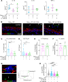
Figure 2. NF-κB is involved in GC-induced proinflammatory cytokine production by the human cervix.
Human cervical tissue explants were incubated without or with MS11OpaCEA or MS11ΔOpa (MOI ~ 10) in the absence or presence of the NF-κB inhibitor Bay11-7082 (3 μM) for 24 hours. (A) Representative CFM images of ectocervical tissue sections stained for NF-κB p65, GC, and nuclei. White dashed lines outline the epithelium, arrows indicate the 135μm depth of the subepithelium, and arrowheads point to GC attached to the epithelium. Scale bar: 50 μm. (B) The mean fluorescent intensity (MFI) (± SD) of NF-κB p65 in the nuclei of individual epithelial and subepithelial cells in the ectocervix (Ecto), transformation zone (TZ), and endocervix (Endo). Data were generated from cervical tissues from 3 to 4 individuals, 2 independent analyses per cervix, and 35–200 epithelial and 45–170 subepithelial cells per analysis per cervical region. (C) The heatmap shows relative mRNA levels of NF-κB–related genes by NanoString Immunology panel and compared MS11OpaCEA- or ΔOpa-inoculated with no GC control tissues from 3 individuals are shown. (D) Nuclear MFI of NF-κB p65 in individual epithelial and subepithelial cells from the ectocervical tissue explants inoculated without or with MS11OpaCEA in the absence or presence of Bay11-7082. Data points represent individual nuclei. Average values (± SD) were generated from ectocervical tissues from 3 individuals, 3 independent analyses per cervix, and 40 nuclei per region per analysis. (E) Cytokine concentrations in the supernatants of ectocervical tissue explants by Luminex Magpix and ELISA. Shown are the ratios of cytokine concentrations (± SD) secreted by explants inoculated with MS11OpaCEA in the presence of Bay11-7082 compared with the vehicle control DMSO. Data points represent individual analysis. *P < 0.05, **P < 0.01, ***P < 0.001, ****P < 0.0001, by 1-way ANOVA (BD) and unpaired Student’s t test (E).
Figure 3
Figure 3. Proinflammatory cytokine reduction by NF-κB inhibition does not affect GC colonization of the ectocervix and ectocervical epithelial shedding.
Human ectocervical tissue explants were inoculated without or with MS11OpaCEA in the absence and presence of the NF-κB inhibitor Bay11-7082 (3 μM) for 24 hours (MOI ~ 10) and cryopreserved. Tissue sections were stained for GC by a polyclonal antibody, DNA by Hoechst, and F-actin by phalloidin and analyzed using CFM. (A) Quantification of GC colonization by the percentage of luminal epithelial cells associated with GC (left) and by fluorescence intensity (FI) of GC staining per μm2 of the luminal surface (right). Arrows point to individual luminal epithelial cells, and the arrowhead points to a GC-associated luminal epithelial cell. White dashed lines outline the luminal surface area where GC FI was measured. (B) The average percentages (± SD) of luminal epithelial cells associated with GC and the average value of GC FI per μm2 of the luminal surface from ectocervical tissues of 3 individuals with 3 independent analyses per cervix and 3–10 randomly acquired images per analysis. Data points represent each independent analysis. (C) Quantification of epithelium shedding by the percentage of remaining epithelium thickness (left, the length of the dashed line vertical against the epithelium) and cell layers based on the FI line profile of F-actin staining on the dashed line (right), compared with no GC control. (D) The average percentages (± SD) of the remaining epithelial thickness (left) and cell layers (right) from ectocervical tissues of 3 individuals with 3 independent analyses per cervix, 3–10 randomly acquired images per analysis, and 3 line profiles per image. Data points represent each independent analysis. There are no significant differences by unpaired Student’s t test.
Figure 4
Figure 4. IL-10 neutralization and IL-10 receptor–blocking antibodies reduce GC colonization by increasing ectocervical epithelial cell shedding.
Human ectocervical tissue explants were incubated without and with MS11OpaCEA in the absence and presence of anti–IL-10 (10 μg/mL) or IL-10Rα antibody (5 μg/mL) for 24 hours (MOI ~ 10). The culture supernatants were collected, and tissues were cryopreserved. (A) The concentrations of IL-10, IL-1β, and TNF-α in the supernatants were measured by Luminex Magpix. Data points represent individual cervixes. n = 3–4. (B) Representative images of ectocervical tissue sections stained for GC, DNA, and F-actin. Arrowheads point to shed epithelial cells. Scale bar: 10 μm. (C and D) Quantification of GC colonization by the percentage (± SD) of luminal epithelial cells associated with GC (C) and by FI (± SD) of GC staining per μm2 of the luminal surface (D). Shown are the average values from 2 to 4 cervixes, 3 independent analyses per cervixes, and 4–8 randomly acquired images per analysis. Data points represent independent analysis. (E and F) Quantification of epithelium shedding by the percentages (± SD) of remaining epithelium thickness (E) and cell layers (F) in tissue explants inoculated with MS11OpaCEA in the presence of IL-10 or IL-10Rα antibody, compared with tissue explants inoculated with MS11OpaCEA without IL-10 or IL-10Rα antibody. Shown are the average values from 2 to 4 cervixes and 3 independent analyses per cervix. Data points represent independent analysis. (G and H) Disruption of epithelial cell-cell junctions was determined by the FI ratio (FIR) of E-cadherin (E-cad) at the cell-cell border relative to the cytoplasm (H) using the average FI of 3 data points (green dots) along line profiles (G). Shown are the average FIR (± SD) from 2 to 4 cervixes with 3 independent analyses per cervix, 4–10 randomly acquired images per analysis, and 3 line profiles per image. Data points represent individual line profiles. *P < 0.05, **P < 0.01, ***P < 0.001, ****P < 0.001, by 1-way ANOVA.
Figure 5
Figure 5. GC-induced IL-10 but not IL-1β and TNF-α secretion by human cervical tissue explants requires CEACAM1 signaling.
Supernatants of cervical tissue explants inoculated with MS11OpaCEA in the absence or presence of the SHP1/2 inhibitor NSC-87877 (20 μM) for 24 hours were collected. The cytokine concentrations of IL-10 (A), IL-1β (B), TNF-α (C), and TGF-β (D) were measured by Luminex Magpix or ELISA (TGF-β) and normalized to the luminal surface area and the supernatant volume of each explant. Shown are the average concentration (± SD) from 4 to 7 cervixes. Data points represent individual cervixes. *P < 0.05, by unpaired Student’s t test.

References

    1. CDC. Sexually transmitted infection surveillance 2022. https://www.cdc.gov/std/statistics/2022/default.htm#:~:text=Overall%2C%20in%202022%2C%20more%20than,in%20the%20past%20five%20years. Accessed November 14, 2024.
    1. Elder H, et al. Factors associated with delays in presentation and treatment of gonorrhea, Massachusetts 2015–2019. Sex Transm Dis. 2024;51(3):146–155. doi: 10.1097/OLQ.0000000000001917. - DOI - PMC - PubMed
    1. Detels R, et al. The incidence and correlates of symptomatic and asymptomatic Chlamydia trachomatis and Neisseria gonorrhoeae infections in selected populations in five countries. Sex Transm Dis. 2011;38(6):503–509. doi: 10.1097/OLQ.0b013e318206c288. - DOI - PMC - PubMed
    1. Darville T. Pelvic inflammatory disease due to Neisseria gonorrhoeae and Chlamydia trachomatis: immune evasion mechanisms and pathogenic disease pathways. J Infect Dis. 2021;224(12 suppl 2):S39–S46. doi: 10.1093/infdis/jiab031. - DOI - PMC - PubMed
    1. Xu SX, Gray-Owen SD. Gonococcal pelvic inflammatory disease: placing mechanistic insights into the context of clinical and epidemiological observations. J Infect Dis. 2021;224(12 suppl 2):S56–S63. doi: 10.1093/infdis/jiab227. - DOI - PMC - PubMed

MeSH terms