. 2024 Dec 3;121(49):e2410607121.
doi: 10.1073/pnas.2410607121. Epub 2024 Nov 25.

AtFH5 recruits and transports the arabinogalactan protein AGP23 to maintain the tip growth of pollen tube

Affiliations

AtFH5 recruits and transports the arabinogalactan protein AGP23 to maintain the tip growth of pollen tube

Jiang Li et al. Proc Natl Acad Sci U S A. .

Abstract

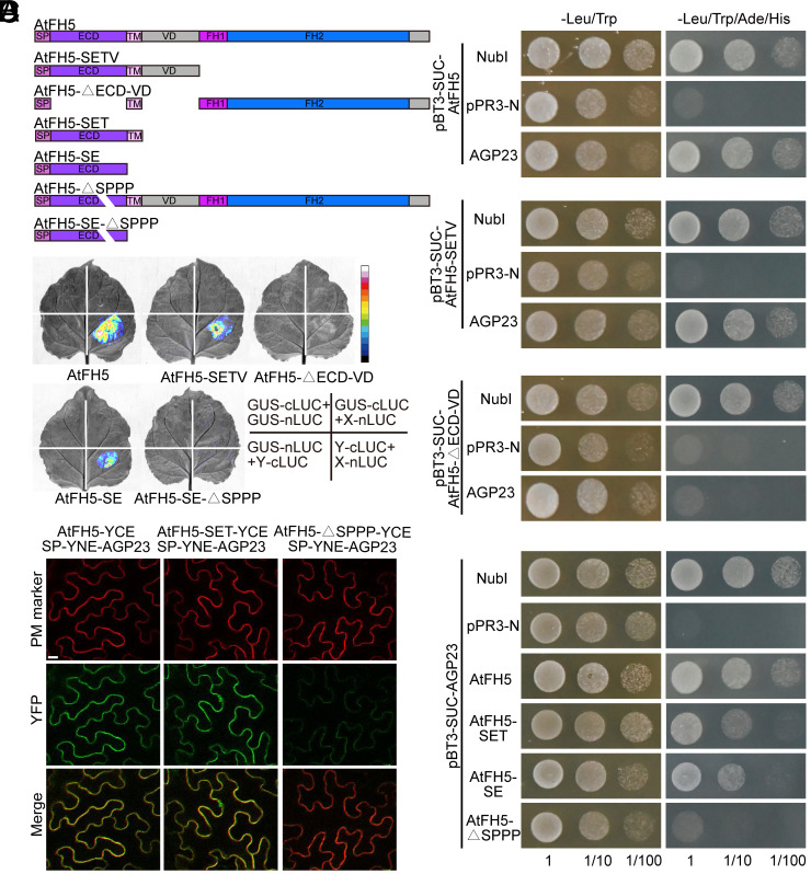
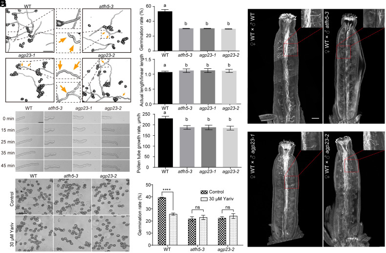
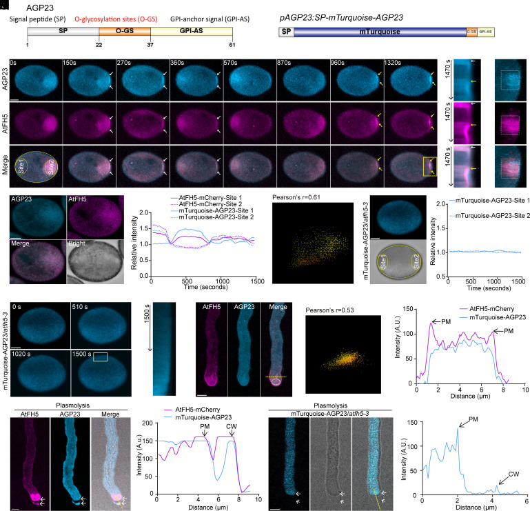
Actin cytoskeleton drives the targeted transport of cell wall components to sustain the tip growth of pollen tubes for double fertilization; however, the underlying mechanism remains largely unknown. Arabidopsis formin 5 (AtFH5), an actin-nucleating protein, localizes at secretory vesicles and mediates actin polymerization-based vesicle trafficking in pollen. Here, we demonstrate that AtFH5 determines the recruitment and transport of cell wall components in AtFH5-labeled vesicles during the tip growth of pollen tubes. Through a screen of interacting proteins of AtFH5, we identify many cell wall-related proteins, with arabinogalactan protein 23 (AGP23) occupying the highest frequency. AtFH5 interacts with AGP23 via its N-terminal extracellular domain (ECD) and jointly regulate the pollen germination and tube growth process. Further observations reveal that AGP23 co-localizes with AtFH5 at moving vesicles, germination sites, and pollen tube tips, suggesting that AGP23 is delivered by AtFH5-labeled vesicles. Deletion of the ECD of AtFH5 interrupts the dynamic localization and cell-wall connection of AGP23 in pollen grains and tubes. Cytological and genetic evidence shows that AGP23 and AtFH5 work in the same pathway to modulate cell wall composition. Together, our data uncover a role of formin in directing the sorting and deposition of cell wall components via secretory vesicle trafficking during pollen germination and tube growth.

Keywords: AGP23; AtFH5; cell wall; pollen; tip growth.


Conflict of interest statement

Competing interests statement:The authors declare no competing interest.

Figures

Fig. 1.
Fig. 1.
The N-terminal of AtFH5 interacts with an arabinogalactan protein AGP23. (A) Full-length and different types of truncated AtFH5. SP, signal peptide; ECD, extracellular domain; TM, transmembrane domain; VD, variable domain; FH1 and FH2, Formin Homology domains. AtFH5-SETV (SP+ECD+TM+VD); AtFH5-△ECD-VD (SP+TM+FH1+FH2); AtFH5-SET (SP+ECD+TM); AtFH5-SE (SP+ECD); AtFH5-△SPPP (full-length AtFH5 with the EXT motif deletion); AtFH5-SE-△SPPP (AtFH5-SE with the EXT motif deletion); (B) Y2H assays showing that AtFH5 interacts with AGP23 via its ECD and EXT motif. The protein+NubI pair is used as a positive control, and the protein+pPR3-N pair is used as a negative control. (C) Split-LCAs showing that the interaction between AGP23 and AtFH5 needs the ECD and EXT motif. The GUS-cLUC+GUS-nLUC, GUS-cLUC+X-nLUC, and GUS-nLUC+Y-cLUC pairs are used as negative controls. (D) Bimolecular fluorescence complementation assays showing that the interaction between AGP23 and AtFH5 is weakened due to the EXT motif deletion. PM marker indicates the PM in tobacco leaf cells.
Fig. 2.
Fig. 2.
The agp23 and atfh5-3 mutants displayed similar defects during pollen germination and tube growth. (A) Observation of pollen germination in wild type, atfh5-3, agp23-1, and agp23-2. Arrows indicate the turns of pollen tubes. Bar, 100 μm. (B) Pollen germination rate in wild type and mutants. (C) The curvature degree of pollen tubes represented by the ratio between actual length and linear length of pollen tubes. (D) Observation of pollen tube growth in wild type, atfh5-3, agp23-1, and agp23-2. Bar, 20 μm. (E) Pollen tube growth rate in wild type and mutants. Values in B, C, and E are means ± SD and the lowercases indicate P < 0.05, one-way ANOVA. (F) Semi-in vivo ovule targeting assay showing that pollen tubes from atfh5-3, agp23-1, and agp23-2 are able to grow into WT embryo sacs. Bar, 200 μm. (G) Observation of pollen germination in the wild type, atfh5-3, and agp23-2 after Yariv treatment. Bar, 20 μm. (H) Pollen germination rate with or without Yariv treatment. Values are mean±SD; ****P < 0.0001; ns, not significant; two-sided Student’s t test.
Fig. 3.
Fig. 3.
AtFH5 recruits AGP23 to secretory vesicles, the germination site, and pollen tube tip. (A) The domain analysis of AGP23. SP, signal peptide; O-GS, O-glycosylation sites; GPI-AS, glycosylphosphatidylinositol-anchor signal. (B) The pAGP23:SP-mTurquoise-AGP23 vector expressing an AGP23 fusion protein within which mTurquoise is located between SP and O-GS. (C) Representative time-lapse images of AtFH5-mCherry and mTurquoise-AGP23 during pollen germination. The white and yellow arrows indicate the prospective germination site during the first and the second time of association with the PM of AtFH5-mCherry and mTurquoise-AGP23, respectively. Bar, 5 μm. (D) A representative image showing the bright field image of a pollen grain. Bar, 5 μm. (E) Relative fluorescent intensity of AtFH5-mCherry and mTurquoise-AGP23 at site1 and site2 in C. (F) Kymograph analysis of AtFH5-mCherry and mTurquoise-AGP23 at the germination site indicated by the yellow box in C. The white and yellow arrows indicate the time point when the first and the second time of association with the PM of AtFH5-mCherry and mTurquoise-AGP23, respectively. (G) A 3D reconstruction analysis of the image of 1,320 s in C with a view facing the germination site. (H) Co-localization analysis of mTurquoise-AGP23 and AtFH5-mCherry at the germination site. The analyzed region was indicated with white boxes in G. (I) A representative image of mTurquoise-AGP23 in an atfh5 pollen grain showing the analyzed region of site1 and site2. Bar, 5 μm. (J) Relative fluorescent intensity of mTurquoise-AGP23 in atfh5-3 at site1 and site2 in I. (K) Representative time-lapse images of mTurquoise-AGP23 in atfh5-3 during pollen germination. Bar, 5 μm. (L) Kymograph analysis of mTurquoise-AGP23 in atfh5-3 at the germination site indicated by the yellow box in K. (M) A representative image showing AtFH5-mCherry and mTurquoise-AGP23 in a growing pollen tube. Bar, 5 μm. (N) Co-localization analysis of mTurquoise-AGP23 and AtFH5-mCherry at pollen tube tip indicated with a white oval in M. (O) Fluorescence intensity of AtFH5-mCherry and mTurquoise-AGP23 along the yellow line in M. The arrows indicate the PM. (P) Observation of AtFH5-mCherry and mTurquoise-AGP23 of a growing pollen tube after plasmolysis. Bar, 5 μm. (Q) Fluorescence intensity of AtFH5-mCherry and mTurquoise-AGP23 along the yellow line in P. The arrows indicate the PM and cell wall (CW), respectively. (R) Observation of mTurquoise-AGP23 in atfh5-3 of a growing pollen tube after plasmolysis. Bar, 5 μm. (S) Fluorescence intensity of mTurquoise-AGP23 along the yellow line in R. The arrows indicate the PM and CW, respectively.
Fig. 4.
Fig. 4.
The dynamic localization of AGP23 relies on the ECD and the EXT motif of AtFH5. (A) The pAtFH5:gAtFH5-△ECD-GFP vector expressing a truncated AtFH5 with the ECD deletion and fused with GFP; The pAtFH5:gAtFH5-△SPPP-GFP vector expressing a truncated AtFH5 with the EXT motif deletion and fused with GFP. (B) Observation of pollen germination in the wild type, atfh5-3, the pAtFH5:gAtFH5-△ECD-GFP/atfh5-3 line, and the pAtFH5:gAtFH5-△SPPP-GFP/atfh5-3 line. Bar, 200 μm. (C) Pollen germination rate in wild type, atfh5-3, the pAtFH5:gAtFH5-△ECD-GFP/atfh5-3 line, and the pAtFH5:gAtFH5-△SPPP-GFP/atfh5-3 line. (D) Observation of pollen tube growth in the wild type, atfh5-3, the pAtFH5:gAtFH5-△ECD-GFP/atfh5-3 line, and the pAtFH5:gAtFH5-△SPPP-GFP/atfh5-3 line. Bar, 20 μm. The arrow heads indicate the turns of pollen tubes. (E) The growth rate of pollen tubes in the wild type, atfh5-3, the pAtFH5:gAtFH5-△ECD-GFP/atfh5-3 line, and the pAtFH5:gAtFH5-△SPPP-GFP/atfh5-3 line. (F) The curvature degree of pollen tubes in the wild type, atfh5-3, the pAtFH5:gAtFH5-△ECD-GFP/atfh5-3 line, and the pAtFH5:gAtFH5-△SPPP-GFP/atfh5-3 line. Values in C, E, and F are means ± SD and the lowercases indicate P < 0.05, one-way ANOVA. (G) Representative time-lapse images of AtFH5-△ECD-GFP and mCherry-AGP23 during pollen germination. Bar, 5 μm. (H) Relative fluorescent intensity of AtFH5-△ECD-GFP and mCherry-AGP23 at site1 and site2 in G. (I) Kymograph analysis of AtFH5-△ECD-GFP and mCherry-AGP23 at the germination site indicated by the white band in G. (J) Observation of AtFH5-△ECD-GFP and mCherry-AGP23 of a growing pollen tube after plasmolysis. Bar, 5 μm. (K) Fluorescence intensity of AtFH5-△ECD-GFP and mCherry-AGP23 along the yellow line in J. The arrows indicate the PM and CW, respectively. (L) Representative time-lapse images of AtFH5-△SPPP-GFP and mCherry-AGP23 during pollen germination. Bar, 5 μm. (M) Relative fluorescent intensity of AtFH5-△SPPP-GFP and mCherry-AGP23 at site1 and site2 in L. (N) Kymograph analysis of AtFH5-△SPPP-GFP and mCherry-AGP23 at the germination site indicated by the white band in L. (O) Observation of AtFH5-△SPPP-GFP and mCherry-AGP23 of a growing pollen tube after plasmolysis. Bar, 5 μm. (P) Fluorescence intensity of AtFH5-△SPPP-GFP and mCherry-AGP23 along the yellow line in O. The arrows indicate the PM and CW, respectively.
Fig. 5.
Fig. 5.
AtFH5 and AGP23 work together to regulate cell wall composition and the proposed working model. (A) Immunolabeling of pollen tubes of wild type, atfh5-3, agp23-2, and the atfh5-3 agp23-2 double mutant using the LM2 antibody. Bar, 5 μm. (B) Quantitative analysis of the relative fluorescence intensity of LM2. (C) Immunolabeling of pollen tubes of wild type, atfh5-3, agp23-2, and atfh5-3 agp23-2 using the JIM7 antibody. Bar, 5 μm. (D) Quantitative analysis of the relative fluorescence intensity of JIM7. (E) Immunolabeling of pollen tubes of wild type, atfh5-3, agp23-2, and atfh5-3 agp23-2 using the JIM5 antibody. Bar, 5 μm. (F) Quantitative analysis of the relative fluorescence intensity of JIM5. (G) Aniline blue staining of pollen tubes in wild type, atfh5-3, agp23-2, and atfh5-3 agp23-2. Bar, 5 μm. The white dotted lines indicate the analysis region of the fluorescence intensity in A, C, E, and G. (H) Quantitative analysis of the relative fluorescence intensity of Aniline blue. Values in B, D, F, and H are means ± SD and the lowercases indicate P < 0.05, one-way ANOVA. (I–K) The proposed working model for AtFH5-mediated recruitment, transport, and deposition of AGP23 during pollen germination and tube growth. First, AtFH5 localizes at secretory vesicles and promotes actin polymerization to drive vesicle trafficking during pollen germination; meanwhile, AtFH5 recruits cell wall components, like AGP23, to these vesicles via its ECD and the EXT motif (I and J). Second, the AtFH5-labeled vesicles carry AGPs to the cell surface destination and determine their disposition at cell wall of growing tips (J and K). Third, loss of AtFH5, ECD, or the EXT motif disrupts the transport and deposition of AGP23, and finally alters cell wall composition and the tip growth of pollen tubes (J and K).

Similar articles

References

    1. Hepler P. K., Rounds C. M., Winship L. J., Control of cell wall extensibility during pollen tube growth. Mol. Plant 6, 998–1017 (2013). - PMC - PubMed
    1. Cascallares M., et al. , A complex journey: Cell wall remodeling, interactions, and integrity during pollen tube growth. Front. Plant Sci. 11, 599247 (2020). - PMC - PubMed
    1. Hao G. J., et al. , Vesicle trafficking in pollen tubes. FEBS Lett. 596, 2231–2242 (2022). - PubMed
    1. Fu Y., The cytoskeleton in the pollen tube. Curr. Opin. Plant Biol. 28, 111–119 (2015). - PubMed
    1. Ruan H. Q., Li J., Wang T., Ren H. Y., Secretory vesicles targeted to plasma membrane during pollen germination and tube growth. Front. Cell Dev. Biol. 8, 615447 (2021). - PMC - PubMed

MeSH terms

LinkOut - more resources