. 2024 Dec 3;121(49):e2403810121.
doi: 10.1073/pnas.2403810121. Epub 2024 Nov 25.

AgRP neurons mediate activity-dependent development of oxytocin connectivity and autonomic regulation

Affiliations

AgRP neurons mediate activity-dependent development of oxytocin connectivity and autonomic regulation

Jessica E Biddinger et al. Proc Natl Acad Sci U S A. .

Abstract

During postnatal life, leptin specifies neuronal inputs to the paraventricular nucleus of the hypothalamus (PVH) and activates agouti-related peptide (AgRP) neurons in the arcuate nucleus of the hypothalamus. Activity-dependent developmental mechanisms impact refinement of sensory circuits, but whether leptin-mediated postnatal neuronal activity specifies hypothalamic neural projections is largely unexplored. Here, we used chemogenetics to manipulate the activity of AgRP neurons during a discrete postnatal critical period and evaluated the development of AgRP inputs to the PVH and descending efferent outflow to the dorsal vagal complex (DVC). In leptin-deficient mice, targeting of AgRP neuronal outgrowth to PVH oxytocin neurons was reduced, and despite the lack of leptin receptors found on oxytocin neurons in the PVH, oxytocin-containing connections to the DVC were also impaired. Activation of AgRP neurons during early postnatal life not only normalized AgRP inputs to the PVH but also oxytocin outputs to the DVC in leptin-deficient mice. Blocking AgRP neuron activity during the same postnatal period reduced the density of AgRP inputs to the PVH of wild type mice, as well as the density of oxytocin-containing DVC fibers, and these innervation deficits were associated with dysregulated autonomic function. These findings suggest that leptin-mediated AgRP neuronal activity is required for the development of PVH connectivity and represents a unique activity-dependent mechanism for specification of neural pathways involved in the hypothalamic integration of autonomic responses.

Keywords: autonomic; hypothalamus; leptin; neural development; paraventricular hypothalamic nucleus.


Conflict of interest statement

Competing interests statement:The authors declare no competing interest.

Figures
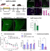
Fig. 1.
Fig. 1.
Postnatal activity of AgRP neurons specifies axon outgrowth and regulates autonomic output. Schematic of experimental paradigm. Mice received daily CNO (1.0 mg/kg i.p.) injections from P4 to P14, and neuroanatomical experiments and tests of autonomic function were conducted in adolescence and adulthood unless denoted otherwise (A). Confocal images illustrating ARH AgRP-Cre-hM4Di mCitrine expression and localization of leptin-induced cFos at P10 (B). Confocal images illustrating ARH leptin-induced cFos-immunoreactive nuclei in mCitrine-labeled neurons in AgRP-Cre-hM4Di mice that had received i.p. injection of saline or CNO 1 h prior to leptin injection (C). Postnatal DREADD-mediated AgRP neuronal inhibition results in significantly decreased AgRP fiber density in the PVH at P30, compared with control AgRP-Cre-hM4Di mice that received postnatal i.p. saline injections (DF). Early postnatal AgRP neuron inhibition resulted in impaired thermoregulatory responses when exposed to a cold environment in adulthood (G), as well as dysregulated GI transit time in response to multiple GI metabolic stimuli (H) and altered intestinal permeability (I). Two-way repeated-measures ANOVA with multiple comparisons was used to compare groups in the cold challenge assay, unpaired t tests were used to compare data between two groups. Differences between groups were considered statistically significant at P < 0.05. Asterisk denotes statistically significant comparisons.
Fig. 2.
Fig. 2.
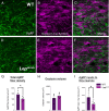
Leptin specifies targeting of AgRP projections to PVH oxytocin neurons. Confocal images depicting AgRP-immunoreactive axons (green) and Oxytocin-Cre-SynTom fluorescence (magenta) in Oxy-Cre-SynTom-WT (AC) and leptin-deficient Oxy-Cre-SynTom-OB mice (DF) in the PVH at P30. Density of total AgRP-immunoreactive axons within the PVH was significantly decreased in leptin-deficient mice (A, D, and G). No differences were observed in oxytocin volume (B, E, and H). AgRP-immunoreactive axons targeting Oxytocin-Cre neurons were also significantly decreased in leptin-deficient mice compared with WT controls (C, F, and I).
Fig. 3.
Fig. 3.
Leptin signaling in oxytocin and oxytocin receptor-expressing neurons in the PVH and DVC. Stereotaxic injection of Cre-dependent AAV-tdTomato virus into the PVH of adult Oxytocin-Cre mice labels projections of neurons expressing Oxytocin-Cre (A). Confocal images depicting representative injection site of AAV-tdTomato virus (red) into Oxytocin-Cre-expressing neurons and oxytocin-immunolabeled neurons (green) in the caudal PVH (BD). TdTomato-labeled Oxytocin-Cre axonal inputs (red) are shown at three levels of the dorsomedial medulla after AAV-mediated recombination. Neurons (gray) are immunolabeled with the pan-neuronal marker HuC/D (EG). Confocal images of oxytocin (red) and cFos (green) immunolabeling in the PVH of mice injected with i.p. saline or leptin at P16 (H and I). Representative confocal images of oxytocin-immunolabeled neurons (blue) and LepRb-Cre-tdTom fluorescence (red) in caudal regions of the PVH at P18 taken with 20× (J) and high-magnification 63× objectives, respectively (K and L). Confocal image of OxyR-expressing neurons in the DVC at P30, visualized by OxyR-Venus fluorescence (M). Representative image of pSTAT3-immunoreactivity (red) in the DVC of OxyR-Venus mice injected i.p. with leptin (N). Merged image showing pSTAT3-immunoreactive nuclei in response to i.p. leptin injection and DVC OxyR-Venus labeled neurons (O). Abbreviations: PVH, paraventricular nucleus of the hypothalamus; DVC, dorsal vagal complex; AP, area postrema; DMX, dorsal motor nucleus of the vagus nerve; Gr, Gracile nucleus; NTSm, medial subnucleus of the solitary tract; NTSdm, dorsomedial subnucleus of the solitary tract; 3V, third ventricle; 4V, fourth ventricle; cc, central canal; ts, solitary tract.
Fig. 4.
Fig. 4.
Postnatal ontogeny of DVC oxytocin terminals is impaired in leptin-deficient mice. Density and distribution of DVC oxytocin terminals at postnatal ages P8, P16, P30, and P60 visualized by genetic targeting of fluorescent synaptophysin-tdTomato fusion protein (red) to neurons expressing Oxytocin-Cre (AH). In WT mice (Oxy-Cre-SynTom-WT), Oxy-Cre-SynTom axons are observed in the caudal DVC as early as P8 (A), significantly increase in density by P16 (B), and reach adult-like levels by P30 (C), which are maintained at P60 (D). In leptin-deficient mice (Oxy-Cre-SynTom-Lepob/ob), a significant decrease in Oxy-Cre-SynTom labeled terminals emerged in the DVC at P16 (B, F, and J) and was maintained at P30 (C, G, and K) and P60 d of age (D, H, and L). No changes were detected in the number of Oxytocin-Cre-SynTom labeled neurons in the PVH at P16 or P30 (MQ). Boxes indicate location of Region of interest (ROIs) used for quantitative analysis. Unpaired t-tests were used to test for significant differences between genotypes at each age examined. Asterisk denotes P-values < 0.05. Error bars indicate mean ± SEM; circles represent individual values. Images are maximum intensity projections from confocal image stacks collected through 30um-thick sections. Abbreviations: AP, area postrema; Gr, Gracile nucleus; NTS, nucleus of the solitary tract; cc, central canal.
Fig. 5.
Fig. 5.
Early postnatal AgRP neuronal activity specifies descending oxytocin projections to the DVC. Maximum intensity projection confocal images of oxytocin-immunolabeled neurons (red) in caudal PVH regions known to contain preautonomic neurons in AgRP-Cre-hM4Di mice at P30 that received daily postnatal injection of either saline or CNO i.p. from P4 to P14 (A, B, and G). Early postnatal DREADD-mediated AgRP neuronal inhibition led to a significant reduction in the density of oxytocin-immunolabeled inputs to the DVC at P30 (C, D, and H) and adult animals at P60 or older (E, F, and I).
Fig. 6.
Fig. 6.
Early postnatal activation of AgRP neurons in leptin-deficient AgRP-Cre-hM3Dq-Lepob/ob mice restores impaired AgRP and oxytocin projections in Lepob/ob mice. Representative images of AgRP (green) and oxytocin (magenta) immunolabeling in the PVH of AgRP-Cre-hM3Dq-Lepob/ob mice that received saline (A and B) or CNO (C and D) daily from P4 to P14. DREADD-mediated early postnatal AgRP neuronal activation in AgRP-Cre-hM3Dq-Lepob/ob mice that received daily CNO showed significantly elevated densities of AgRP-immunoreactive terminals closely apposed to oxytocin neurons in the PVH in adulthood, compared with those observed in Lepob/ob mice that received saline (E). Density of oxytocin-immunoreactive fibers and terminals in the DVC was significantly higher in AgRP-Cre-hM3Dq-Lepob/ob mice that were treated postnatally with CNO (FH). No change was observed in PVH oxytocin neuron number in any of the groups tested (I). Schematic of proposed activity-dependent mechanism of action: During postnatal days (P4 to P14 leptin levels surge in neonatal mice and activate (depolarize; red plus symbol) AgRP neurons in the ARH. This early postnatal AgRP neuronal activation is required for normal development of AgRP axonal projections to the PVH where they innervate oxytocin neurons. Development of neural projections from oxytocin neurons to the DVC is also dependent on AgRP neuronal activity through a transneuronal mechanism of activity-dependent neural development, with downstream effects on autonomic function (J). Boxes on 20× images correspond to adjacent higher magnification images, and denote location of ROIs used for analysis. Confocal images taken with 20× (A, C, F, and G) and high- magnification (B and D) objectives, respectively. Abbreviations: Agi, AgRP-Cre-hM4Di; AgQO, AgRP-Cre-hM3Dq; PVH, paraventricular nucleus of the hypothalamus; ARH, arcuate nucleus of the hypothalamus; DVC, dorsal vagal complex; AP, area postrema; DMX, dorsal motor nucleus of the vagus nerve; NTS, nucleus of the solitary tract; 3V, third ventricle; cc, central canal.

Update of

References

    1. Rosenbaum M., Leibel R. L., 20 years of leptin: Role of leptin in energy homeostasis in humans. J. Endocrinol. 223, T83–T96 (2014). - PMC - PubMed
    1. Elson A. E., Simerly R. B., Developmental specification of metabolic circuitry. Front. Neuroendocr. 39, 38–51 (2015). - PMC - PubMed
    1. Skowronski A. A., Leibel R. L., LeDuc C. A., Neurodevelopmental programming of adiposity: Contributions to obesity risk. Endocr. Rev. 45, 253–280 (2023). - PMC - PubMed
    1. Donato J., Programming of metabolism by adipokines during development. Nat. Rev. Endocrinol. 19, 385–397 (2023). - PubMed
    1. Zeltser L. M., Feeding circuit development and early-life influences on future feeding behaviour. Nat. Rev. Neurosci. 19, 302–316 (2018). - PMC - PubMed

MeSH terms

LinkOut - more resources