. 2024 Dec 3;121(49):e2414333121.
doi: 10.1073/pnas.2414333121. Epub 2024 Nov 25.

Global hinge sites of proteins as target sites for drug binding

Affiliations

Global hinge sites of proteins as target sites for drug binding

Haotian Zhang et al. Proc Natl Acad Sci U S A. .

Abstract

Hinge sites of proteins play a key role in mediating conformational mechanics. Among them, those involved in the most collective modes of motion, also called global hinges, are of particular interest, as they support cooperative rearrangements that are often functional. Yet, the utility of targeting global hinges for modulating function remains to be established. We present here a systematic study of a series of proteins resolved in drug-bound forms to examine the probabilistic occurrence of spatial overlaps between hinge sites and drug-binding pockets. Our analysis reveals a high propensity of drug binding to hinge sites compared to random. Notably, one-third of currently approved drugs are colocalized with hinge sites. These mechanosensitive sites are predictable by simple models such as the Gaussian Network Model. Their targeting thus emerges as a viable strategy for developing a new class of drugs that would exploit and modulate the target proteins' intrinsic dynamics, and potentially alleviate drug-resistance when used in combination with orthosteric or allosteric drugs.

Keywords: Gaussian Network Model; collective motions; drug binding sites; ensemble analysis; protein dynamics.


Conflict of interest statement

Competing interests statement:The authors declare no competing interest.

Figures

Fig. 1.
Fig. 1.
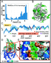
Schematic description of the methodology for evaluating the overlap between global hinge sites and drug-binding sites. Data for PARP1 catalytic domain are shown for illustration. Gaussian Network Model (GNM) analysis yields h hinges in modes 1 and 2, s of which overlap with the b drug-binding residues. The ribbon diagram shows in space-filling the hinge residues in mode 1 (orange) and 2 (yellow) along with drug-binding sites (in green; Fig. 2).
Fig. 2.
Fig. 2.
Results for PARP1 catalytic domain ensemble (A). Normalized distribution of residue displacements along mode 1 (Top) and 2 (Bottom) predicted by SignDy. The solid curve represents the average mode profile, and the light blue shade displays the SD based on 175 homologs. Hinge sites (green dots), drug-binding sites (red stars), and their overlaps (magenta diamonds, labeled) are indicated. (B) Hinge residues from mode 1 (orange spheres), and 2 (yellow spheres) line the drug-binding pocket. Drugs from multiple structures are overlaid in green sticks. The diagram is color-coded by the motions along mode 1, blue and cyan referring to opposite direction motions (see Upper Right scale).
Fig. 3.
Fig. 3.
Results for Angiotensin-converting enzyme ACE and its structural homologs. (A) RMSD distribution of 514 structural homologs with respect to the reference structure (PDB: 2X92, chain A). (B) Positions of small molecules bound to these structures, superposed on the reference structure (C) Mode 1 evaluated using SignDy. The average curve is in dark blue, and the results within one and two SD from the average are indicated by the dark and light blue shades. (D and E) Location of hinge residues, and close-up view of the coordination of drugs (superposed from multiple homologs) by these global hinge residues, shown in orange (mode 1), yellow (mode 2), and wheat (mode 3) spheres.
Fig. 4.
Fig. 4.
Results for dimeric targets. Dimers may bind drugs at the interface of the two monomers (as in PR and PA) or at active sites within monomers (as in COX2, PDE5, PPARg, and HIV-1 RT). Mode 1 hinge sites, usually located at the interface, are not involved in binding the drugs located within the monomers. Instead, hinges from mode 2 (yellow) or 3 (wheat) to colocalize with the drugs.
Fig. 5.
Fig. 5.
Results for RT ensemble. Panel (A) displays the different subdomains of RT p66 subunit in different colors, and the p51 subunit in light blue. FDA-approved drugs (green sticks) bound to the palm subdomain are coordinated by mode 3 hinge residues (wheat spheres). (B) RT first 3 mode shapes. The palm is highly constrained in all modes (near y = 0 line), as a hinge region. (C) Closeup (and rotated) view of interactions at the drug-binding site. Hinges are shown in magenta. (D) thumb–connection interface also binds inhibitors (red sticks). Two hinge residues (magenta sticks), identified in mode 1, interact with the inhibitors.
Fig. 6.
Fig. 6.
Statistical analysis shows the high propensity of hinges among drug-binding sites. (A) Enrichment of hinge residues at drug-binding sites, compared to other regions. (B) Histogram of the fraction of hinge residues at drug-binding sites.
Fig. 7.
Fig. 7.
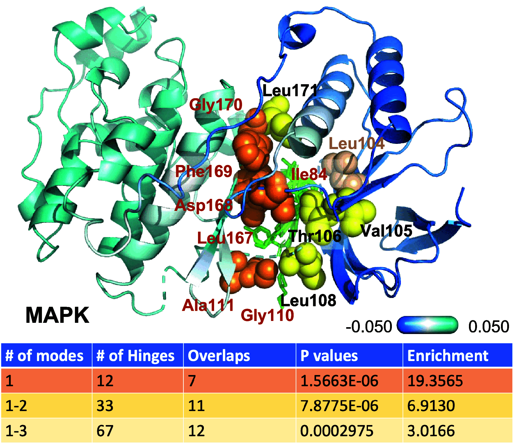
Results for MAPK14. The diagram shows the hinge residues from mode 1 (orange), 2 (yellow), and 3 (wheat) lining the drug-binding pocket. Their residue labels are orange, black, and wheat, respectively. A series of drugs (from multiple structures structurally aligned against the reference), are shown in green sticks. The ribbon diagram is colored by the normalized displacements along mode 1. The Lower panel lists the number of hinge residues in mode 1, 1-2, and 1-3, their overlap with drug-binding residues, corresponding hypergeometric scores, and enrichment at drug-binding sites.

Similar articles

Cited by

References

    1. Nussinov R., Tsai C. J., The different ways through which specificity works in orthosteric and allosteric drugs. Curr. Pharm. Des. 18, 1311–1316 (2012). - PMC - PubMed
    1. Lu S., Shen Q., Zhang J., Allosteric methods and their applications: Facilitating the discovery of allosteric drugs and the investigation of allosteric mechanisms. Acc. Chem. Res. 52, 492–500 (2019). - PubMed
    1. Wagner J. R., et al. , Emerging computational methods for the rational discovery of allosteric drugs. Chem. Rev. 116, 6370–6390 (2016). - PMC - PubMed
    1. Hardy J. A., Wells J. A., Searching for new allosteric sites in enzymes. Curr. Opin. Struc. Biol. 14, 706–715 (2004). - PubMed
    1. Wells J. A., McClendon C. L., Reaching for high-hanging fruit in drug discovery at protein-protein interfaces. Nature 450, 1001–1009 (2007). - PubMed

LinkOut - more resources