Case Reports
. 2024 Dec 24;103(12):e210084.
doi: 10.1212/WNL.0000000000210084. Epub 2024 Nov 25.

Sulcal Hyperintensity as an Early Imaging Finding in Cerebral Amyloid Angiopathy-Related Inflammation

Affiliations
Case Reports

Sulcal Hyperintensity as an Early Imaging Finding in Cerebral Amyloid Angiopathy-Related Inflammation

Larysa Panteleienko et al. Neurology. .

Abstract

Background and objectives: Cerebral amyloid angiopathy-related inflammation (CAA-ri) is a subtype of CAA with distinct clinical and radiologic features. Existing diagnostic criteria require the presence of characteristic asymmetrical white matter hyperintensity (WMH), together with classical hemorrhagic neuroimaging markers of CAA. There are limited data for other diagnostic neuroimaging markers of CAA-ri.

Methods: This is a case series from a specialist hospital intracerebral hemorrhage service.

Results: We describe 4 patients with CAA-ri who had regions of sulcal hyperintensity, with or without gyral swelling at clinical presentation, but did not fulfill current diagnostic criteria because of the absence of typical asymmetric WMH on brain MRI. All 4 patients were subsequently diagnosed with CAA-ri; three later developed asymmetric WMHs with disease relapse, and 2 had pathologically proven CAA-ri; 1 patient had both.

Discussion: Regions of sulcal hyperintensity, sometimes with associated gyral swelling, can be an early imaging finding in CAA-ri. These neuroimaging markers could potentially improve the accuracy of existing diagnostic criteria for CAA-ri to allow earlier diagnosis and treatment without biopsy in patients with atypical presentations.


Conflict of interest statement

L. Panteleienko is supported in her work at the UCL Queen Square Institute of Neurology and National Hospital for Neurology and Neurosurgery, Queen Square, by a Council for At-Risk Academics (CARA-UCL) Academic Sanctuary Fellowship. G. Banerjee is an NIHR Clinical Lecturer and additionally receives research funding from Alzheimers Research UK and the Stroke Association. M.S. Zandi receives funding from the UCL/UCLH Biomedical Research Centre, and has received honoraria for one lecture each for each of the 4 mentioned in the last 4 years: Cygnet Healthcare; UCB Pharma and GSK. Go to Neurology.org/N for full disclosures.

Figures

Figure 1
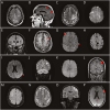
Figure 1. Representative MRI Findings for the Cases Described
Case 1 (A–D): MRI at initial presentation showed sulcal hyperintensity (SH) on fluid attenuated inversion recovery (FLAIR) images (A), leptomeningeal contrast enhancement (B, red arrowheads); MRI at relapse 5 months after initial presentation showed asymmetric white matter hyperintensity (WMH), over the left parietal and occipital lobes (C), in addition to multiple cortical and subcortical foci of restricted diffusion on DWI sequences (D). Case 2 (E–H): MRI at presentation showed SH over the left frontal lobe on FLAIR images (E), leptomeningeal contrast enhancement (F, red arrowheads), lobar cerebral microbleeds (CMB, red arrowheads), and areas of cortical superficial siderosis (cSS), mostly over the left frontal lobe on SWI (G), and multiple subcortical DWI lesions (H). Case 3 (I–L): MRI at initial presentation showed a subtle area of cortical swelling and SH in the left parietal region (I, red arrowhead); MRI at relapse showed a new patch of the WMH in the left temporoparietal region (J), associated with a cluster of CMB (K). The follow-up image 2 years later showed leptomeningeal enhancement in the left temporoparietal region (L, red arrowhead). Case 4 (M–P): MRI at initial presentation showed SH with gyral swelling on the FLAIR image over the right frontal lobe and occipital lobes bilaterally (M); MRI at relapse 9 months later showed areas of WMH with mild parenchymal swelling on T2 (N) and FLAIR (O) images primarily affecting the left perirolandic region, and leptomeningeal contrast enhancement (P, red arrowheads).

References

    1. Auriel E, Charidimou A, Gurol ME, et al. . Validation of clinicoradiological criteria for the diagnosis of cerebral amyloid angiopathy-related inflammation. JAMA Neurol. 2016;73(2):197-202. doi:10.1001/jamaneurol.2015.4078 - DOI - PubMed
    1. Melzer N, Harder A, Gross CC, et al. . CD4(+) T cells predominate in cerebrospinal fluid and leptomeningeal and parenchymal infiltrates in cerebral amyloid β-related angiitis. Arch Neurol. 2012;69(6):773-777. doi:10.1001/archneurol.2011.2441 - DOI - PubMed
    1. Boncoraglio GB, Piazza F, Savoiardo M, et al. . Prodromal Alzheimer's disease presenting as cerebral amyloid angiopathy-related inflammation with spontaneous amyloid-related imaging abnormalities and high cerebrospinal fluid anti-Aβ autoantibodies. J Alzheimers Dis. 2015;45(2):363-367. doi:10.3233/JAD-142376 - DOI - PubMed
    1. Theodorou A, Tsibonakis A, Pateras IS, et al. . Multiple cerebral microinfarcts: an uncommon presentation of Cerebral Amyloid Angiopathy-related inflammation. Neurol Res Pract. 2023;5(1):28. doi:10.1186/s42466-023-00253-9 - DOI - PMC - PubMed
    1. Theodorou A, Palaiodimou L, Malhotra K, et al. . Clinical, neuroimaging, and genetic markers in cerebral amyloid angiopathy-related inflammation: a systematic review and meta-analysis. Stroke. 2023;54(1):178-188. doi:10.1161/STROKEAHA.122.040671 - DOI - PubMed

Publication types

MeSH terms