

Penetrating intracranial injury from a pencil in a pediatric patient: illustrative case
- PMID: 39586095
- PMCID: PMC11605530
- DOI: 10.3171/CASE24494
Penetrating intracranial injury from a pencil in a pediatric patient: illustrative case
Abstract
Background: Intracranial penetrating injuries from a pencil are exceptionally rare. The most common mechanism is a child running while holding a pencil. Potential consequences of intracranial pencil injury include direct trauma to brain structures, vascular injury, and intracranial abscess formation.
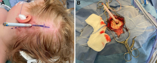
Observations: A 3-year-old girl was at daycare and had fallen while running with a pencil. Computed tomography showed a pencil penetrating the left parietal bone through the left temporal lobe, terminating in the posterior limb of the internal capsule. Cerebral angiography was performed prior to the removal of the pencil to rule out vascular injury. Angiography of the left carotid artery revealed slight irregularity in the left M2 but no active extravasation. The patient was then taken to the operating room to have the pencil removed. Postoperatively, she did well and was discharged home after 6 days with no neurological deficits.
Lessons: Pencils are rare causes of intracranial injury in children. Definitive vascular imaging prior to pencil removal to rule out vascular injury and minimize the risk of hemorrhage after removal is recommended. Intraoperative irrigation and debridement, followed by antibiotics, are recommended to avoid abscess formation. Follow-up vascular imaging is recommended to rule out pseudoaneurysm. https://thejns.org/doi/10.3171/CASE24494.
Keywords: foreign object; pediatrics; pencil; penetrating intracranial injury.
Figures


References
-
- Bursick DM, Selker RG. Intracranial pencil injuries. Surg Neurol. 1981;16(6):427-431. - PubMed
-
- Gupta A, Chacko A, Anil MS, Karanth SS, Shetty A. Pencil in the brain: a case of temporal lobe abscess following an intracranial penetrating pencil injury. Pediatr Neurosurg. 2011;47(4):307-308. - PubMed
-
- Horner FA, Berry RG, Frantz M. Broken pencil points as a cause of brain abscess. N Engl J Med. 1964;271(7):342-345. - PubMed
-
- Sharif G, Roberts J, Phillips S. Transnasal penetrating brain injury with a ball-pen. Br J Neurosurg. 2000;14(2):159-160. - PubMed
LinkOut - more resources
Full Text Sources
