. 2024 Dec:78:103439.
doi: 10.1016/j.redox.2024.103439. Epub 2024 Nov 20.

Podocyte SIRPα reduction in diabetic nephropathy aggravates podocyte injury by promoting pyruvate kinase M2 nuclear translocation

Affiliations

Podocyte SIRPα reduction in diabetic nephropathy aggravates podocyte injury by promoting pyruvate kinase M2 nuclear translocation

Yang Chen et al. Redox Biol. 2024 Dec.

Abstract

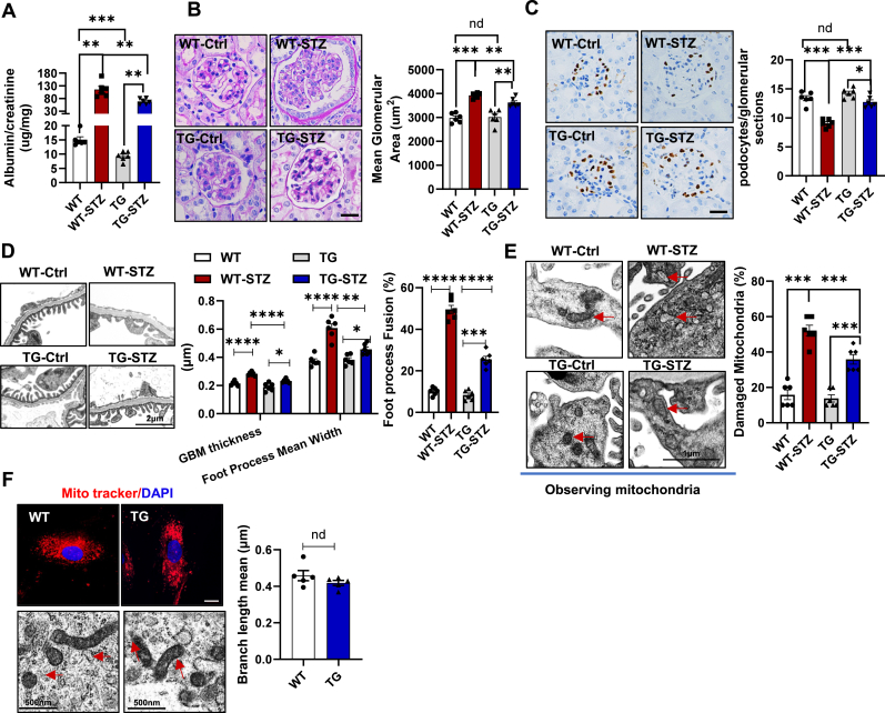
Podocyte injury is a critical event in the pathogenesis of diabetic nephropathy (DN). Hyperglycemia, oxidative stress, inflammation, and other factors contribute to podocyte damage in DN. In this study, we demonstrate that signaling regulatory protein alpha (SIRPα) plays a pivotal role in regulating the metabolic and immune homeostasis of podocytes. Deletion of SIRPα in podocytes exacerbates, while transgenic overexpression of SIRPα alleviates, podocyte injury in experimental DN mice. Mechanistically, SIRPα downregulation promotes pyruvate kinase M2 (PKM2) phosphorylation, initiating a positive feedback loop that involves PKM2 nuclear translocation, NF-κB activation, and oxidative stress, ultimately impairing aerobic glycolysis. Consistent with this mechanism, shikonin ameliorates podocyte injury by reducing PKM2 nuclear translocation, preventing oxidative stress and NF-κB activation, thereby restoring aerobic glycolysis.

Keywords: Aerobic glycolysis; NF-κB; Oxidative stress; PKM2; Podocyte injury; SIRPα.


Conflict of interest statement

Declaration of competing interest The authors declare the following financial interests/personal relationships which may be considered as potential competing interests: Limin Li reports financial support was provided by the National Natural Science Foundation of China (32170897). Limin Li reports a relationship with the National Natural Science Foundation of China (32170897) that includes: funding grants. If there are other authors, they declare that they have no known competing financial interests or personal relationships that could have appeared to influence the work reported in this paper.

Figures

Image 1
Graphical abstract
Fig. 1
Fig. 1
Reduction of SIRPα in Podocytes of Diabetic Nephropathy. (A) Schematic representation of wild type (WT-STZ) and CKO mice (CKO-STZ) induced by streptozotocin (STZ). (B) Analysis of the urinary albumin-to-creatinine ratio in WT-STZ and CKO-STZ mice. (C, D) Periodic Acid-Schiff (PAS) staining and WT1 immunohistochemistry (scale bar, 25 μm). Glomerular area was quantified based on PAS staining (C) (n = 6, 50 glomeruli randomly counted per mouse). Podocyte number was quantified based on WT1 immunohistochemistry results (D) (n = 6, 50 glomeruli randomly counted per mouse). (E) Ultrastructural analysis of podocytes in WT-STZ and CKO-STZ mice. Glomerular basement membrane (GBM) thickness, podocyte foot process width (GBM length/number of foot processes), percentage of podocyte foot process fusion (foot process fusion length/GBM length), and damaged mitochondria in podocytes were quantitatively analyzed based on podocyte ultrastructure (n = 6, 3 fields randomly selected per mouse). (F, G) Mitochondrial morphology in primary mouse podocytes (MitoTracker, scale bar, 15 μm) and mitochondrial ultrastructure in mouse podocytes. The mitochondrial network was reconstructed using ImageJ, and the mean mitochondrial branch length was measured (F) (n = 5). Mitochondrial ultrastructure was quantified, and mitochondrial area was analyzed at the same order of magnitude (G).
Fig. 2
Fig. 2
Podocyte-Specific Transgenic SIRPα protects against hyperglycemia-induced podocyte injury. (A) Urinary albumin-to-creatinine ratio in WT, WT-STZ, TG, and TG-STZ mice (n = 6). (B) PAS staining of renal paraffin sections (scale bar, 25 μm). Glomerular area was quantified (n = 6, 50 glomeruli randomly counted per mouse). (C) Podocyte number was quantitatively analyzed based on WT1 immunohistochemistry (n = 6, 50 glomeruli randomly counted per mouse; scale bar, 25 μm). (D) Ultrastructural analysis of podocytes, including glomerular basement membrane (GBM) thickness, podocyte foot process width (GBM length/number of foot processes), percentage of podocyte foot process fusion (foot process fusion length/GBM length). (E) Damaged mitochondria in podocytes were quantitatively assessed (n = 6, 3 fields randomly selected per mouse). (F) Mitochondrial morphology in primary mouse podocytes (MitoTracker, scale bar, 15 μm) and mitochondrial ultrastructure. The mitochondrial network was reconstructed using ImageJ, and the mean mitochondrial branch length was measured (n = 5).
Fig. 3
Fig. 3
SIRPα rescues high glucose-induced oxidative stress and increases glycolytic flux in podocytes. (A) Gene Set Enrichment Analysis (GSEA) enrichment profiles of oxidative stress (NES = 1.80, FDR<0.01, P < 0.001). RT-qPCR was used to verify the expression of related genes (n = 6). (B) The total ROS levels in primary podocytes from WT and CKO mice (n = 3). (C) The OCR and ATP levels in podocytes from WT and CKO mice (n = 6). (D) The mRNA levels of pyruvate metabolism-related genes were detected (n = 6). (E) The pyruvate kinase activity in podocytes from WT and CKO mice (n = 3). Mito-TEMPO (10 μM, 24 hours) was used to clearance of ROS. (F–H) Levels of mito-ROS (F), OCR and ATP (G), pyruvate kinase activity (H) in podocytes. (IM) TG and WT podocytes were treated with 30 mM glucose for 48 h. Oxidative stress related genes (I), total ROS (J, n=3), OCR and ATP levels (K), pyruvate metabolism related genes (L, n = 6), and pyruvate kinase activity (M, n = 3) in HG-induced WT and TG podocytes were detected.
Fig. 4
Fig. 4
SIRPα deficiency facilitates PKM2 nuclear translocation by promoting PKM2 phosphorylation. (A) Co-immunoprecipitation of SHP1 with SIRPα and PKM2. (B) Co-immunoprecipitation of PKM2 with SHP1. (C) Phosphorylation of PKM2 was induced in SIRPα knockdown podocytes (SIRPα siRNA). (D) Phosphorylation of PKM2 was inhibited in SIRPα overexpressing podocytes (OE-SIRPα). (E) Protein levels of SIRPα, pPKM2, PKM2 and podocin in HPC treated with serum from healthy persons and DN patients (n = 5). (F) The expression and distribution of SIRPα (red), and pPKM2 (green) were determined by immunofluorescence staining in renal tissues of DN patients. Scale bar, 25 μm. Three experiments were repeated independently. (G) The expression and distribution relationship between SIRPα and PKM2 were determined in primary podocytes from WT, TG, CKO mice with or without 30 mM HG treatment. Nucleus (blue, DAPI), F-actin (red), and PKM2 (green). Three experiments were repeated independently. (H) SIRPα, pPKM2, and PKM2 were measured in podocytes obtained from WT, TG, CKO, WT-STZ, TG-STZ, and CKO-STZ mice (Scale bar, 15 μm, n = 6). The level of pPKM2 was calculated using Image Pro Plus 6.0.
Fig. 5
Fig. 5
Deficiency of SIRPα results in formation a positive feedback loop between nuclear PKM2, NF-κB activation, and oxidative stress. (A) KEGG secondary classification of signal pathway, sorted by enrichment scores (fc > 1.5, P < 0.05). (B, C) Proteins levels of SIRPα, pP65, P65, and IL-6 were detected by Western blotting (n = 3). (D, E) Levels of Nox4 mRNA in podocytes (n = 3). (F–I) Podocytes isolated from CKO mice were treated with (KO-Shikonin) or without 1 μM shikonin (KO-Ctrl) for 24 hours (n = 3). PKM2 distribution (F, Scale bar, 15 μm), protein level of pP65, P65 and IL-6 (G), total ROS production (H), mRNA level of Nox4 (I) in podocytes were determined. (JL) NOX4 inhibitor GLX351322 (GLX, 10 μM, 24 hours) and Mito-TEMPO (10 μM, 24 hours) were used to inhibit the production or clearance of ROS in podocytes. PKM2 distribution (J, Scale bar, 15 μm), total ROS production (K), and protein levels of pP65, P65 and IL-6 were evaluated (L). (M, N) The primary CKO podocytes were transfected with control siRNA (siCtrl) or IL-6 siRNA (siIL-6). The nuclear distribution of PKM2 (M, Scale bar, 15 μm), and total ROS production were determined (n = 3) (N, Scale bar, 15 μm).
Fig. 6
Fig. 6
Shikonin alleviates hyperglycemia induced podocyte injury in CKO mice via inhibiting PKM2 nuclear translocation. (A) Schematic diagram of shikonin administration on STZ-induced DN experimental mice. WT and CKO mice were given STZ at 8 weeks of age for constructing experimental DN model, and shikonin was administrated every two days. After four weeks of administration, biochemical and pathological indicators of the mice were measured to evaluate the effect of shikonin on renal disease progression. (B) Levels of urinary albumin/creatinine (n = 6). (C) PAS staining of renal sections and glomerular area was counted (n = 6, and 50 glomeruli were counted randomly per mouse, Scale bar, 25 μm). (D) Immunohistochemical staining of WT1 to calculate the number of podocytes (n = 6, and 50 glomeruli were counted randomly per mouse, Scale bar, 25 μm). (E) The expression and nuclear distribution (blue, DAPI) relationship between SIRPα (purple), Nephrin (red), and PKM2 (green) were determined through immunofluorescence staining. Scale bar, 15 μm. (F) The protein levels of pP65, P65 and IL-6 in renal cortex of mice.

References

    1. Wu L., Liu C., Chang D.-Y., et al. The attenuation of diabetic nephropathy by annexin A1 via regulation of lipid metabolism through the AMPK/PPARα/CPT1b pathway. Diabetes. 2021;70(10):2192–2203. doi: 10.2337/db21-0050. [published Online First: Epub Date]|. - DOI - PubMed
    1. Kim Y., Lim J.H., Kim M.Y., et al. The adiponectin receptor agonist AdipoRon ameliorates diabetic nephropathy in a model of type 2 diabetes. J. Am. Soc. Nephrol. 2018;29(4):1108–1127. doi: 10.1681/ASN.2017060627. [published Online First: Epub Date]|. - DOI - PMC - PubMed
    1. Wu M., Yang Z., Zhang C., et al. Inhibition of NLRP3 inflammasome ameliorates podocyte damage by suppressing lipid accumulation in diabetic nephropathy. Metabolism. 2021;118 doi: 10.1016/j.metabol.2021.154748. [published Online First: Epub Date]|. - DOI - PubMed
    1. Fu J., Shinjo T., Li Q., et al. Regeneration of glomerular metabolism and function by podocyte pyruvate kinase M2 in diabetic nephropathy. JCI Insight. 2022;7(5) doi: 10.1172/jci.insight.155260. [published Online First: Epub Date]|. - DOI - PMC - PubMed
    1. Fu Y., Sun Y., Wang M., et al. Elevation of JAML promotes diabetic kidney disease by modulating podocyte lipid metabolism. Cell Metabol. 2020;32(6):1052–10562. doi: 10.1016/j.cmet.2020.10.019. [published Online First: Epub Date]|. - DOI - PubMed

Publication types

MeSH terms