. 2024 Dec:110:105452.
doi: 10.1016/j.ebiom.2024.105452. Epub 2024 Nov 24.

NF-κB c-Rel is a critical regulator of TLR7-induced inflammation in psoriasis

Affiliations

NF-κB c-Rel is a critical regulator of TLR7-induced inflammation in psoriasis

Angela Rose Liu et al. EBioMedicine. 2024 Dec.

Abstract

Background: Nuclear factor kappa B (NF-κB) c-Rel is a psoriasis susceptibility locus, however mechanisms underlying c-Rel transactivation during disease are poorly understood. Inflammation in psoriasis can be triggered following Toll-like Receptor 7 (TLR7) signalling in dendritic cells (DCs), and c-Rel is a critical regulator of DC function. Here, we studied the mechanism of TLR7-induced c-Rel-mediated inflammation in DCs.

Methods: The overall expression of c-Rel was analysed in skin sections from patients with psoriasis in human transcriptomics datasets as well as the imiquimod-induced psoriasis mouse model. The function of c-Rel in DCs following TLR7 stimulation was determined by c-Rel CRISPR/Cas9 knockout DC2.4 immortalised cells and primary bone marrow derived dendritic cells from c-Rel knockout C57BL6/J mice.

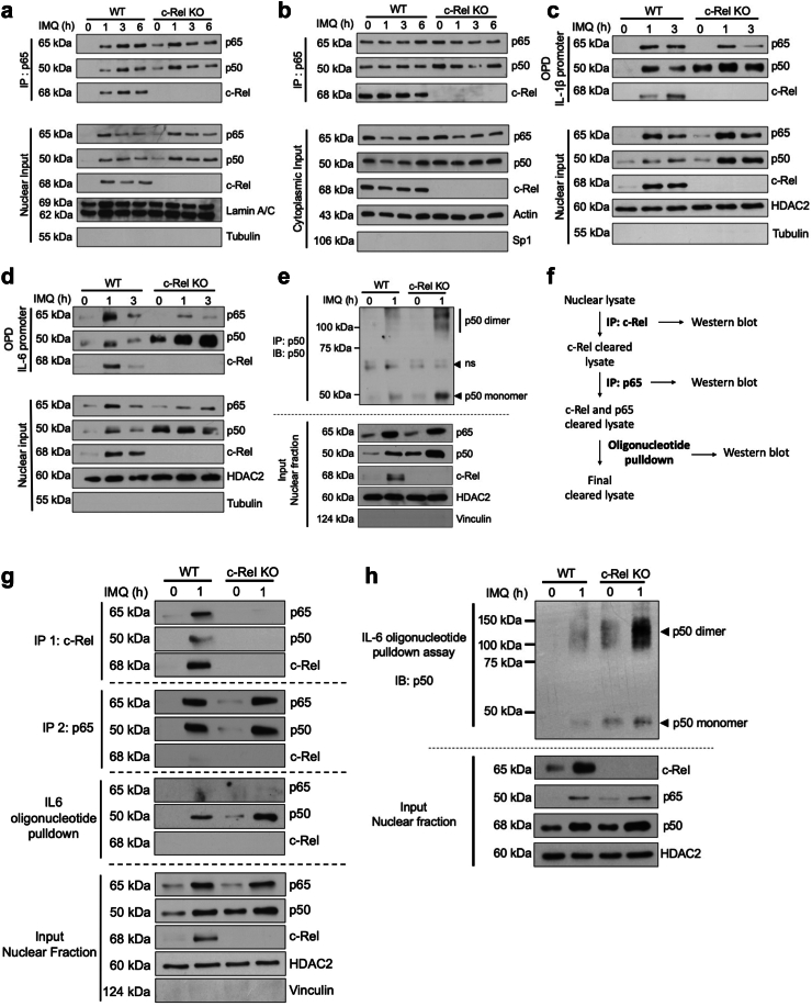
Findings: c-Rel is highly expressed in lesional skin of patients with psoriasis and TLR7-induced psoriatic lesions in mice. c-Rel deficiency protected mice from the disease, and specifically compromised TLR7-induced, and not TLR9- or TLR3-induced, inflammation in dendritic cells. Mechanistically, c-Rel deficiency disrupted activating NF-κB dimers and allowed binding of inhibitory NF-κB homodimers to the IL-1β and IL-6 promoters thus inhibiting their expression. This functionally compromises the ability of c-Rel deficient DCs to induce Th17 polarisation, which is critical in psoriasis pathogenesis.

Interpretation: Our findings reveal that c-Rel is a key regulator of TLR7-mediated dendritic cell-dependent inflammation, and that targeting c-Rel-dependent signalling could prove an effective strategy to dampen excessive inflammation in TLR7-related skin inflammation.

Funding: A complete list of funding sources that contributed to this study can be found in the Acknowledgements section.

Keywords: Inflammation; NF-κB c-Rel; Psoriasis; TLR7; Transcription.


Conflict of interest statement

Declaration of interests KDC is Vice President-Elect of the American Academy of Dermatology and Treasurer of the International Eczema Council. The authors declare no further potential conflicts of interest.

Figures

Fig. 1
Fig. 1
c-Rel expression is increased in the skin of human patients with psoriasis and IMQ-induced psoriasis-like mouse model. (a) c-Rel levels in different public GEO datasets of healthy, nonlesional, and psoriatic lesional skin biopsies (GSE13355, GSE14905). (b, c) c-Rel and (c) p65 levels in psoriatic lesions in patients treated with different biologics (GSE117468, GSE11903). Box and whisker plots tails are defined as 1.5 times interquartile range (IQR) from the first and third quartiles of each dataset. (a–c) Each point is representative of mean expression with 95% CI (One-way ANOVA, Tukey’s multiple comparisons test). (d) Representative immunohistochemistry staining of c-Rel in control or IMQ-treated dorsal skin of C57BL/6J mice at day 5, n = 3 (magnification: 60×, scale bar = 50 μm).
Fig. 2
Fig. 2
c-Rel deficiency protects mice from TLR7-induced psoriasiform skin pathology but not from systemic inflammation. (a) Schematic of daily topical application of IMQ to the dorsal skin of WT and c-Rel KO mice. (b) Representative images of the dorsal areas of WT and c-Rel KO mice following 5 d IMQ treatment or controls (actual unscaled photograph). (c) Daily scores of erythema and scaling of the shaved dorsal skin on a scale from 0 to 4. The assumption of sphericity was violated (Geisser-Greenhouse epsilon = 0.62) and the Geisser-Greenhouse correction was applied. Each point is representative of the mean score with 95% CI (Two-way ANOVA, mixed-effects analysis with Tukey’s multiple comparisons test, n = 6). (d) Histological sections of the dorsal skin were stained with H&E for acanthosis measurement (n = 4, scale bar = 50 μm). (e) Average thickness of four separate measurements were taken from four different locations in the epidermis of each skin section (magnification: 20×) (Two-way ANOVA, Sidak’s multiple comparisons test, n = 4). (f) Expression of IL-1β, IL-17A, and IL-17F in the dorsal skin sections of control and IMQ treated WT and c-Rel KO mice relative to the housekeeping gene L32. Each data point is the mean of technical duplicates per mouse and represented as means with 95% CI (Two-way ANOVA, Sidak’s multiple comparisons test, n = 4–5). (g) Daily body weight changes of control and IMQ treated WT and c-Rel KO mice represented as means with 95% CI (Two-way ANOVA, mixed-effects analysis with Tukey’s multiple comparisons test, n = 6). (h) Representative spleens of indicated mice (left, scale bar = 10 mm) and their lengths (right) were measured and represented as means with 95% CI (Two-way ANOVA, Sidak’s multiple comparisons test, n = 6).
Fig. 3
Fig. 3
c-Rel is critical for TLR7-induced proinflammatory gene expression in DC2.4 cells. (a) Total lysates of WT and c-Rel KO DC2.4 cells were analysed by Western blotting for TLR7 and c-Rel expression in WT and c-Rel KO DC2.4 cells. Actin was used as a loading control. (b–d) WT and c-Rel KO DC2.4 cells were stimulated with IMQ for 0 h, 0.5 h and 3 h. Expression of proinflammatory genes (b) IL-1β, IL-6, ICAM1, TNFα, CXCL2, IP10, A20, (c) c-Fos and (d) IFNβ were determined by quantitative real-time PCR. Data in (b–d) are shown as technical duplicates from at least three separate experiments and represented as means with 95% CI (Two-way ANOVA, Sidak’s multiple comparisons test). (e) Dorsal skin sections from control and IMQ treated WT C57BL/6 mice were stained for CD11c, c-Rel and TLR7 (n = 4, scale bar = 300 μm, zoom scale bar = 30 μm). (f) WT DC2.4 cells were pretreated with indicated amounts of PTX for 0.5 h. IMQ was added as indicated for an additional 0.5 h. Cytoplasmic and nuclear extracts were prepared and analysed by Western blotting with antibodies for p65, p50, and c-Rel. Actin and hnRNPA1 were used as loading controls and Sp1 and tubulin were used as purity controls for cytoplasmic and nuclear fractions, respectively. (g) WT DC2.4 cells were pretreated with 500 μg/mL PTX for 0.5 h then treated with IMQ for an additional 3 h. Data are represented as the average fold change over cells not treated with IMQ and plotted as technical duplicates from four separate experiments (two-tailed unpaired Student’s t test). All values were normalised to the housekeeping gene L32, and expression of target genes was determined using the ΔΔCt method.
Fig. 4
Fig. 4
c-Rel is critical for proinflammatory gene expression following TLR7, but not TLR3 or TLR9 agonists and both c-Rel and p65 are required for IMQ-induced inflammatory gene expression. (a and b) WT and c-Rel KO DC2.4 cells were treated with (a) CL307 (200 ng/mL) and (b) loxoribine (2 mM) for 0 h, 0.5 h, and 3 h. (c) WT and c-Rel KO DC2.4 cells were stimulated with CpG (1 μM) or poly (I:C) (12.5 μg/mL) for 3 h. (d) WT, p65 KO, and p50 KO DC2.4 cells were treated with IMQ for 0 h, 0.5 h, and 3 h. (a–d) Gene expression of IL-1β and IL-6 were determined by quantitative real-time PCR using the ΔΔCt method. All values were normalised to the housekeeping gene L32. Data are shown as technical duplicates from at least three separate experiments and represented as means with 95% CI (Two-way ANOVA, (a–c) Sidak’s multiple comparisons test, and (d) Tukey’s multiple comparisons test).
Fig. 5
Fig. 5
c-Rel deficiency enriches p50 containing NF-κB dimers binding to IL-1β and IL-6 promoters. (a–h) WT and c-Rel KO DC2.4 cells were stimulated with IMQ for the indicated time points. Nuclear and cytoplasmic fractions were prepared. (a, b) Nuclear and cytoplasmic extracts subjected to immunoprecipitation (IP) with an anti-p65 antibody (top panels), and lysate input for the amounts of respective proteins in each sample (bottom panels) were analysed by Western blotting (IB) with antibodies against indicated proteins. Lamin A/C and actin were used as loading controls and tubulin and Sp1 were used as the nuclear and cytoplasmic fraction purity controls, respectively. (c and d, g and h). Top: 100 μg of the nuclear fractions were utilised in a pull-down assay using biotinylated (c) IL-1β promoter oligonucleotide or (d, g-h) IL-6 promoter oligonucleotide. (c–e, g and h) Bottom: Amounts of indicated proteins in the nuclear extracts. HDAC2 was used as a loading control. Tubulin or vinculin were used as a nuclear fraction purity control. (e) Nuclear fractions were subjected to immunoprecipitation (IP) with an anti-p50 antibody. Protein complexes were crosslinked in 4% formaldehyde before analysis with Western blotting (IB) with antibody against p50. (f) Schematic of workflow for sequential immunoprecipitation. (g) Sequential immunoprecipitation with indicated antibodies was performed on 100 μg of nuclear lysates. Three rounds of immunoprecipitation with an anti-p65 antibody was performed before proceeding to oligonucleotide pulldown assay with biotinylated IL-6 promoter oligo. (h) Nuclear fraction was used in an oligonucleotide pulldown assay using biotinylated IL-6 oligonucleotide. Samples were crosslinked in 4% formaldehyde before analysis with Western blotting (IB) with antibody against p50. (a–e, g and h) Data is representative of three independent experiments.
Fig. 6
Fig. 6
c-Rel is critical for the production of proinflammatory genes in primary BMDCs. (a) Total lysates of WT and c-Rel KO BMDCs were probed to assess TLR7 expression. Actin was used as a loading control. (b) WT and c-Rel KO BMDCs were collected and stimulated with IMQ for 0 h, 0.5 h, 3 h and analysed for the expression of IL-1β and IL-6. (c) WT and c-Rel KO BMDCs were stimulated with IMQ for 8 h and assessed for protein expression of IL-1β and IL-6 via intracellular flow cytometry. (d) WT and c-Rel KO BMDCs were pretreated for 0.5 h with 500 μg/mL PTX and then treated with IMQ for an additional 3 h. Data are represented as the average fold change over cells not treated with IMQ. (e) WT and c-Rel KO BMDCs were stimulated with CpG (1 μM) and poly (I:C) (12.5 μg/mL) for 3 h. (b–e) Expression of target genes were determined by quantitative real-time PCR using the ΔΔCt method. Each data point is representative of the mean of technical replicates per mouse with 95% CI (Two-way ANOVA, Sidak’s multiple comparisons test, n = 5–8).
Fig. 7
Fig. 7
c-Rel deficient BMDCs have a decreased ability to polarise naïve T cells to Th17 cells. (a) Schematic of the workflow for IMQ-treated BMDC coculture with naïve WT T cells. (b–e) WT and c-Rel KO BMDCs treated with IMQ for 20 h or control BMDCs were cocultured with naïve WT T cells under (b and c) Th17 polarising and (d and e) Th0 polarising conditions for 3 days. Cells were assessed via flow cytometry for CD4+IL17A+ population. (c, e) Each data point represents the percentage of CD4+IL17A+ cells following co-culture under indicated polarising conditions from one mouse (Two-way ANOVA, Sidak’s multiple comparisons test, n = 6–7). (f and g) WT and c-Rel KO BMDCs treated as above were assessed for surface expression of (f) CD80 and (g) CD86 via flow cytometry. Representative histograms (n = 6) are shown (f left, g left). Each data point represents the MFI of the indicated protein from one mouse (f right, g right) (Two-way ANOVA, Sidak’s multiple comparisons test, n = 5–6).
Fig. 8
Fig. 8
Schematic summary of the key findings from this study. (Left) Stimulation of dendritic cells with TLR7 seems to induce p65/c-Rel dimers, which may play a critical role in the transcription of inflammatory genes relevant in psoriasis. (Right) Absence of c-Rel results in enhanced binding of the repressive p50 homodimers to the DNA following TLR7 stimulation, inhibiting the transcription of inflammatory cytokines, thereby protecting the mice from psoriasis.

References

    1. Boehncke W.H., Schön M.P. Psoriasis. Lancet. 2015;386(9997):983–994. - PubMed
    1. Jariwala S.P. The role of dendritic cells in the immunopathogenesis of psoriasis. Arch Dermatol Res. 2007;299(8):359–366. - PMC - PubMed
    1. Lowes M.A., Suárez-Fariñas M., Krueger J.G. Immunology of psoriasis. Annu Rev Immunol. 2014;2014(32):227–255. - PMC - PubMed
    1. Hayden M.S., Ghosh S. NF-κB in immunobiology. Cell Res. 2011;21(2):223–244. - PMC - PubMed
    1. Carmody R.J., Ruan Q., Liou H.C., Chen Y.H. Essential roles of c-rel in TLR-induced IL-23 p19 gene expression in dendritic cells. J Immunol. 2007;178(1):186–191. - PubMed

MeSH terms