. 2025 Jan 1;444(1):114356.
doi: 10.1016/j.yexcr.2024.114356. Epub 2024 Nov 23.

The cystogenic effects of ouabain in autosomal dominant polycystic kidney disease require cell caveolae

Affiliations

The cystogenic effects of ouabain in autosomal dominant polycystic kidney disease require cell caveolae

Jordan Trant et al. Exp Cell Res. .

Abstract

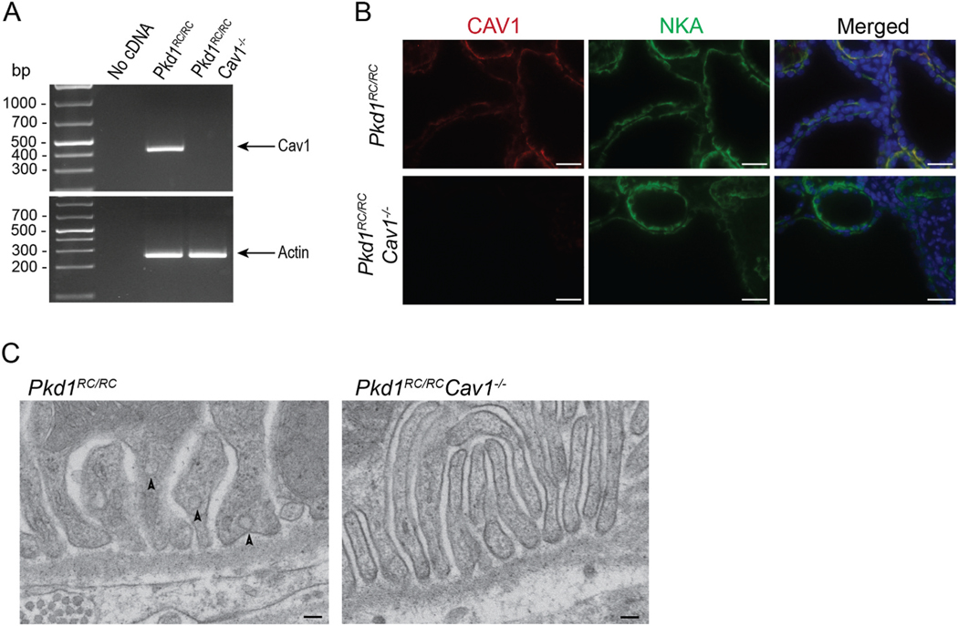
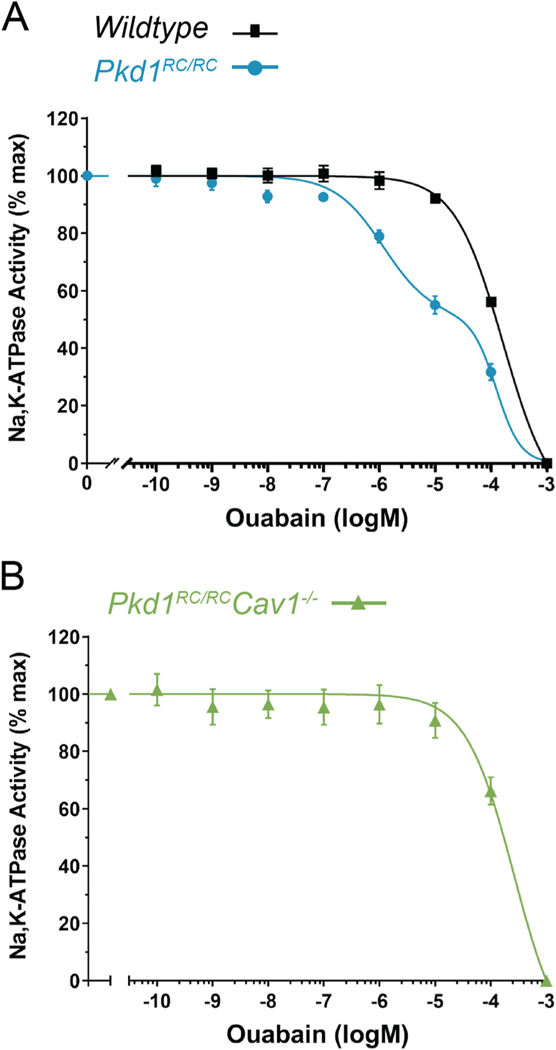
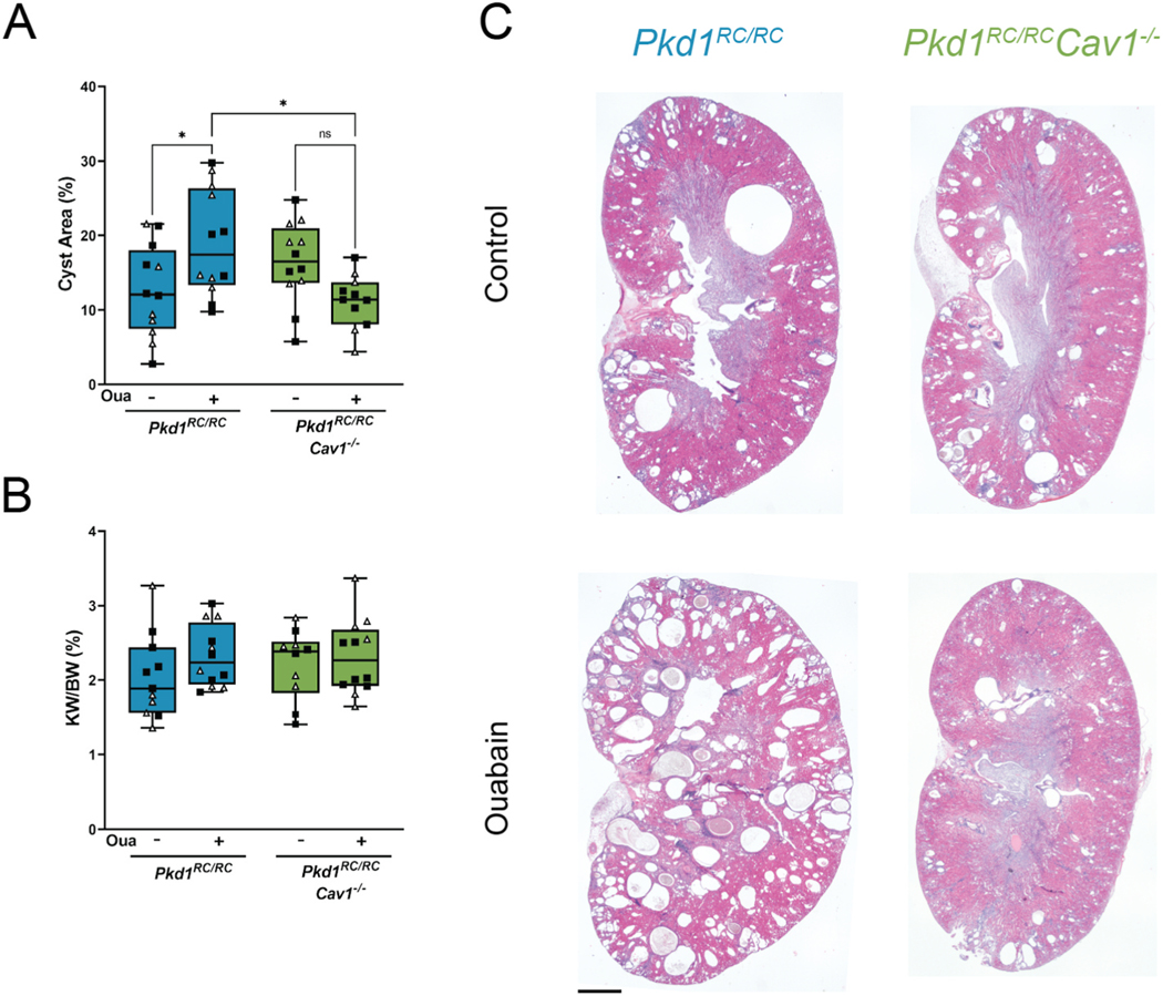
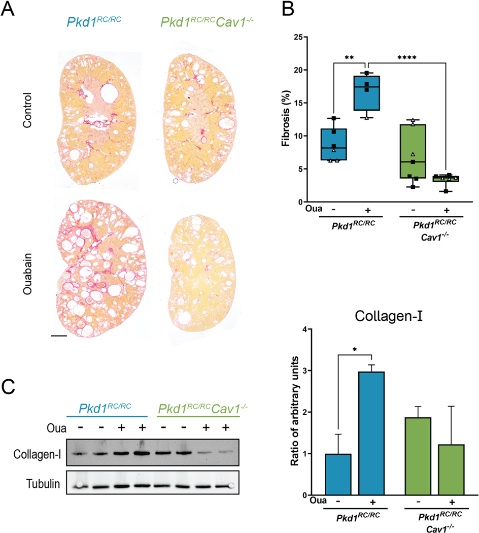
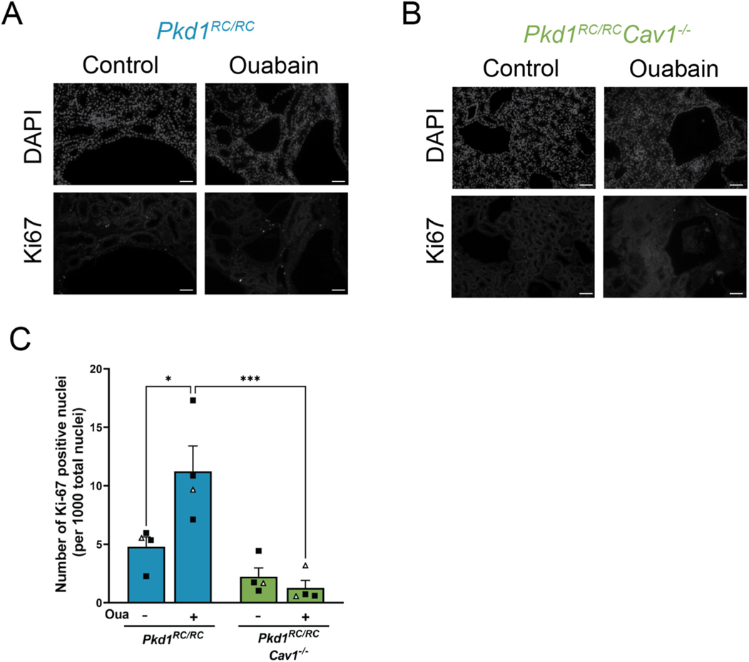
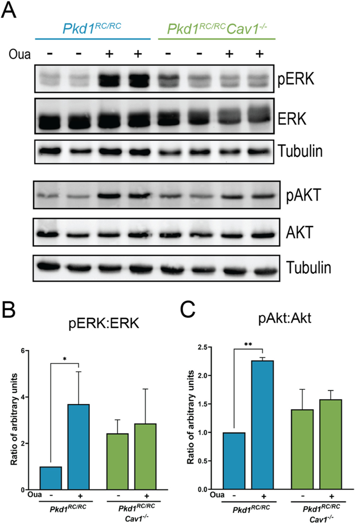
We have previously shown that the hormone ouabain is a circulating factor which can accelerate the progression of autosomal dominant polycystic kidney disease (ADPKD). At physiologic concentrations, ouabain increases cyst area and fibrosis in kidneys from ADPKD but not wildtype mice. These effects are due to an increased affinity for ouabain by its receptor, Na,K-ATPase (NKA), in the kidneys of ADPKD mice which leads to over-activation of NKA signaling function. Previous studies suggested that ouabain's stimulation of NKA signal transduction is mediated by NKA located within cell caveolae. Here, we determined whether caveolae are involved in the ouabain-induced progression of ADPKD cysts. We generated an ADPKD mouse with a global knockout of the main structural component of caveolae, caveolin-1 (CAV1), which we confirmed lacks caveolae in the kidney. When given physiological amounts of ouabain for 5 months, Pkd1RC/RCCav1-/- mice did not exhibit any changes in cyst progression, contrasting with the Pkd1RC/RC mice which showed a significant increase in cystic area and kidney fibrosis. Also, measures of ouabain-induced cell proliferation, including the number of Ki67-positive nuclei and phosphorylation of the extracellular regulated kinase (ERK) and protein kinase B (Akt), did not increase in the Pkd1RC/RCCav1-/- mice compared with the Pkd1RC/RC mice. Moreover, the abnormally increased affinity for ouabain of NKA in Pkd1RC/RC mice was restored to wildtype levels in the Pkd1RC/RCCav1-/- mice. This work highlights the role of caveolae in ouabain-induced NKA signaling and ADPKD cyst progression.

Keywords: Autosomal dominant polycystic kidney disease; Caveolae; Na,K-ATPase.


Conflict of interest statement

Declaration of Competing Interest The authors declare that they have no known competing financial interests or personal relationships that could have appeared to influence the work reported in this paper.

Figures

Fig. 1.
Fig. 1.
Characterization of Pkd1RC/RCCav1−/− mouse model. (A) Polymerase chain reaction (PCR) demonstrating the presence and absence of caveolin-1 cDNA in Pkd1RC/RC and Pkd1RC/RCCav1−/− mice, respectively. (B) Immunofluorescence of CAV1 and NKA staining in the kidney tubules from Pkd1RC/RC (top row) and Pkd1RC/RCCav1−/− (bottom row) mice. Scale bar represents 50 μm. (C) Transmission electron microscopy demonstrating the presence and absence of caveolae in kidney tubules in Pkd1RC/RC (left) and Pkd1RC/RCCav1−/− (right) mice, respectively. Photomicrographs are from collecting duct cells from kidneys from each model. Scale bar represents 100 nm.
Figure 2.
Figure 2.
CAV1 knockout abolishes the abnormal ouabain affinity component of NKA in Pkd1RC/RC mouse kidneys. (A) Ouabain affinity of NKA in WT (black squares, black curve) and Pkd1RC/RC (blue circles, blue curve) mouse kidney homogenates. (B) Ouabain affinity of NKA in Pkd1RC/RCCav1−/− (green triangles, green curve) mouse kidney homogenates. Pkd1RC/RC mouse kidneys exhibit a biphasic dose response to ouabain with a low- and high-affinity response. WT and Pkd1RC/RCCav1−/− mouse kidneys show a monophasic curve with a relatively low affinity for ouabain which have similar IC50. Data for panel (A) is reformatted for comparison from a previous publication [22].
Fig.3.
Fig.3.
Ouabain is unable to increase cyst development and growth in Pkd1RC/RCCav1−/− mice. (A) Cystic index of Pkd1RC/RC and Pkd1RC/RCCav1−/− mice. (B) Kidney weight to body weight ratio (KW/BW) of Pkd1RC/RC and Pkd1RC/RCCav1−/− mice. (C) Representative kidney sections of Pkd1RC/RC and Pkd1RC/RCCav1−/− mice stained with hematoxylin and eosin (H&E). Values are ± SEM of 12 animals per group, 6 male (black squares) and 6 female (white triangles). Scale bar represents 1000 μm *p < 0.05; **p < 0.01.
Fig. 4.
Fig. 4.
Ouabain increases cell proliferation in Pkd1RC/RC but not Pkd1RC/RCCav1−/− mice. (A–B) Representative immunofluorescent images of total nuclei (top) and Ki67-positive nuclei (bottom) in Pkd1RC/RC (A) and Pkd1RC/RCCav1−/− (B) mice. (C) Quantification of the average of total Ki67-positive nuclei from kidneys from both groups. Values are ±SEM of 4 animals per group, mixed male (black squares) and female (white triangles). *p < 0.05, ***p < 0.001.
Fig. 5.
Fig. 5.
Ouabain is unable to increase fibrosis in Pkd1RC/RCCav1−/− mice. (A) Representative kidney sections of Pkd1RC/RC and Pkd1RC/RCCav1−/− mice stained with picrosirius red. (B) Extent of kidney fibrosis in Pkd1RC/RC and Pkd1RC/RCCav1−/− mice as determined by collagen deposition in the kidney. Values are ±SEM of 4–6 animals per group, mixed male (black squares) and female (white triangles). (C) Representative Western blots and quantification of collagen-I and tubulin. Values are ±SEM from 3 animals per group, mixed male and female. Scale bar represents 1000 μm *p < 0.05, ***p < 0.001.
Fig. 6.
Fig. 6.
Ouabain-induced phosphorylation of ERK and Akt in Pkd1RC/RC mouse kidneys is abolished in Pkd1RC/RCCav1−/− mice. (A–C) Ouabain increases the phosphorylation of ERK and Akt in Pkd1RC/RC but not Pkd1RC/RCCav1−/− mice. Values are ±SEM from 3 animals per group, mixed male and female. *p < 0.05, **p < 0.01.

Similar articles

References

    1. Ghata J, Cowley BD Jr., Polycystic kidney disease, Compr. Physiol 7 (2017) 945–975. - PubMed
    1. Bergmann C, Guay-Woodford LM, Harris PC, Horie S, Peters DJM, Torres VE, Polycystic kidney disease, Nat Rev Dis Primers 4 (2018) 50. - PMC - PubMed
    1. Cornec-Le Gall E, Alam A, Perrone RD, Autosomal dominant polycystic kidney disease, Lancet 393 (2019) 919–935. - PubMed
    1. Cloutier M, Manceur AM, Guerin A, Aigbogun MS, Oberdhan D, Gauthier-Loiselle M, The societal economic burden of autosomal dominant polycystic kidney disease in the United States, BMC Health Serv. Res 20 (2020) 126. - PMC - PubMed
    1. Grantham JJ, Cook LT, Wetzel LH, Cadnapaphornchai MA, Bae KT, Evidence of extraordinary growth in the progressive enlargement of renal cysts, Clin. J. Am. Soc. Nephrol 5 (2010) 889–896. - PMC - PubMed

MeSH terms

LinkOut - more resources