

Use of the Thermal Camera to Assess the Perfusion of the Nipple-Areola Complex and Lower Adipoglandular Flap in Post-Explantation Mastopexy
- PMID: 39586858
- PMCID: PMC12069413
- DOI: 10.1007/s00266-024-04403-5
Use of the Thermal Camera to Assess the Perfusion of the Nipple-Areola Complex and Lower Adipoglandular Flap in Post-Explantation Mastopexy
Abstract
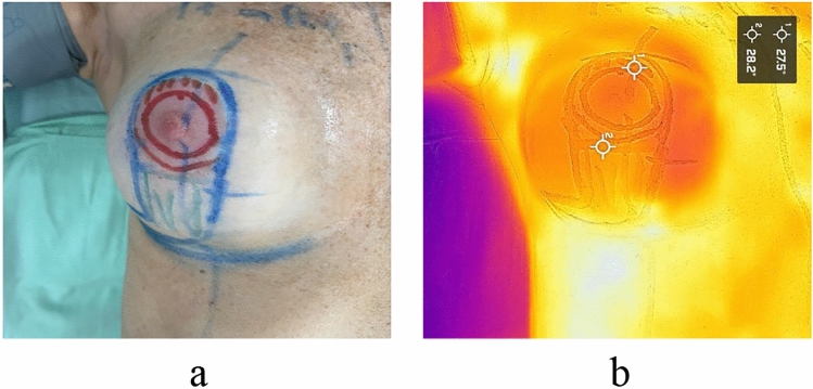
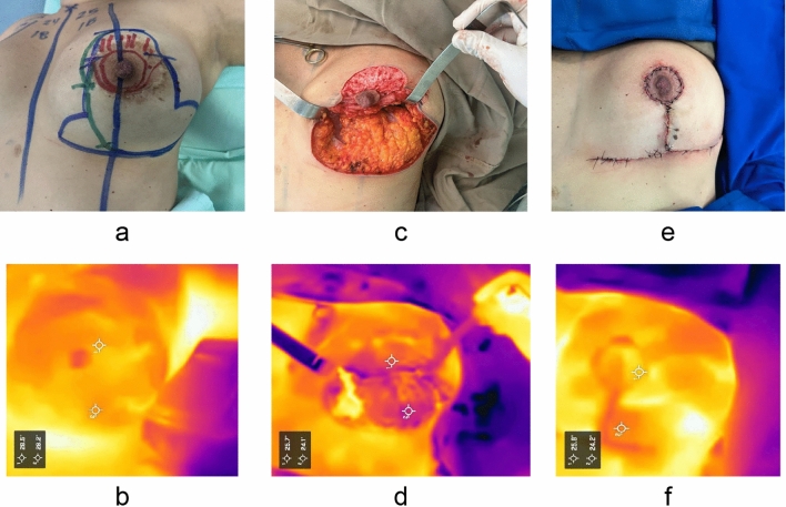
Thermography is a valuable tool for assessing tissue temperature. In recent years, explantation surgery has increased due to the fact that, despite technological advancements, breast implants have a limited lifespan and eventually require explantation, whether for pathological, aesthetic, or personal reasons. Among the complications that can arise in breast surgery are necrosis of the nipple-areola complex (NAC) and fat necrosis of the lower dermoglandular flap, which may affect the outcomes. Considering that the blood supply and perfusion of the breast were altered during the initial surgical procedure, and the technique previously used is often unknown, it is crucial to ensure adequate vascularization of the NAC and the flap. This study demonstrates the usefulness of the thermal camera for intraoperative and postoperative monitoring of the NAC and the lower adipoglandular flap, with the goal of determining tissue perfusion and identifying hypoperfused areas for immediate management, thus providing a new safety parameter in breast surgery.Level of Evidence V This journal requires that authors assign a level of evidence to each article. For a full description of these Evidence-Based Medicine ratings, please refer to the Table of Contents or the online Instructions to Authors www.springer.com/00266 .
Keywords: Breast reconstruction; Breast surgery safety; Perfusion; Thermal image; Thermography.
© 2024. The Author(s).
Conflict of interest statement
Declarations. Conflict of interest: The authors declare no conflicts of interest to disclose. Ethical Approval: All procedures involving human participants were conducted in accordance with the ethical standards of the institutional research committee and with the 1964 Helsinki Declaration and its later amendments or comparable ethical standards. Informed Consent: All patients involved in this study provided informed consent and agreed to the use of their images for medical research.
Figures


References
-
- Pereira N, Hallock GG (2021) Smartphone thermography for lower extremity local flap perforator mapping. J Reconstr Microsurg 37(1):59–66. 10.1055/s-0039-3402032 - PubMed
-
- Scarafoni EE (2021) Revisión sistemática sobre el uso de termografía en la evaluación de colgajos de perforantes. Cirugía Plástica Ibero-Latinoamericana. 47:411–424. 10.4321/S0376-78922021000400012
-
- Coroneos CJ, Selber JC, Offodile ACII, Butler CE, Clemens MW (2019) US FDA breast postapproval studies: long-term outcomes in 99,993 patients. Annal Surg 269(1):30–36. 10.1097/SLA.0000000000002990 - PubMed
-
- Calobrace MB, Mays C (2021) An algorithm for the management of explantation surgery. Clin Plast Surg 48(1):1–16. 10.1016/j.cps.2020.09.005 - PubMed
MeSH terms
LinkOut - more resources
Full Text Sources
Medical
