Review
. 2024 Nov 25;81(1):463.
doi: 10.1007/s00018-024-05499-3.

Regulation of formin INF2 and its alteration in INF2-linked inherited disorders

Affiliations
Review

Regulation of formin INF2 and its alteration in INF2-linked inherited disorders

Leticia Labat-de-Hoz et al. Cell Mol Life Sci. .

Abstract

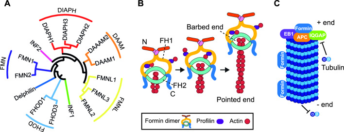
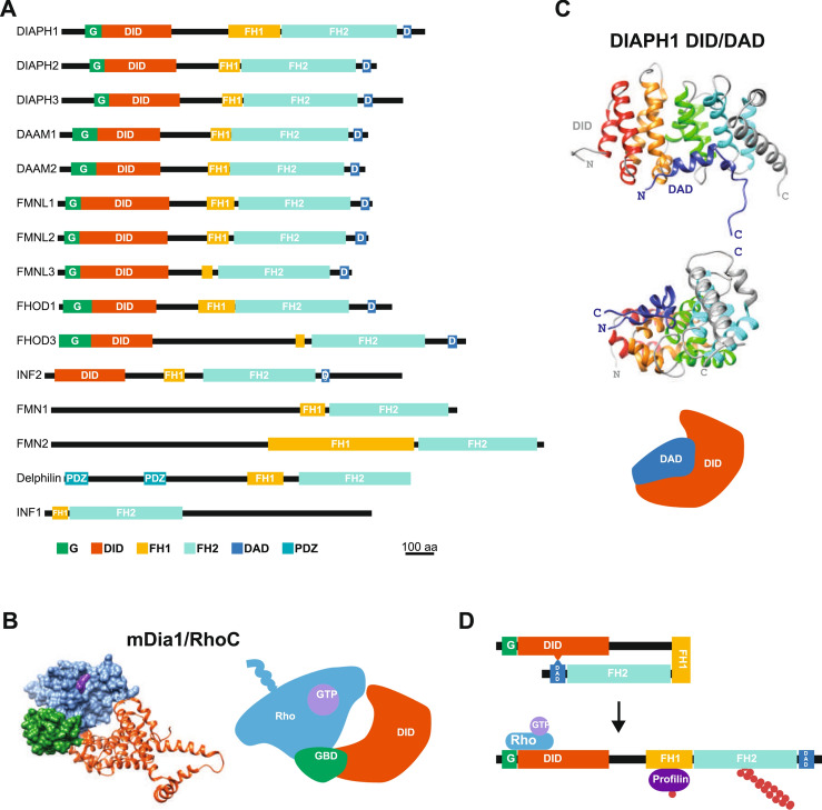
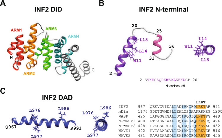
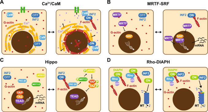
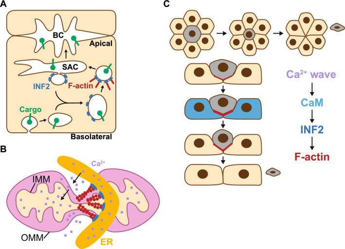
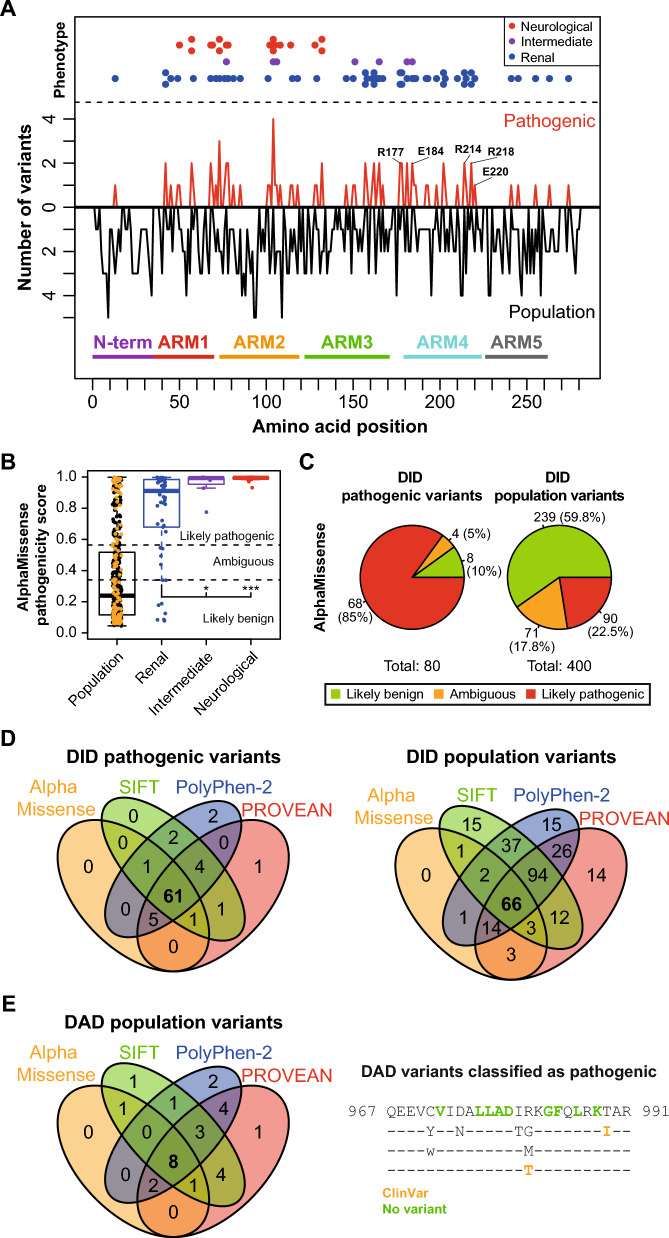
Formins are proteins that catalyze the formation of linear filaments made of actin. INF2, a formin, is crucial for correct vesicular transport, microtubule stability and mitochondrial division. Its activity is regulated by a complex of cyclase-associated protein and lysine-acetylated G-actin (KAc-actin), which helps INF2 adopt an inactive conformation through the association of its N-terminal diaphanous inhibitory domain (DID) with its C-terminal diaphanous autoinhibitory domain. INF2 activation can occur through calmodulin binding, KAc-actin deacetylation, G-actin binding, or association with the Cdc42 GTPase. Mutations in the INF2 DID are linked to focal segmental glomerulosclerosis (FSGS), affecting podocytes, and Charcot-Marie-Tooth disease, which affects Schwann cells and leads to axonal loss. At least 80 pathogenic DID variants of INF2 have been identified, with potential for many more. These mutations disrupt INF2 regulation, leading to excessive actin polymerization. This in turn causes altered intracellular trafficking, abnormal mitochondrial dynamics, and profound transcriptional reprogramming via the MRTF/SRF complex, resulting in mitotic abnormalities and p53-mediated cell death. This sequence of events could be responsible for progressive podocyte loss during glomerular degeneration in FSGS patients. Pharmacological targeting of INF2 or actin polymerization could offer the therapeutic potential to halt the progression of FSGS and improve outcomes for patients with INF2-linked disease.

Keywords: Actin; Charcot–Marie–Tooth disease; Focal segmental glomerulosclerosis; Mitotic catastrophe; Pathogenic variants; Podocytes.


Conflict of interest statement

Declarations. Conflict of interest: The authors declare no competing interests. Ethical approval: Not applicable. Consent to participate: Not applicable. Consent for publication: Not applicable.

Figures

Fig. 1
Fig. 1
The formin family and functions. A Tree of human formins. The tree was constructed using the Muscle algorithm of Mega software (megasoftware.net/home; version 10.1.8), aligning the FH2 sequences of the proteins. The Uniprot protein accession numbers of the corresponding sequences are DIAPH1 (O60610), DIAPH2 (O60879), DIAPH3 (Q9NSV4), DAAM1 (Q9Y4D1), DAAM2 (Q86T65), FMNL1 (O95466), FMNL2 (Q96PY5), FMNL3 (Q8IVF7), FHOD1 (Q9Y613), FHOD3 (Q2V2M9), INF2 (Q27J81), FMN1 (Q68DA7), FMN2 (Q9NZ56), Delphilin (A4D2P6) and INF1 (Q9C0D6). B Actin polymerization by formins. The FH2 domains of a formin dimer nucleate the formation of an actin polymer, assisted by profilin and the formin C-terminal segment. The formin remains bound to the barbed end during elongation. C Model of microtubule stabilization. Formins interact with other proteins, such as the microtubule plus-end tracking proteins EB1 and APC, and the scaffolding protein IQGAP1, to form a complex that caps the growing + end of microtubules and inhibits the addition of new tubulin subunits. In addition, formins bind laterally along microtubules via the FH2 domain, contributing to the stabilization and protection of microtubules against disassembly. APC, adenomatous polyposis coli; EB1, end-binding protein 1; FH1, formin homology 1 domain; FH2, formin homology 2 domain; IQGAP, IQ motif-containing GTPase-activating protein
Fig. 2
Fig. 2
Structure of human formins and regulation of DIAPH activity by Rho family GTPases. A Domain organization of the fifteen human formins. B Structure of the complex of the DID of mDia1 with RhoC (PBD: 8FG1). The illustration shows the interaction of the RhoC with the GTPase-binding domain, which consists of the G region and the N-terminal half of the DID. C Structure of the DID-DAD complex of DIAPH1 (PBD: 8FG1). The illustration depicts the interaction of DID with DAD. D Regulation of DIAPH formins. The DID-DAD interaction maintains the formin in a closed, inactive conformation. Binding of a specific GTP-loaded Rho family GTPase to the N-terminal region of the formin opens the molecule, activating it. The FH1 domain recruits profilin, which supplies the FH2 domain with G-actin for actin filament elongation. DAD, diaphanous autoregulatory domain; DID, diaphanous inhibitory domain; G, GTPase-binding region; GBD, GTPase-binding domain; FH1, formin homology 1 domain; FH2, formin homology 2 domain; PDZ, PSD95/DLG/ZO-1
Fig. 3
Fig. 3
Structure of the regulatory domains of INF2. A Predicted structure of the INF2 DID according to AlphaFold. B Structure of the N-terminal extension of INF2 (amino acids 2–36) as determined by NMR of the isolated peptide (top, left panel; PBD: 9FJW). The orthogonal projection of the first helix, which contains the CaM-binding site, is also shown (top, right panel). Alignment of the amino acid sequence of the N-terminal extension of INF2 and a consensus CaM and centrin-binding site (bottom panel). The critical residues W11, L14 and L18 involved in CaM and centrin binding are indicated. φ indicates a hydrophobic amino acid, and X represents any amino acid. C Longitudinal and orthogonal views of the α-helical structure of the INF2 WH2/DAD sequence as determined by NMR of the isolated INF2 967–991 peptide (left panel, PBD: 9G7T). Alignment of the WH2/DAD sequence of INF2 (Q27J81) with the DAD of mDia1 (O8808) and the WH2 of the indicated panel of proteins: WASP (P42768), N-WASP (O00401), WAVE1 (Q92558) and WAVE2 (Q9Y6W5). The three conserved L residues in the WH2 motif are indicated in the structure (top panels). In the alignment, the actin-binding LKKT motif is shaded in orange, and all other amino acid identities are shaded in blue. ARM, armadillo repeat
Fig. 4
Fig. 4
Proposed mechanisms of INF2 regulation. INF2 adopts an inactive conformation through interaction between DID and the DAD, facilitated by CAP/KAc-actin. INF2 activation occurs via the binding of Ca2+/CaM (or centrin) to the CaM and centrin-binding site of INF2, which is present in the N-terminal extension. Other proposed mechanisms of activation involve deacetylation of KAc-actin by a KDAC (potentially HDAC6), binding of G-actin to the DAD, and association of the Rho family Cdc42 GTPase with the DID. The latter association requires mediation by as yet unidentified protein(s). CaM, calmodulin; CAP, cyclase-associated protein; CBS, calmodulin and centrin-binding site; DAD, diaphanous autoregulatory domain; DID, diaphanous inhibitory domain; FH1, formin homology 1 domain; FH2, formin homology 2 domain; KA, lysine-acetylated actin; KDAC, lysine deacetylase
Fig. 5
Fig. 5
Signaling pathways in which INF2 has been implicated. A Ca2+/CaM signaling. Increased cytosolic Ca2+ levels, whether from external entry or intracellular release, activate INF2 through the binding of Ca2+/CaM to the N-terminal extension. This activation leads to massive actin polymerization, resulting in the formation of a characteristic F-actin ring around the nuclear envelope. Elevated Ca2+ levels in the nucleus also activate INF2, resulting in the transient formation of thin actin filaments. B MRTF-SRF signaling. INF2-mediated actin polymerization depletes G-actin from the cytosol, allowing MRTF to enter the nucleus and associate with the transcription factor SRF. The MRTF/SRF complex directs the transcription of target genes. C Hippo pathway. Activated INF2 polymerizes specialized actin filaments, facilitating the recruitment of PKC βII, which phosphorylates the transcription factors YAP and TAZ. This phosphorylation prevents the nuclear translocation of these proteins and their association with TEAD, thereby inhibiting the transcriptional activation of target genes. D Rho-DIAPH signaling. The DID of INF2 interacts with the DAD of DIAPH formins, inhibiting actin polymerization via DIAPH. Additionally, INF2 and DIAPH collaborate in stabilizing microtubules in response to Rho signaling
Fig. 6
Fig. 6
Cellular functions of INF2. A INF2 facilitates the transcytotic transport of basolateral cargo proteins to the apical membrane of hepatic cells. BC, bile canaliculus; SAC, subapical compartment. B INF2 promotes mitochondrial fission by inducing constriction of the outer (OMM) and inner (IMM) mitochondrial membranes. ER, endoplasmic reticulum. C In response to a wave of increased Ca2+ levels triggered by a nearby apoptotic or cancerous cell (in gray), this cell is extruded by surrounding cells in an INF2 expression-dependent manner. The schematic illustrates XY (top) and XZ (bottom) views of the cell monolayer during the extrusion process. CaM, calmodulin
Fig. 7
Fig. 7
Distribution and analysis of human INF2 DID variants. A The number of human INF2 variants reported as pathogenic (red) and those annotated in public databases but not yet studied for disease implications (black) are shown relative to their position in the INF2 sequence. The predominant phenotypes (renal alone, renal plus neurological/neurological alone, or intermediate) are indicated (top panel). “Intermediate” refers to mutations that result in only FSGS in some patients but in FSGS combined with CMTD in others. The N-terminal extension and the armadillo repeat (ARM) arrangement of the DID are also illustrated (bottom panel). B Boxplots displaying the pathogenicity scores assigned by AlphaMissense to INF2 DID mutations associated with renal-only, renal plus neurological or neurological-only, and intermediate phenotypes, as well as those variants annotated in public databases with not known disease implications. Variants found in ClinVar are highlighted in orange. *, p < 0.05; *** p < 0.001; Mann–Whitney test. Variants are categorized as likely pathogenic, likely benign, or ambiguous based on AlphaMissense thresholds. C AlphaMissense pathogenicity prediction for 80 INF2 DID variants reported to be pathogenic (left panel) and 400 DID variants annotated in public databases without no disease involvement (right panel). D Venn diagram illustrating the overlap in pathogenicity predictions for 80 human INF2 variants reported as pathogenic and 400 variants annotated in public databases, based on the AlphaMissense, SIFT, PolyPhen-2, and PROVEAN algorithms. E Venn diagram showing pathogenicity predictions from the AlphaMissense, SIFT, PolyPhen-2, and PROVEAN algorithms for the 32 human INF2 WH2/DAD variants annotated in public databases (left panel). The eight mutations classified as likely pathogenic by all four predictors are indicated (right panel). The two variants from this group annotated in ClinVar are shown in orange. INF2 DAD residues with no substitutions are highlighted in green in the wt INF2 sequence. Only mutations predicted as “likely pathogenic” by AlphaMissense, “deleterious” by PROVEAN, “damaging” by SIFT, or “probably damaging” by PolyPhen-2 were considered for both panels (D, E). ARM, armadillo repeat
Fig. 8
Fig. 8
Effect of the pathogenic mutations on the regulatory interactions of INF2. A Pathogenic mutations in INF2 disrupt the binding of Ca2+/CaM to the calmodulin and centrin-binding site, the CAP/KAc-actin complex, and the DAD of DIAPH to the DID. CaM, calmodulin; CAP, cyclase-associated protein; CBS, calmodulin and centrin-binding site; DAD, diaphanous autoregulatory domain; DID, diaphanous inhibitory domain; FH1, formin homology 1 domain; FH2, formin homology 2 domain; KA, lysine-acetylated actin. B Model of podocyte loss during glomerular degeneration due to pathogenic INF2. Over time, the expression of pathogenic INF2 in podocytes results in progressive dysregulation of critical processes. These include impaired polarized transport of slit diaphragm proteins, dysregulated mitochondrial fission, and altered MRTF/SRF complex-mediated transcription. As a consequence, podocytes depolarize and attempt division, encountering severe mitotic abnormalities that lead to mitotic catastrophe and death, either immediately or after generating cells with nuclear aberrations

References

    1. Schönichen A, Geyer M (2010) Fifteen formins for an actin filament: a molecular view on the regulation of human formins. Biochim Biophys Acta - Mol Cell Res 1803:152–163. 10.1016/j.bbamcr.2010.01.014 - PubMed
    1. Chesarone MA, DuPage AG, Goode BL (2010) Unleashing formins to remodel the actin and microtubule cytoskeletons. Nat Rev Mol Cell Biol 11:62–74. 10.1038/nrm2816 - PubMed
    1. Goode BL, Eck MJ (2007) Mechanism and function of formins in the control of actin assembly. Annu Rev Biochem 76:593–627. 10.1146/annurev.biochem.75.103004.142647 - PubMed
    1. Bartolini F, Gundersen GG (2010) Formins and microtubules. Biochim Biophys Acta Mol Cell Res 1803:164–173. 10.1016/j.bbamcr.2009.07.006 - PMC - PubMed
    1. DeWard AD, Alberts AS (2008) Microtubule stabilization: formins assert their independence. Curr Biol 18:R605–R608. 10.1016/j.cub.2008.06.001 - PubMed

MeSH terms