. 2024 Nov 25;15(1):10201.
doi: 10.1038/s41467-024-54309-8.

Autoantibodies immuno-mechanically modulate platelet contractile force and bleeding risk

Affiliations

Autoantibodies immuno-mechanically modulate platelet contractile force and bleeding risk

Oluwamayokun Oshinowo et al. Nat Commun. .

Abstract

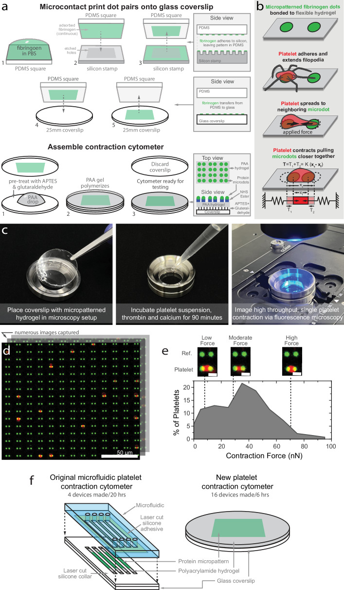
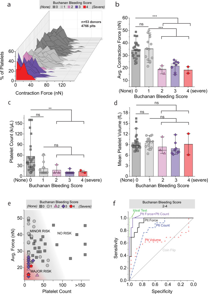
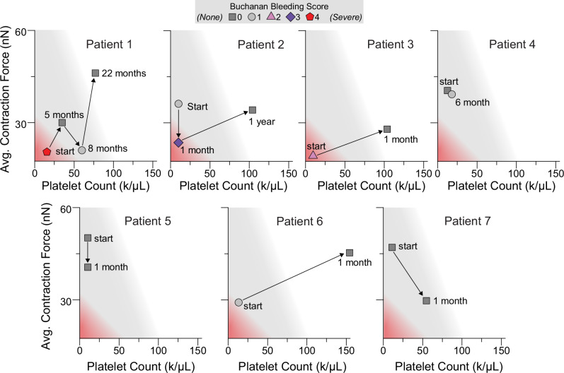
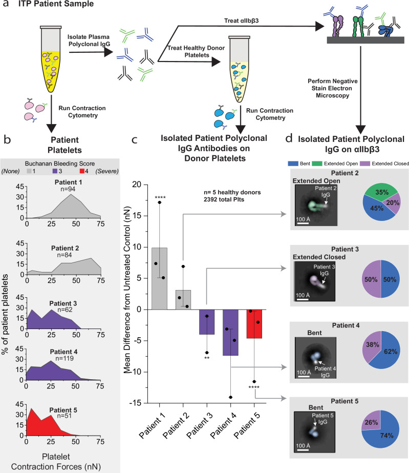
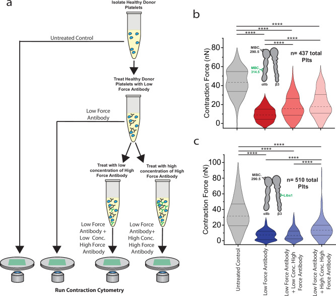
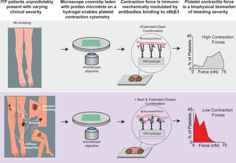
Altered mechanotransduction has been proposed as a putative mechanism for disease pathophysiology, yet evidence remains scarce. Here we introduce a concept we call single cell immuno-mechanical modulation, which links immunology, integrin biology, cellular mechanics, and disease pathophysiology and symptomology. Using a micropatterned hydrogel-laden coverslip compatible with standard fluorescence microscopy, we conduct a clinical mechanobiology study, specifically focusing on immune thrombocytopenia (ITP), an autoantibody-mediated platelet disorder that currently lacks a reliable biomarker for bleeding risk. We discover that in pediatric ITP patients (n = 53), low single platelet contraction force alone is a "physics-based" biomarker of bleeding (92.3% sensitivity, 90% specificity). Mechanistically, autoantibodies and monoclonal antibodies drive increases and decreases of cell force by stabilizing integrins in different conformations depending on the targeted epitope. Hence, immuno-mechanical modulation demonstrates how antibodies may pathologically alter mechanotransduction to cause clinical symptoms and this phenomenon can be leveraged to control cellular mechanics for research, diagnostic, and therapeutic purposes.


Conflict of interest statement

Competing interests: The authors declare no competing interests.

Figures

Fig. 1
Fig. 1. Simplified platelet contraction cytometer enables any lab to perform high-throughput measurements of single-cell forces toward clinical translation.
a Our cellular contraction cytometer has been simplified to comprise a microscope coverslip functionalized with a polyacrylamide hydrogel micropatterned with fluorescent extracellular matrix proteins. b Individual platelets adhere, spread, and contract against the micropatterned fibrinogen microdot pairs on the hydrogel. The microdots act akin to a spring, which enables straightforward calculations of platelet contractile force at the single cell level using measurements of the microdot size and spacing. c This micropatterned coverslip is adaptable for use with any fluorescence microscope. d The large array of fibrinogen microdot pairs enables the collection of numerous images and, therefore, allows measurements of platelet contraction forces to be captured. Scale bar is 50 μm. e The histogram is representative of a healthy adult, with a distribution of low, moderate, and highly contractile platelets. Scale bar denotes 4 μm. f Our micropatterned hydrogel-laden microscope coverslip-based cellular contraction cytometry technology allows for platelet contraction cytometers to be produced at a rate of 3.33 devices/h, a stark improvement from our original microfluidic system, which had a production rate of 0.2 devices/h. The microscope image in e is representative of at least 20 images collected for each of the 53 patient samples and 30 healthy donors, and the histogram in f is representative of 30 histograms collected for healthy donors.
Fig. 2
Fig. 2. Platelet contraction force is a potential clinical biophysical biomarker for bleeding risk in immune thrombocytopenia (ITP).
a ITP patients (n = 53) with bleeding scores of 2–4 consistently lack highly contractile platelets as compared to patients with little or no bleeding. b Statistically, the average platelet forces of patients with moderate-to-heavy bleeding were all significantly lower than patients with no bleeding symptoms (n = 53) c Platelet count, which is currently the main clinical biomarker used in ITP, no matter how low, is not significantly associated with Buchanan bleeding scores, as a substantial population of patients have extremely low platelet counts (<20 K/μL) but little to no bleeding (n = 53). d Mean platelet volume, a surrogate marker of platelet size and immaturity, has no association with bleeding, as patients with higher bleeding scores of 2–4 possess both low and high platelet volumes (n = 53). As such, platelet force best stratifies low bleeding score patients from higher bleeding score patients. e When platelet force is considered in the context of platelet count and clinical bleeding severity score, platelet force and platelet count synergistically stratify patients by clinical severity, whereas patients with platelet counts <40 K/μL and platelet forces below 25.24 nN have an increased likelihood of having higher bleeding scores (n = 53). f Historically, platelet count and platelet volume have been proposed as biomarkers for bleeding in ITP. To compare these benchmarks to platelet force, we performed an ROC analysis, and found that platelet force alone has a high diagnostic value for predicting bleeding scores of 2–4, as demonstrated by an area under the curve (AUC) of 0.95 (CI: 0.89–1.0), exceeding both platelet count 0.82 (CI: 0.71–0.93) and platelet volume 0.64 (CI: 0.42–0.87). An improved biomarker with an AUC of 0.99 (CI:0.98–1) can be achieved by combining platelet contractile force and platelet count. Statistical significance was determined by one way ANOVA with Tukey’s post hoc test for pairwise comparisons. Whiskers for 2b–d denote mean, 10th and 90th percentiles. *P ≤ 0.05, **P ≤ 0.01, ***P ≤ 0.001 and specific p-values are shown in Supplementary Table 2. Source data are provided as a Source Data file.
Fig. 3
Fig. 3. Changes in platelet contractile force are associated with the onset or resolution of bleeding symptoms in patients over time.
For several patients, we examined their platelets at multiple time points and observed that platelet forces as well as platelet counts can change over time. Moreover, these longitudinal changes in platelet contractile force were associated with the clinical bleeding score. Higher bleeding scores occurred when the patient’s platelet counts were <40 K/uL and platelet forces were <25 nN, and lower bleeding scores coincided with either an increase in platelet count, platelet force, or both. These data highlight the synergistic relationship between platelet count and platelet force. Additionally, leveraging a mixed mean model, and keeping platelet count constant while adjusting for patient and visit, we found that platelet contractile force is 10.79 nN less during instances where a patient’s bleeding score is between 2–4 versus when the bleeding score is either 0 or 1. Colored zones match those established in Fig. 2e. Source data are provided as a Source Data file.
Fig. 4
Fig. 4. ITP Patient polyclonal IgG platelet-associated (PA) antibodies are directed towards epitopes the integrin αIIbβ3 and modulate platelet force in a confirmation-dependent manner.
a We performed contraction cytometry on the platelets from ITP patients and extracted polyclonal IgG antibodies from the plasma for further analysis. b In those patients, the extent of clinical bleeding severity scores correlated with the lack of highly contractile platelets. c We found that isolated IgG antibodies (10 μg/mL) from those ITP patients with bleeding modulated the forces of platelets from healthy donors (n = 3,3,2,3,3 donors/condition with 379, 369, 338, 393, 390 antibody treated platelets, respectively and 532 untreated control platelets analyzed as shown in Supplementary Fig. 9). d As platelet contractile force is transmitted via the αIIbβ3 integrin, we used negative stain electron microscopy to study those specific antibody-integrin interactions. We found that autoantibodies from ITP patients with higher bleeding scores stabilized integrins in the bent and extended closed conformations. For (c), all antibody-treated platelets were compared to the untreated condition, and statistical significance was determined by mixed effects model, to account for within-subject variation, followed by the Benjamini-Hochberg’s posthoc test to account for multiple comparisons. Antibody treated platelet contraction force difference for (c) is shown as mean ± the minimum and maximum differences from the untreated control mean. *P ≤ 0.05, **P ≤ 0.01, ****P ≤ 0.0001 and specific p-values are shown in Supplementary Table 5. Source data are provided as a Source Data file.
Fig. 5
Fig. 5. Immuno-mechanical modulation by autoantibodies directed to specific αIIbβ3 integrin epitopes is associated with increases and decreases in platelet contractile force.
a To investigate the relationship between conformation- and epitope-dependent platelet contractile force modulation, we used a panel of well-characterized monoclonal antibodies (2.5 μg/mL) directed toward different epitopes of αIIbβ3. b Our data show that exposure to monoclonal antibodies alone can modulate average platelet force by ~ ±15 nN (55%). Moreover, a pattern became evident in which antibodies that bind closer to the tail region increase platelet contractile force, whereas those that bind toward the head region decrease force (n = 3,3,3,4,2,3,3,3,2,3 donors/condition with 434, 514, 450, 421, 213, 331, 176, 144, 123, 192 antibody treated platelets, respectively and 1139 untreated control platelets analyzed as shown in Supplementary Fig. 10). c Consistent with our electron microscopy data from patient polyclonal autoantibodies, negative stain electron microscopy showed that antibodies that cause a decrease in contractile force bound mostly to the bent conformation and extended closed formation of the integrin, although this data show that there is only a minor decrease in force when all integrins are stabilized in the extended closed conformation. Antibodies that stabilized integrins in an extended open conformation were associated with increased forces. For (b), all antibody-treated platelets were compared to the untreated condition, and statistical significance was determined by mixed effects model, to account for within-subject variation, followed by the Benjamini-Hochberg’s posthoc test to account for multiple comparisons. Antibody treated platelet contraction force difference for (b) is shown as mean ± the minimum and maximum differences from the untreated control mean. *P ≤ 0.05, **P ≤ 0.01, ****P ≤ 0 .0001 and specific p-values are shown in Supplementary Table 5. Source data are provided as a Source Data file.
Fig. 6
Fig. 6. Exposure to antibodies that induce “high” platelet contractile force partially restore the contractile forces of platelets first treated with “low” contractile force antibodies.
a To investigate whether high force antibodies have therapeutic potential to increase platelet forces, we measured the contraction force of healthy donor platelets (untreated control, n = 278 platelets). We next treated a subset of the healthy donor platelets with 2.5 µg/mL of the most potent low force antibody MBC 290.5 (n = 235 platelets) and measured the contractile force. Finally, we took a subset of the MBC 290.5 treated platelets and then treated with a high force antibody (Libs 1 or MBC 314.5) at a low concentration (0.25 µg/mL) or a high concentration (2.5 µg/mL). b MBC 314.5 significantly increased the contraction force of MBC 290.5 treated platelets at both low (+111%, n = 107 platelets) and high (+154%, n = 106 platelets) concentrations. c Libs 1 significantly increased the contraction force of MBC 290.5 treated platelets at a high concentration (+61%, n = 120 platelets) but not at a low concentration (n = 101 platelets). Violin plots show the median (dotted line) and the 25th and 75th percentile (solid lines) of contractile platelets. For (b, c), statistical significance was determined by mixed effects model, to account for within-subject variation, followed by the Benjamini-Hochberg’s posthoc test to account for multiple comparisons. ****P ≤ 0.0001 and specific p-values are shown in Supplementary Table 6. Source data are provided as a Source Data file.
Fig. 7
Fig. 7. Force cytometry reveals single platelet contraction force as a potential clinical biophysical biomarker for bleeding severity modulated by autoantibodies targeted to the αIIbβ3 platelet integrin.
Our micropatterned hydrogel-laden microscope coverslip-based cellular contraction cytometry technology demonstrates a direct link between platelet biophysics and a patient’s bleeding phenotype. In the case of ITP, we found that a patient’s bleeding score directly correlates with a decrease in single platelet contraction. Mechanistically, we discovered that a patient’s autoantibodies targeted against the αIIbβ3 integrin “immuno-mechanically” modulate platelet contractile forces (increasing or decreasing) in a conformation- and epitope-dependent manner. Antibodies that bind to the bent or extended-closed integrin conformation decrease platelet contractile force whereas antibodies that bind to the extended-open integrin conformation increase force. Therefore, platelet contractility is associated inversely with bleeding severity in that patient platelets with low contraction forces are associated with an increased risk of a bleeding phenotype, whereas patients with highly contractile platelets will likely be asymptomatic.

References

    1. Discher, D. E., Janmey, P. & Wang, Y. l. tissue cells feel and respond to the stiffness of their substrate. Science310, 1139–1143 (2005). - PubMed
    1. Engler, A. J., Sen, S., Sweeney, H. L. & Discher, D. E. Matrix elasticity directs stem cell lineage specification. Cell126, 677–689 (2006). - PubMed
    1. Hahn, C. & Schwartz, M. A. Mechanotransduction in vascular physiology and atherogenesis. Nat. Rev. Mol. Cell Biol.10, 53–62 (2009). - PMC - PubMed
    1. Seo, J. H. & Yui, N. The effect of molecular mobility of supramolecular polymer surfaces on fibroblast adhesion. Biomaterials34, 55–63 (2013). - PubMed
    1. Bennett, M. et al. Molecular clutch drives cell response to surface viscosity. Proc. Natl Acad. Sci. USA115, 1192 (2018). - PMC - PubMed

Publication types