. 2024 Nov 26;15(11):858.
doi: 10.1038/s41419-024-07249-6.

Microglial cannabinoid receptor type II stimulation improves cognitive impairment and neuroinflammation in Alzheimer's disease mice by controlling astrocyte activation

Affiliations

Microglial cannabinoid receptor type II stimulation improves cognitive impairment and neuroinflammation in Alzheimer's disease mice by controlling astrocyte activation

Akira Sobue et al. Cell Death Dis. .

Abstract

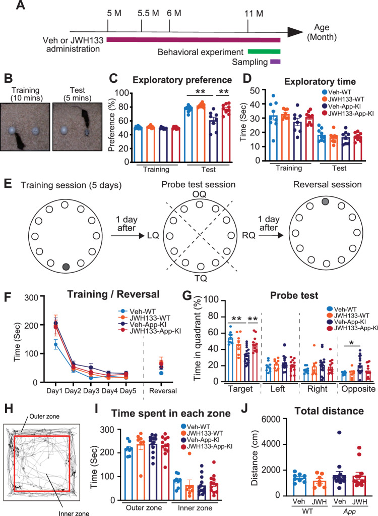
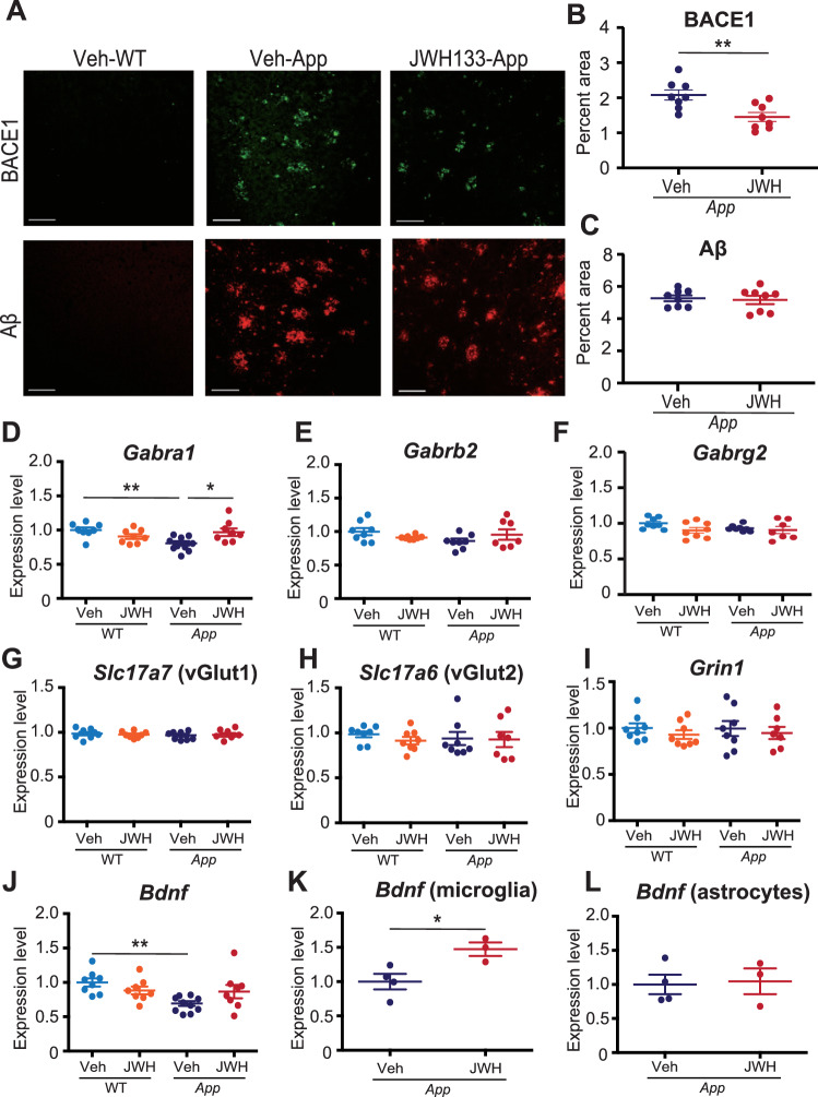
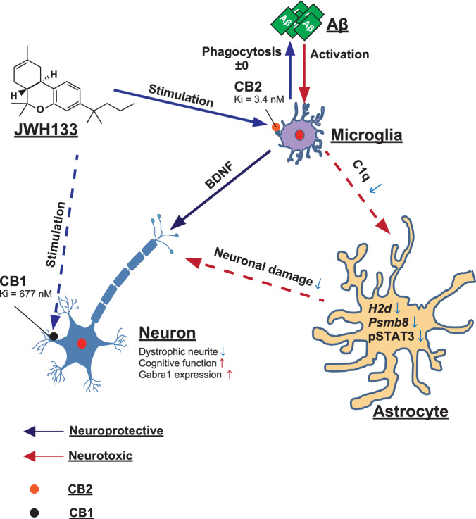
Alzheimer's disease (AD) is the most common form of dementia and is characterized by the accumulation of amyloid β (Aβ) and phosphorylated tau. Neuroinflammation, mainly mediated by glial activation, plays an important role in AD progression. Although there is growing evidence for the anti-neuroinflammatory and neuroprotective effects of the cannabinoid system modulation, the detailed mechanism remains unclear. To address these issues, we analyzed the expression levels of cannabinoid receptor type II (Cnr2/Cb2) in AppNL-G-F/NL-G-F mice and human AD precuneus, which is vulnerable to amyloid deposition in AD, and the effects of JWH 133, a selective CB2 agonist, on neuroinflammation in primary glial cells and neuroinflammation and cognitive impairment in AppNL-G-F/NL-G-F mice. The levels of Cnr2/Cb2 were upregulated in microglia isolated from the cerebral cortex of AppNL-G-F/NL-G-F mice. CNR2 expression was also increased in RNAs derived from human precuneus with advanced AD pathology. Chronic oral administration of JWH 133 significantly ameliorated the cognitive impairment of AppNL-G-F/NL-G-F mice without neuropsychiatric side effects. Microglia and astrocyte mRNAs were directly isolated from the mouse cerebral cortex by magnetic-activated cell sorting, and the gene expression was determined by quantitative PCR. JWH 133 administration significantly decreased reactive astrocyte markers and microglial C1q, an inducer for the reactive astrocytes in AppNL-G-F/NL-G-F mice. In addition, JWH133 administration inhibited the expression of p-STAT3 (signal transducer and activator of transcription 3) in astrocytes in AppNL-G-F/NL-G-F mice. Furthermore, JWH 133 administration suppressed dystrophic presynaptic terminals surrounding amyloid plaques. In conclusion, stimulation of microglial CB2 ameliorates cognitive dysfunction in AppNL-G-F/NL-G-F mice by controlling astrocyte activation and inducing beneficial neuroinflammation, and our study has implications that CB2 may represent an attractive therapeutic target for the treatment of AD and perhaps other neurodegenerative diseases involving neuroinflammation.


Conflict of interest statement

Competing interests: The authors declare no competing interests. Ethics approval and consent to participate: The experiments using human brains were approved by the Ethics Committees at Nagoya University (approval number #328) and Tokyo Metropolitan Institute (approval number # R21-145). Informed consent was obtained from their families. The experiments using genetically modified mice were approved by the Animal Care and Use Committee and the recombinant DNA experiment committee of Nagoya University (approval numbers R240025 and R240026, and #143, respectively). All procedures were conducted in accordance with the Declaration of Helsinki.

Figures

Fig. 1
Fig. 1. Expression of Cb2/CB2 mRNA was commonly upregulated in microglia isolated from AppNL-G-F/NL-G-F mice and precuneus of the patients with AD.
A Schematic overview of gene expression analysis of microglia and astrocytes isolated from AppNL-G-F/NL-G-F. B Cnr2 gene expression was analyzed using RNA sequencing (RNA-Seq) in isolated microglia (WT: n = 4 and AppNL-G-F/NL-G-F: n = 4). WT, wild-type; App, AppNL-G-F/NL-G-F. Data are presented as means ± standard error of the mean (SEM). **q < 0.01. C Quantitative PCR analysis to determine the expression levels of Cnr2 mRNA in glial cells isolated from 2, 4, and 8-month-old AppNL-G-F/NL-G-F mice and WT mice. Data are presented as means ± SEM. N.D.: not detectable. **p < 0.01 (two-way ANOVA). D Human brain samples were selected for analysis based on the Braak staging as follows: control brain (non-AD) defined as Braak stage (senile plaque: SP): 0–A, Braak stage (neurofibrillary tangle: NFT): 0–I; mild-AD brain defined as Braak stage (SP): C, Braak stage (NFT): III–IV; and advanced AD brain defined as Braak stage (SP): C and Braak stage (NFT): V-VI. Schematic overview of the gene expression analysis of CNR2 in the precuneus of non-AD (n = 12), mild-AD (n = 11), and advanced-AD (n = 11). E Quantitative PCR analysis to determine the expression levels of CNR2 mRNA in the precuneus of non-AD (n = 12), mild-AD (n = 11), and advanced-AD (n = 11). Data are presented as means ± SEM. *p < 0.05, **p < 0.01 (two-way ANOVA).
Fig. 2
Fig. 2. Microglial CB2 stimulation suppresses both microglial and astrocytic activation in vitro.
A A schematic protocol for JWH 133 treatment in primary microglia. B, C Expression levels of mRNAs in JWH 133-treated primary microglia determined by quantitative PCR. Relative expression levels for Tnf (B) and Cxcl10 (C) were determined and are presented as means ± SEM. n = 6, each. *p < 0.05, **p < 0.01 (two-way ANOVA). D A schematic protocol for microglial condition medium (MCM) treatment of primary astrocytes. E, F Expression levels of mRNAs in MCM-treated primary astrocytes determined by quantitative PCR. Relative expression levels for Psmb8 (E) and H2d (F) were determined and are presented as means ± SEM. n = 6, each. *p < 0.05, **p < 0.01 (two-way ANOVA).
Fig. 3
Fig. 3. JWH 133 administration ameliorated the cognitive impairments in AppNL-G-F/NL-G-F mice.
A Experimental timeline for JWH 133 administration and analysis of AppNL-G-F mice. B An experimental protocol for the novel object recognition test. C, D Effects of JWH 133 on performance in the novel object recognition test in WT and AppNL-G-F/NL-G-F mice. Exploratory preference (%) and time are plotted as means ± SEM [Veh-WT (n = 9), JWH 133-WT (n = 8), Veh-AppNL-G-F/NL-G-F (n = 9), JWH 133-AppNL-G-F/NL-G-F (n = 9)]. *p < 0.05 and **p < 0.01 (two-way ANOVA). E A schematic protocol for the Barnes maze test. In the training session (E left), one hole indicated as black was designated as the target hole with an escape box. A probe test was performed 1 day after the last training session, where the escape box was hidden (E middle). TQ: target quadrant; OQ: opposite quadrant; RQ: right quadrant; LQ: left quadrant. In the reversal session (E right), the target hole (gray) was relocated to the position opposite to the original position 1 day after the probe test. F, G Effects of JWH 133 on the performance of WT and AppNL-G-F/NL-G-F mice in the Barnes maze test. Values are presented means ± SEM [Veh-WT (n = 9), JWH 133-WT (n = 8), Veh-AppNL-G-F/NL-G-F (n = 15), JWH 133-AppNL-G-F/NL-G-F (n = 10)]. *p < 0.05 and **p < 0.01 (training: repeated measures three-way ANOVA, Probe test and Reversal: two-way ANOVA). H Image showing the travel path recorded for 5 min in the open field test arena. I, J Locomotor activity and anxiety were evaluated in an open field test as the time spent in each zone (I) and total distance traveled (J). Values are presented as means ± SEM. [Veh-WT (n = 8), JWH-WT (n = 7), Veh-AppNL-G-F/NL-G-F (n = 13), and JWH-AppNL-G-F/NL-G-F (n = 12)]. (two-way ANOVA).
Fig. 4
Fig. 4. Chronic oral administration of JWH 133 ameliorates neuroinflammation in AppNL-G-F/NL-G-F mice.
AD Representative immunofluorescent images demonstrating the expression of IBA1 (red, A) or GFAP (red, C) in the cerebral cortices of Veh-/JWH 133-administered WT (left panels) and AppNL-G-F/NL-G-F mice (right panels) at 12 months old. Scale bars: 50 μm. Percentages of the area immunopositive for IBA1 (B) or GFAP (D) are quantified. Values are presented as means ± SEM (B and D). [n = 7–8. Seven sections per mouse were quantified.] *p < 0.05 and **p < 0.01 (two-way ANOVA) (B and D). E Expression levels of mRNAs in microglia isolated from JWH 133-administered WT and AppNL-G-F/NL-G-F mice determined using quantitative PCR. Relative expression levels of DAM marker (Cd11c) and complementary marker (C1q) are plotted as the mean ± SEM. [Veh-WT (n = 6), JWH 133-WT (n = 6), Veh-AppNL-G-F/NL-G-F (n = 6), and JWH 133- AppNL-G-F/NL-G-F (n = 6)]. *p < 0.05 and **p < 0.01 (two-way ANOVA). F Expression levels of mRNAs in astrocytes isolated from JWH 133-administered WT and AppNL-G-F/NL-G-F mice determined using quantitative PCR. Relative expression levels of reactive astrocytic markers (H2d and Psmb8) are plotted as means ± SEM. [Veh-WT (n = 5-6), JWH 133-WT (n = 6), Veh-AppNL-G-F/NL-G-F (n = 6), and JWH 133-AppNL-G-F/NL-G-F (n = 6)]. *p < 0.05 and **p < 0.01 (two-way ANOVA). G, H Representative immunofluorescence images demonstrating the expression of GFAP (green), pSTAT3 (red), or IBA1 (white) in the cerebral cortices of Veh-WT (top panels), Veh-AppNL-G-F/NL-G-F mice (middle panels) and JWH 133-AppNL-G-F/NL-G-F mice (bottom panels). Arrowheads indicate pSTAT3- and GFAP-positive astrocytes (dashed rectangle areas have been magnified in the far right panels). Scale bars: 50 μm (magnified images) and 25 μm (others) (G). Percentages of pSTAT3 / GFAP cells are quantified. Values are presented as means ± SEM. [n = 7. Seven sections per mouse were quantified.] *p < 0.05 (Student’s t-test) (H).
Fig. 5
Fig. 5. JWH 133 administration suppresses dystrophic presynaptic terminals surrounding amyloid plaques but not amyloid deposition.
A Representative micrographs showing BACE1 (green) and Aβ (red) in the cerebral cortices of Veh- or JWH 133-administered WT or AppNL-G-F/NL-G-F mice. Scale bar: 100 μm. B, C Percentage of the area immunopositive for BACE1 (B) or Aβ (C) in the cerebral cortices of Veh- or JWH 133-administered AppNL-G-F/NL-G-F mice. Values are represented as means ± SEM. [n = 8. Seven sections per mouse were quantified.] *p < 0.05 and **p < 0.01 (Student’s t-test). D–I Expression levels of mRNAs in the cerebral cortices from JWH 133-administered WT and AppNL-G-F/NL-G-F mice were determined using quantitative PCR. Relative expression levels of GABAergic markers (Gabra1, Gabrb2 and Gabrg2) are plotted as means ± SEM (D–F). Relative expression levels of glutamatergic markers (Slc17a7, Slc17a6 and Grin1) are plotted as means ± SEM (G–I). [Veh-WT (n = 8), JWH 133-WT (n = 8), Veh-AppNL-G-F/NL-G-F (n = 8), and JWH 133-AppNL-G-F/NL-G-F (n = 7)]. *p < 0.05 and **p < 0.01 (two-way ANOVA) (D–I). J–L Expression levels of mRNAs in the cerebral cortices and glial cells isolated from JWH 133-administered WT and AppNL-G-F/NL-G-F mice determined using quantitative PCR. Relative expression levels of Bdnf are plotted as means ± SEM (J: cerebral cortex, K: microglia, and L: astrocytes). [Cortex: Veh-WT (n = 8), JWH 133-WT (n = 8), Veh-AppNL-G-F/NL-G-F (n = 10), and JWH 133-AppNL-G-F/NL-G-F (n = 8), microglia/astrocyte: Veh-AppNL-G-F/NL-G-F (n = 4) and JWH 133-AppNL-G-F/NL-G-F (n = 3)]. *p < 0.05 and **p < 0.01 (two-way ANOVA or Student’s t-test).
Fig. 6
Fig. 6. Schematic illustration of the role of microglial CB2 in ameliorating cognitive decline by regulating neuroinflammation in AD mice.
JWH 133 binds to CB2 with greater affinity than CB1 and acts as a potent CB2 selective agonist. Stimulation of microglial CB2 reduces the release of C1q, which an inducer of reactive astrocytes, and induces the release of BDNF from microglia. Regulated astrocytic activation ameliorates cognitive dysfunction in AppNL-G-F/NL-G-F mice by protecting neuronal functions.

Similar articles

Cited by

References

    1. 2023 Alzheimer’s disease facts and figures. Alzheimers Dement 2023,19:1598–695. - PubMed
    1. Sobue A, Komine O, Yamanaka K. Neuroinflammation in Alzheimer’s disease: microglial signature and their relevance to disease. Inflamm Regen. 2023;43:26. - PMC - PubMed
    1. Streit WJ, Mrak RE, Griffin WS. Microglia and neuroinflammation: a pathological perspective. J Neuroinflammation. 2004;1:14. - PMC - PubMed
    1. De Strooper B, Karran E. The cellular phase of Alzheimer’s disease. Cell. 2016;164:603–15. - PubMed
    1. Heneka MT, Carson MJ, Khoury JE, Landreth GE, Brosseron F, Feinstein DL, et al. Neuroinflammation in Alzheimer’s disease. Lancet Neurol. 2015;14:388–405. - PMC - PubMed

MeSH terms

Substances