. 2025 Jun;30(6):2265-2283.
doi: 10.1038/s41380-024-02833-w. Epub 2024 Nov 25.

Multimodal beneficial effects of BNN27, a nerve growth factor synthetic mimetic, in the 5xFAD mouse model of Alzheimer's disease

Affiliations

Multimodal beneficial effects of BNN27, a nerve growth factor synthetic mimetic, in the 5xFAD mouse model of Alzheimer's disease

Maria Kokkali et al. Mol Psychiatry. 2025 Jun.

Abstract

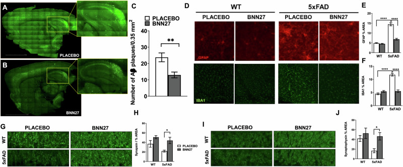
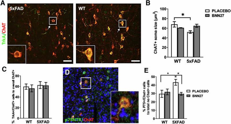
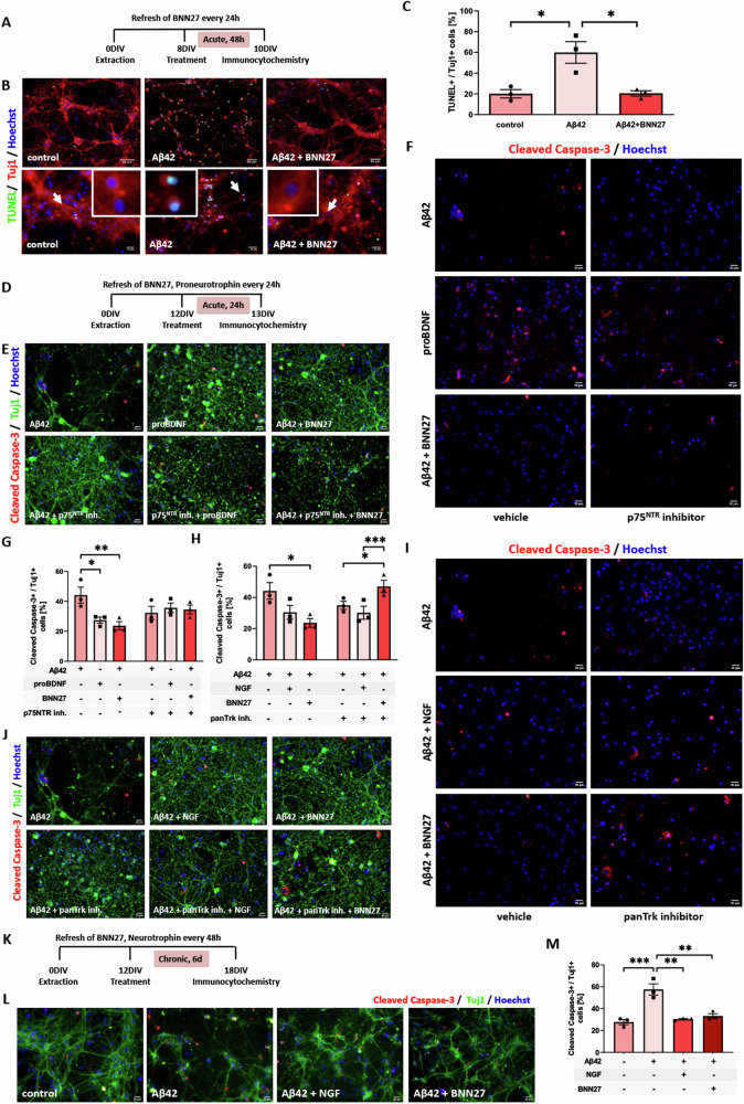
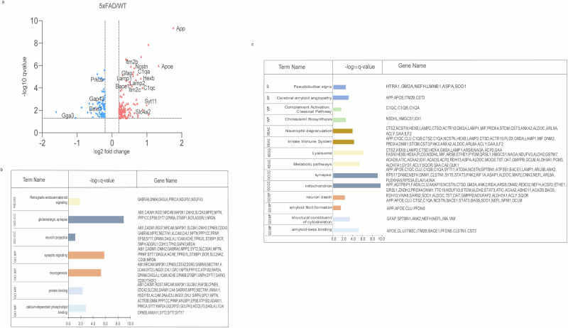
Alzheimer's Disease (AD) is an incurable and debilitating progressive, neurodegenerative disorder which is the leading cause of dementia worldwide. Neuropathologically, AD is characterized by the accumulation of Aβ amyloid plaques in the microenvironment of brain cells and neurovascular walls, chronic neuroinflammation, resulting in neuronal and synaptic loss, myelin and axonal failure, as well as significant reduction in adult hippocampal neurogenesis. The hippocampal formation is particularly vulnerable to this degenerative process, due to early dysfunction of the cholinergic circuit. Neurotrophic factors consist major regulatory molecules and their decline in AD is considered as an important cause of disease onset and progression. Novel pharmacological approaches are targeting the downstream pathways controlled by neurotrophins, such as nerve growth factor (NGF) receptors, TrkA and p75NTR, which enhance hippocampal neurogenic capacity and neuroprotective mechanisms, and potentially counteract the neurotoxic effects of amyloid deposition. BNN27 is a non-toxic, newly developed 17-spiro-steroid analog, penetrating the blood-brain-barrier (BBB) and mimicking the neuroprotective effects of NGF, acting as selective activator of its receptors, both TrkA and p75NTR, thus promoting survival of various neuronal cell types. Our present research aims at determining whether and which aspects of the AD-related pathology, BNN27 is able to alleviate, exploring the cellular and molecular AD components and link these changes with improvements in the cognitive performance of an animal AD model, the 5xFAD mice. Our results clearly indicate that BNN27 administration significantly reduced amyloid-β load in whole brain of the animals, enhanced adult hippocampal neurogenesis, restored cholinergic function and synaptogenesis, reducing inflammatory activation and leading to significant restoration of cognitive functions. BNN27 may represent a new lead multimodal molecule with neuroprotective, neurogenic and anti-neuroinflammatory actions for developing druggable anti-Alzheimeric agents. Proteomics data are available via ProteomeXchange with the identifier PXD044699.


Conflict of interest statement

Competing interests: All authors, except Achille Gravanis, declare that they have not any competing financial interests in relation to the work described. Dr Achille Gravanis is the co-founder of spin-off Bionature EA LTD, proprietary of compound BNN27 (patented with the WO 2008/ 1555 34 A2 number at the World Intellectual Property Organization). Gravanis A is co-founder of Bionature E.A. Ltd. The BNN compounds are proprietary and patented by the Bionature E.A. Ltd ( http://www.bionature.net ) (Patent Number: WO2008/155534 A2). Institutional Review Board Statement: The study was conducted according to the guidelines of the Declaration of Helsinki, and approved by the Ethics Committee of FORTH (protocol code 262272 and date of approval 29 October 2018, FORTH Institute animal license: EL91-BIObr-01 and EL91-BIOexp-02). All primary cells derived from animals that were grouped housed in the Animal House of the Institute of Molecular Biology and Biotechnology (IMBB-FoRTH, Heraklion, Greece), in a temperature-controlled facility on a 12-h light/dark cycle, fed by standard chow diet and water ad libitum. All research activities strictly adhered to the EU adopted Directive 2010/63/EU on the protection of animals used for scientific purposes. All procedures were performed under the approval of Veterinary Directorate of Prefecture of Heraklion (Crete) and carried out in compliance with Greek Government guidelines and the guidelines of FORTH ethics committee and were performed in accordance with approved protocols from the Federation of European Laboratory Animal Science Associations (FELASA) and Use of Laboratory animals [License number: EL91-BIOexp-02), Approval Code: 360667, Approval Date: 29/11/2021 (active for 3 years)].

Figures

Fig. 1
Fig. 1. Histopathological assessment of the BNN27 treated animals.
Evaluation of the Aβ plaque load within the hippocampus of the 5xFAD animals [A, B] showed that the BNN27 treatment significantly decreased (PStudent;s t test = 0.0042) the formation of Αβ plaques in the area of the hippocampus [C] (n = 9). Immunostaining for astrogliosis (GFAP, top-panel) and microgliosis (IBA1, bottom panel) in the hippocampus of placebo and BNN27 treated animals [D]. There was an overall effect of the treatment [PTwo-way ANOVA <0.0001] and genotype [PTwo-way ANOVA <0.0001] in the astrogliosis and microgliosis. In the 5xFAD animal there is increased activation of the astrocytes [PBonferroni posttests<0.0001] and the microglia [PBonferroni posttests<0.0001] compare to the WT animals [E, F]. Moreover, in the 5xFAD animals BNN27 treatment significantly reduced astrogliosis [PBonferroni posttests<0.0001] and microgliosis in the hippocampus [PBonferroni posttests<0.0001] (n = 7–9). Immunostaining for Synapsin I in the CA2 mossy fibers of the hippocampus of placebo and BNN27 treated animals [G]. There was an overall effect of the treatment (PTwo-way ANOVA = 0.0138) in SYNAPSIN I. Moreover, in the 5xFAD animals BNN27 treatment significantly (PBonferroni posttests <0.05) increased SYNAPSIN I staining in the mossy fibers of the hippocampus (n = 6–9) [H]. Immunostaining for Synaptophysin in the CA2 mossy fibers of the hippocampus of placebo and BNN27 treated animals [I] There was an overall effect of the treatment (PTwo-way ANOVA = 0.0139) and genotype (PTwo-way ANOVA = 0.049) in SYNAPTOPHYSIN staining. Moreover, in the 5xFAD animals BNN27 treatment significantly (PBonferroni posttests <0.05) increased SYNAPTOPHYSIN staining in the mossy fibers of the hippocampus [J]. Graphs showing mean ± SΕΜ; n = 6–9 *P < 0.05, **P < 0.01, ***P < 0.0001, ****P < 0.0001.
Fig. 2
Fig. 2. Reduced cholinergic atrophy in the basal forebrain of the BNN27 treated mice.
Representative image of TrkA immunostained cholinergic neurons (ChAT+) in the basal forebrain (BF) [A]. In the 5xFAD placebo mice there was a significant reduction in ChAT positive neurons soma size (P Bonferroni posttests = 0.0485) when compared with the WT placebo animals [B]. No change was observed in the percentage of double positive TrkA+/Chat+ neurons between genotypes or treatments [C]. Representative images of p75NTR immunostained cholinergic neurons (ChAT+) in the basal forebrain [D]. There was a significant increase in the percentage p75NTR +/ChAT+ neurons at the BF of the 5xFAD animals compared to the WT (PBonferroni posttests = 0.0317). Moreover, in the 5xFAD animals, BNN27 treatment significantly reduced the percentage p75NTR +/ChAT+ neurons (PBonferroni posttests = 0.0198) in the basal forebrain compared to 5xFAD placebo animals [E]. Graphs showing mean ± SΕΜ; n = 4 *P < 0.05, **P < 0.01, ***P < 0.0001, ****P < 0.0001.
Fig. 3
Fig. 3. BNN27 reverses the Aβ40 induced toxicity in the E17.5 immature hippocampal neurons.
A Illustrative graph that shows the ‘acute’ 48h administration of BNN27 and proneurotrophin in the E17.5 mature hippocampal neuronal culture and the initiation of the immunocytochemistry (ICC) experiment. B Representative images of immunofluorescence with TUNEL (green), Tuj1 (red), Hoechst (blue) conditions in the presence of Aβ vs the complete medium (CTR) condition. C Quantification of TUNEL (+) / Tuj1 (+) cells under 48 h of Aβ40 (5 μM) administration to E17.5 hippocampal neurons in the presence and absence of BNN27 (1-way ANOVA, Bonferroni multiple comparison test). p75NTR - not Trk receptors - seem to be implicated in the established effect. D Illustrative graph that shows the ‘acute’ 24h administration of BNN27 and proneurotrophin in the E17.5 mature hippocampal neuronal culture and the initiation of the immunocytochemistry (ICC) experiment. E Immunofluorescence with Cleaved Caspase-3 (green), Hoechst (blue) and [F] Tuj1 (green) conditions in the presence or absence of Aβ [Aβ40, 5 μM)] and/or p75NTR inhibitor [anti-p75 Receptor antibody (MC-192) Abcam], proBDNF and BNN27. G Quantification of TUNEL (+) / Tuj1 (+) cells under a 24 h administration of Aβ, p75NTR inhibitor treated as indicated at a 12DIV hippocampal neuronal cell culture (1-way ANOVA, Bonferroni multiple comparison test). H Quantification of TUNEL (+) / Tuj1 (+) cells under a 24 h administration of Aβ, panTrk inhibitor (AZD-1332, Alomone) treated as indicated at a 12DIV hippocampal neuronal cell culture (1-way ANOVA, Sidak’s multiple comparison test). I,J Immunofluorescence with Cleaved Caspase-3 (green), Hoechst (blue) and Tuj1 (green) conditions in the presence or absence of Aβ [Aβ40, 5 μM)] and/or panTrk inhibitor, NGF and BNN27. BNN27 affects the reciprocal extensive exposure to the Aβ40 toxicity at the E17.5 mature hippocampal neurons K Illustrative graph that shows the exact time periods for drug administrations and the immunocytochemistry (ICC) induction [L] Representative images of Cleaved Caspase-3 (red), Tuj1 (green), Hoechst (blue) staining of hippocampal 12DIV neurons after Aβ induction in the presence or absence of NGF, BNN27. The complete medium (CTR) condition serves as positive control. The images were taken at ×32 magnification (Scale bar, 20 μΜ). M Quantification of Cleaved Caspase-3 (+) / Tuj1 (+) after chronic administration (6 days) of NGF, BNN27 in order to test their ‘long term’ effects in the primary neuronal cell culture, with simultaneous injection of Aβ40 (5 μM) (1-way ANOVA followed by Sidak’s multiple comparison test) Data are expressed as mean ± SEM; *P < 0.05, **P < 0.01, ***P < 0.001, n = 3; Scale bar = 20 μm.
Fig. 4
Fig. 4. BNN27 promotes adult hippocampal neurogenesis in the 5xFAD mice.
Immunostaining for BrdU and NeuN in the dentate gyrus of the hippocampus of placebo and BNN27 treated 5xFAD animals [A, B]. There was an overall effect of the treatment (PTwo-way ANOVA = 0.0007) and genotype (PTwo-way ANOVA = 0.0011) in the number of BrdU+/NeuN+ neurons. Moreover, in the 5xFAD animals BNN27 treatment significantly (PBonferroni posttests = 0.0032) increases the number of BrdU positive neurons in the hippocampus compared to 5xFAD placebo animals [C]. Immunostaining for DCX in the dentate gyrus of the hippocampus of placebo and BNN27 treated animals [D]. There was an overall effect of the treatment (PTwo-way ANOVA = 0.0097) and genotype (PTwo-way ANOVA =0.0007) in the number of DCX+ neurons [2-way ANOVA for treatment and genotype]. Moreover, in the 5xFAD animals BNN27 treatment significantly (PBonferroni posttests = 0.011) increases the number of newly born neurons hippocampus [E]. Graphs showing mean ± SΕΜ; n = 4 *P < 0.05, **P < 0.01, ***P < 0.0001, ****P < 0.0001.
Fig. 5
Fig. 5. BNN27 protects the hippocampal Neural Stem Cells from oligomeric Aβ induced toxicity.
There was no detectable difference on the percentage of the BrdU(+) cells given the presence of BNN27 in the P7 Aβ-introduced NSC culture. A BrdU incorporation (green) evaluation of the Nestin(+) P7 NSCs (red). B Quantification of BrdU(+)/Nestin(+) cells under 48h induction of Aβ at the the P7 NSC hippocampal culture, in the presence or absence of NGF and BNN27 treatment, respectively. The complete condition depicts the positive control [1-way ANOVA for treatment and genotype, Sidak's multiple comparisons Post Hoc tests]. C Staining with CellTox Green Cytotoxicity Assay. D Evaluation of the in vitro cytotoxicity of Aβ42 oligomer as well as the potential 48 h neuroprotective activity of BDNF neurotrophin and BNN27 in the P7 NSCs of hippocampus. The complete medium (CTR) condition serves as positive control. [1-way ANOVA for treatment and genotype, Sidak's multiple comparisons Post Hoc tests]. Both Trk receptors and p75NTR seem to be implicated in the observed result. E Immunofluorescence with CellTox (green) and Hoechst (blue) conditions that shows the 24h administration in the P7 hippocampal neural stem cell culture in the presence or absence of Aβ42 (5 μM) and/or panTrk inhibitor (AZD-1332, Alomone), NGF or BNN27. F Quantification of CellTox (+) / Hoechst (+) cells under a 24 h administration of Aβ and/or panTrk inhibitor, treated as indicated at a P7 hippocampal neural stem cell culture (1-way ANOVA, Tukey multiple comparison test). G Immunofluorescence with CellTox (green) and Hoechst (blue) conditions that shows the 24h administration in the P7 hippocampal neural stem cell culture in the presence or absence of Aβ42 (5 μM) and/or p75NTR inhibitor [anti-p75 Receptor antibody (MC-192) Abcam], proBDNF or BNN27. H Quantification of CellTox (+) / Hoechst (+) cells under a 24 h presence or absence of Aβ and/or p75NTR inhibitor, treated as indicated at a P7 hippocampal neural stem cell culture (1-way ANOVA, Tukey multiple comparison test). Data are expressed as mean ± SEM; *P < 0.05, **P < 0.01, ***P < 0.001, n = 3; Scale bar = 20 μm).
Fig. 6
Fig. 6. Working memory evaluated in the T-maze.
A Graphic representation of the T-maze apparatus. B Working memory was evaluate by measuring the spontaneous alternation of the mice it the apparatus. It was shown that there is an overall significant effect of the treatment [P Two-way ANOVA = 0.0007] and genotype [P Two-way ANOVA = 0.0408] in the spontaneous alternation performance of the mice. Moreover, in the 5xFAD animals BNN27 treatment significantly improved working memory performance (P Bonferroni posttests < 0.01) [Graph showing mean ± SΕΜ; n = 7–9; **P < 0.01).
Fig. 7
Fig. 7. Proteomics analysis shows the Aβ induced negative impact in 5xFAD mice.
a Significantly regulated proteins in 5xFAD/WT comparison (FDR≤ 0.05, log2 fold change ≤ −0.2 or ≥0.2, pink: up-regulated, blue: down-regulated proteins), [b] protein group analysis on significantly down-regulated and [c] on significantly up-regulated proteins in the 5xFAD/WT comparison (enrichment analysis using the whole mouse genome as background).
Fig. 8
Fig. 8. Proteomics analysis shows that BNN27 has global rescue effects on Aβ induced negative impact in 5xFAD mice.
a Venn diagram showing the common proteins that are present in both the 5xFAD+BNN27/WT and 5xFAD/WT comparisons, [b] double volcano blot showing the shift in log2 fold change induced by BNN27 (circle: 5xFAD+BNN27/WT, triangle:5xFAD/WT), [c] all rescued down- (≥10%) and [d] all up- (≤ −10%) rescued regulated proteins in 5xFAD+BNN27/WT compared with 5xFAD/WT shows the rescue effect in fold changes of major AD-induced proteins, in the presence of BNN27 (increase/decrease percentage in log2 fold change of proteins significantly regulated in both sets FDR ≤ 0.05, log2 fold change ≤ −0.2 or ≥0.2).

References

    1. World Alzheimer Report 2022: The Global Economic Impact of Dementia. London, UK: Alzheimer’s Disease International.
    1. Long JM, Holtzman DM. Alzheimer disease: an update on pathobiology and treatment strategies. Cell. 2019;179:312–39. - PMC - PubMed
    1. Wilson DM 3rd, Cookson MR, Van Den Bosch L, Zetterberg H, Holtzman DM, Dewachter I. Hallmarks of neurodegenerative diseases. Cell. 2023;186:693–714. - PubMed
    1. Coyle JT, Price DL, DeLong MR. Alzheimer’s disease: a disorder of cortical cholinergic innervation. Science. 1983;219:1184–90. - PubMed
    1. Arendt T, Bigl V, Tennstedt A, Arendt A. Correlation between cortical plaque count and neuronal loss in the nucleus basalis in Alzheimer's disease. Neuroscience Letters. 1984;48:81–85. - PubMed

MeSH terms