. 2025 May;21(5):746-757.
doi: 10.1038/s41589-024-01777-0. Epub 2024 Nov 25.

Discovery and mechanism of K63-linkage-directed deubiquitinase activity in USP53

Affiliations

Discovery and mechanism of K63-linkage-directed deubiquitinase activity in USP53

Kim Wendrich et al. Nat Chem Biol. 2025 May.

Abstract

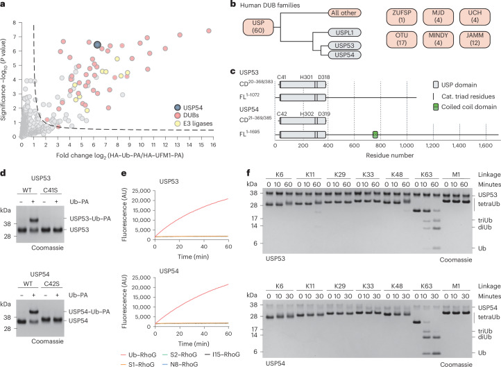
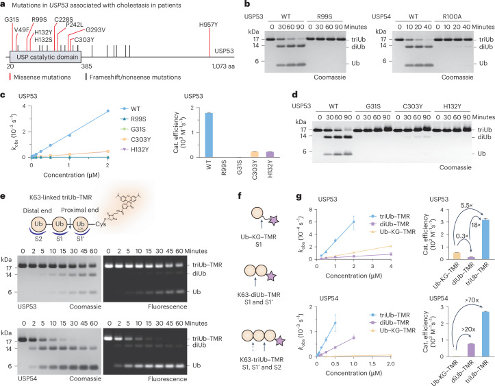
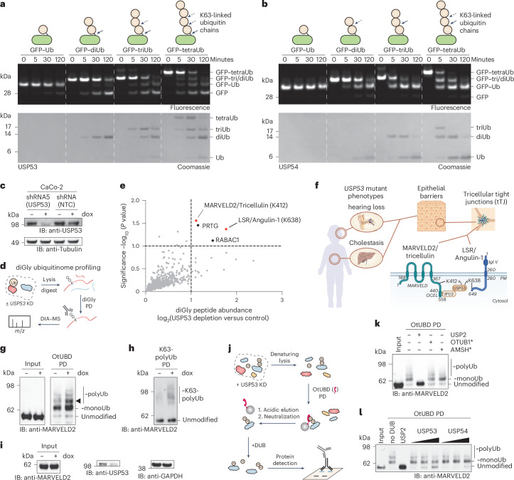
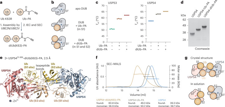
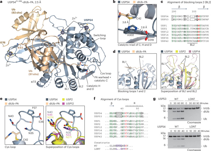
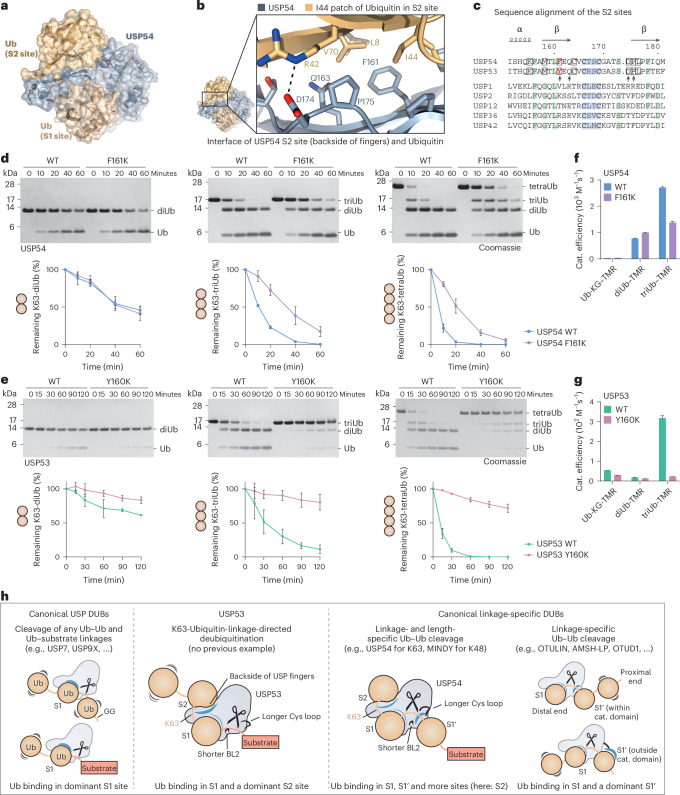
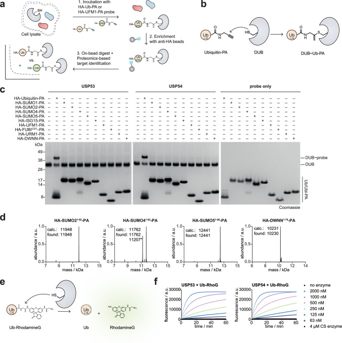
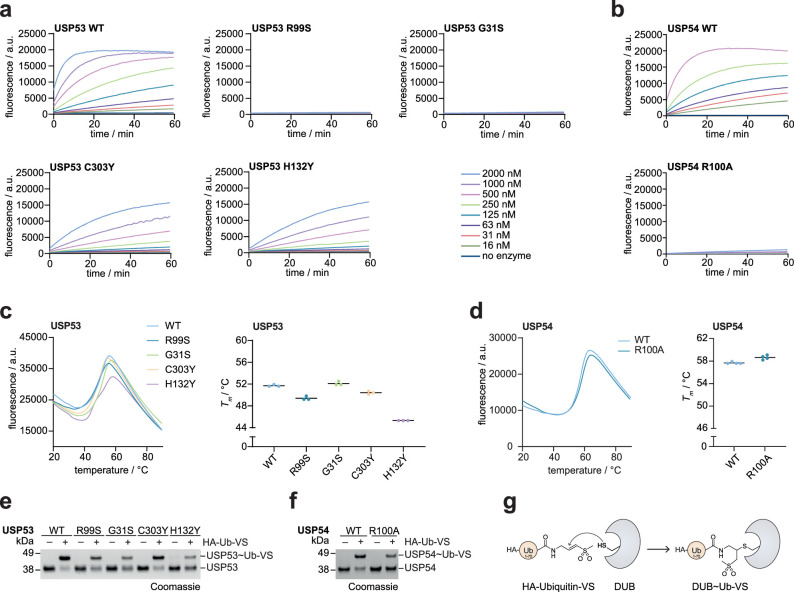
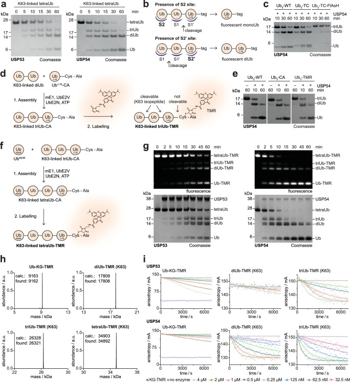
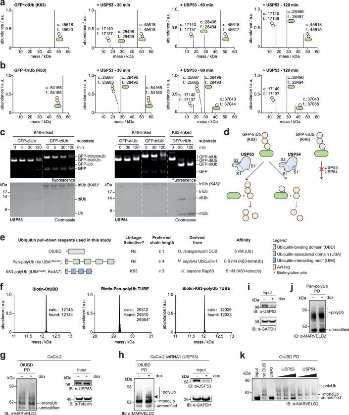
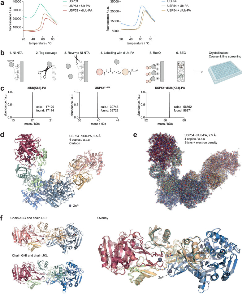
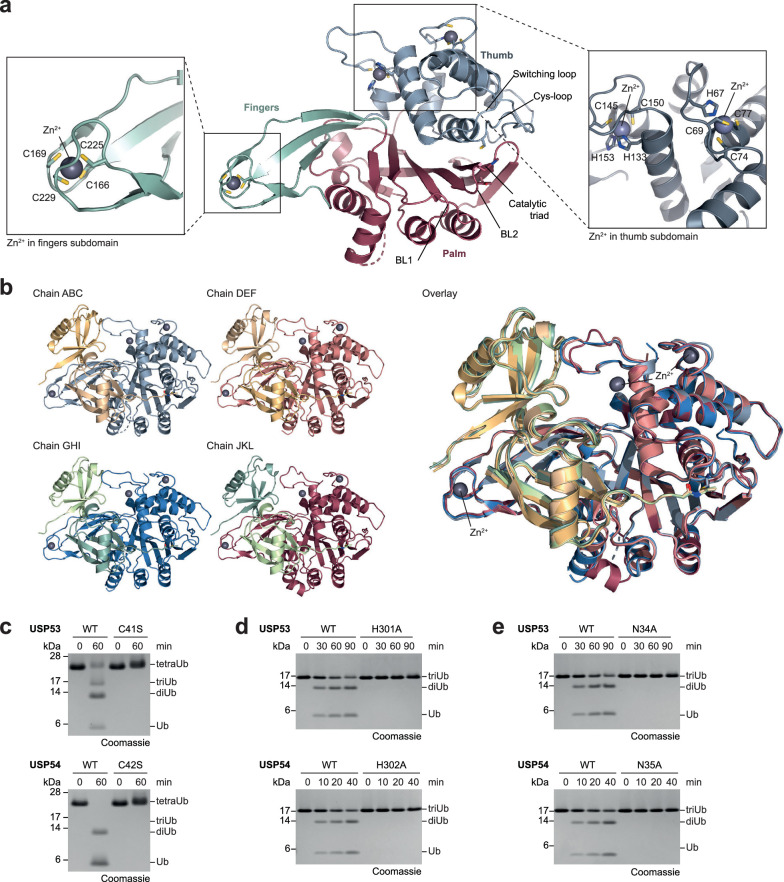
Ubiquitin-specific proteases (USPs) represent the largest class of human deubiquitinases (DUBs) and comprise its phylogenetically most distant members USP53 and USP54, which are annotated as catalytically inactive pseudoenzymes. Conspicuously, mutations within the USP domain of USP53 cause progressive familial intrahepatic cholestasis. Here, we report the discovery that USP53 and USP54 are active DUBs with high specificity for K63-linked polyubiquitin. We demonstrate how USP53 mutations abrogate catalytic activity, implicating loss of DUB activity in USP53-mediated pathology. Depletion of USP53 increases K63-linked ubiquitination of tricellular junction components. Assays with substrate-bound polyubiquitin reveal that USP54 cleaves within K63-linked chains, whereas USP53 can en bloc deubiquitinate substrate proteins in a K63-linkage-dependent manner. Biochemical and structural analyses uncover underlying K63-specific S2 ubiquitin-binding sites within their catalytic domains. Collectively, our work revises the annotation of USP53 and USP54, provides reagents and a mechanistic framework to investigate K63-linked polyubiquitin decoding and establishes K63-linkage-directed deubiquitination as a new DUB activity.


Conflict of interest statement

Competing interests: The authors declare no competing interests.

Figures

Fig. 1
Fig. 1. USP53 and USP54 are active DUBs for K63-linked polyubiquitin.
a, Proteomics analysis of ubiquitin–PA probe-labeled proteins in HeLa cell lysate. The volcano plot shows the enrichment (fold change) of proteins detected in HA pulldowns from lysate treated with HA–ubiquitin–PA probe in comparison to HA–UFM1–PA as a control. Identified proteins are shown as dots, HECT E3 ligases are shown in yellow, DUBs are shown in red and USP54 is shown in blue. b, Human DUB families are shown as boxes with numbers of members given in brackets. USP53 and USP54 are annotated as inactive and comprise the most distantly related catalytic domains within the USP family, with highest similarity to the deSUMOylase USPL1. c, Domain architectures of human full-length (FL) and catalytic domain (CD) constructs of USP53 and USP54 used in this study. d, Probe reactivity assay with recombinant catalytic domains. HA–ubiquitin–PA was incubated with wild-type (WT) USP5320–383 and USP5421–369 or inactive mutants of USP5320–383 and USP5421–385. Probe reactivity was analyzed by SDS–PAGE and Coomassie staining. e, Ubiquitin and Ubl RhoG cleavage assay. USP5320383 (200 nM, top) or USP5421369 (200 nM, bottom) was added to ubiquitin–RhoG (Ub–RhoG), SUMO1–RhoG (S1–RhoG), SUMO2–RhoG (S2–RhoG), NEDD8–RhoG or ISG15–RhoG (I15–RhoG) and the fluorescence was recorded. Data are shown as the average of technical triplicates. f, Gel-based ubiquitin chain cleavage assay. Specifically linked tetraubiquitin chains (2 µM) were incubated with USP5320383 (3 µM) or USP5421369 (300 nM). Samples were taken after the indicated time points and cleavage activity was analyzed by SDS–PAGE and Coomassie staining. diUb, diubiquitin; triUb, triubiquitin; tetraUb, tetraubiquitin. Source data
Fig. 2
Fig. 2. Analysis of USP53 mutations and polyubiquitin chain-length-dependent cleavage.
a, Schematic of human USP53 with amino acid substitutions associated with cholestasis or hearing loss. Single amino acid changes cluster in the catalytic domain. b, Gel-based ubiquitin chain cleavage assay. K63-linked triubiquitin (3 µM) was incubated with USP5320–383 (2 µM, left) or USP5421369 (300 nM, right). Cleavage was analyzed by SDS–PAGE and Coomassie staining. c, Ubiquitin–RhoG cleavage assay. Ubiquitin–RhoG was incubated with USP5320383 (WT or indicated mutants; raw data in Extended Data Fig. 2a) and the fluorescence was recorded. Observed rate constants (kobs, shown as the mean ± s.e.m.) were plotted over enzyme concentrations to obtain catalytic efficiencies (shown as the mean ± s.e.m.). d, Gel-based ubiquitin chain cleavage assay with additional USP53 mutations, shown as in b. e, Fluorescence-based triubiquitin cleavage assay. A K63-linked triubiquitin substrate (3 µM) was used, in which the proximal Ub175-CA was conjugated to maleimide–TAMRA (triubiquitin–TMR). Cleavage by USP5320383 (3 µM) or USP5421369 (300 nM) was analyzed by SDS–PAGE, in-gel fluorescence and Coomassie staining. The major cleavage position is indicated with an arrow. S2, S1 and S1′ ubiquitin-binding sites in the DUB were assigned consistent with the observed cleavage products. f, Schematic of ubiquitin substrates used in g. Cleavage sites are indicated by arrows. Ubiquitin-binding sites, which lead to cleavage when engaged, are given for the respective substrates. TAMRA is shown as a purple star. Structures of the substrates are shown in Extended Data Fig. 3. g, Fluorescence polarization cleavage assays. Substrates were incubated with USP5320383 (top) or USP5421369 (bottom) and fluorescence anisotropy was recorded (raw data in Extended Data Fig. 3i). Observed rate constants (kobs, shown as the mean ± s.e.m.) were plotted over enzyme concentrations to obtain catalytic efficiencies (shown as the mean ± s.e.m.). Source data
Fig. 3
Fig. 3. USP53 shows K63-linkage-directed deubiquitination activity.
a,b, Cleavage assay with isopeptide-linked, K63-linked monoubiquitinated or polyubiquitinated substrates. GFP–ubiquitin substrates (1 µM) were incubated with USP5320–383 (500 nM; a) or USP5421–369 (300 nM; b). In-gel fluorescence was used to visualize GFP species, while Coomassie staining of the same gel was used to visualize ubiquitin chains. Arrows indicate cleavage sites. c, USP53 depletion analysis. CaCo-2 cells stably transduced as indicated were analyzed by western blotting after treatment with doxycycline for 72 h. d, Schematic illustration of the diglycine ubiquitinome profiling to identify USP53 substrates. e, Volcano plot showing log2 fold changes of diglycine (ubiquitinated) peptides upon depletion of USP53 in CaCo-2 cells. Proteins linked to phenotypes similar to USP53 mutations are indicated in red. Diglycine sites were unambiguously identified. f, Model linking USP53-associated phenotypes to USP53-modulated ubiquitination of tricellular tight junction proteins (with domains, ubiquitination sites and residue numbers given). PM, plasma membrane. g, MARVELD2 ubiquitination analysis through OtUBD pulldown. CaCo-2 shRNA5 (USP53) cells were treated as in c, lysates were enriched with the high-affinity ubiquitin binder OtUBD and samples were analyzed by western blotting. The triangle highlights MARVELD2 modified with two ubiquitin moieties, emerging upon USP53 depletion. h, MARVELD2 polyubiquitination analysis with K63-specific tUIMRap80 (Rx3A7) TUBE pulldown. i, Analysis of protein levels for samples processed in h. j. Illustration of the workflow used to generate eluates of OtUBD pulldowns for UbiCRest assays. Acidic conditions allowed the elution of ubiquitinated proteins, with the biotinylated OtUBD reagent being retained on streptavidin beads. k, UbiCRest assay. OtUBD eluates prepared as described in j were treated with the indicated DUBs for 1 h at 37 °C and analyzed by western blot. l, UbiCRest assay as in k with USP5320–383 (0.5, 2 and 5 µM) and USP5421–369 (0.3, 1 and 3 µM), showing en bloc deubiquitination of cellular MARVELD2 by recombinant USP53. The results after 2-h incubation are shown in Extended Data Fig. 4k. Source data
Fig. 4
Fig. 4. A K63-linked diubiquitin–PA probe enabled crystallization of USP54 in complex with ubiquitin.
a, Schematic of the generation of a K63-linked diubiquitin probe, which was enzymatically assembled from ubiquitin K63R and ubiquitin–PA. IEC, ion-exchange chromatography. b, Schematic of catalytic DUB domains after reaction with ubiquitin–PA or diubiquitin–PA probes, illustrating ubiquitin engagement in samples used in c,d. S1′, S1 and S2 ubiquitin-binding sites are labeled. The active site cysteine is depicted as a star. c, Protein stability assessment. USP5320–368 and USP5421–369 were labeled with ubiquitin–PA or diubiquitin–PA probes and the stability of protein samples was analyzed by thermal shift analysis. Melting temperatures (Tm) are shown for technical replicates, indicating the contribution of the S2 site. d, Purified DUB samples. USP5421–369 and USP5421–369 conjugated to ubiquitin–PA and USP5421369 conjugated to K63-linked diubiquitin–PA were analyzed by SDS–PAGE and Coomassie staining. e, Crystal structure of USP54 conjugated to K63-linked diubiquitin–PA. In the observed complex, two USP54 molecules (blue and red) engage two K63-linked diubiquitin–PA molecules (gold and wheat) crosswise. The two isopeptide bonds between the diubiquitin–PA molecules are highlighted and are shown as sticks. f, SEC–MALS experiment of the catalytic domain of USP5421–369 alone (blue), in complex with ubiquitin–PA (brown) or in complex with K63-linked diubiquitin–PA (yellow), demonstrating monomeric species in solution. g, Schematic depiction of the organization of USP54 conjugated to K63-linked diubiquitin–PA in the crystal and in solution. Source data
Fig. 5
Fig. 5. Catalytic activity of USP53 and USP54 depends on a unique Cys loop.
a, Solution arrangement of USP54 conjugated to K63-linked diubiquitin–PA. Cartoon representation depicting USP5421–369 (gray) with ubiquitin moieties bound in its S1 (wheat) and S2 (gold) binding sites. Zinc atoms are shown as gray spheres. b, Close-up view of the catalytic triad. Indicated distances are visualized by dotted lines. c, Sequence alignment of residues forming BL2 in USP DUBs. Catalytic histidines are colored red, while conserved residues are colored green. Residues or gaps in USP54 and USP53, which are unique across the entire human USP family, are highlighted with a box. Residues strongly conserved in the USP DUB family, which led to the initial mischaracterization of USP53 and USP54 as inactive, are highlighted in bold. Secondary-structure elements and numbering are according to USP54. d, Left, close-up view of BL1 and BL2 of USP54. Right, superposition with the corresponding residues in USP2 (PDB 2IBI, yellow) and USP14 (PDB 2AYO, purple), illustrating the atypical shortening of both loops in USP54. e, Left, close-up view of the unique loop of USP54 close to the catalytic cysteine. Right, superposition of the Cys loop with the corresponding residues in USP2 (yellow) and USP12 (PDB 5L8W, magenta). f, Sequence alignment of residues forming the Cys loop in USP DUBs (annotation as in c). Sequences of USP53 and USP54 constructs used to study the Cys loop are shown below with changes marked in violet. g, Gel-based ubiquitin chain cleavage assay. K63-linked triubiquitin chains (3 µM) were incubated with USP5320–383 (3 µM) or USP5421–369 (300 nM) as either WT or Cys loop mutant (labeled as LGNT) (sequences in f) for the indicated time points. Cleavage activity was analyzed by SDS–PAGE and Coomassie staining. Source data
Fig. 6
Fig. 6. A cryptic S2 ubiquitin-binding site in USP53 and USP54 mediates efficient cleavage of K63-linked ubiquitin chains.
a, Structure of USP54 conjugated to K63-linked diubiquitin–PA. b, Interface of ubiquitin and USP54 S2 site. c, Sequence alignment of residues forming the S2 site in USP54 and USP53. Cys-x-x-Cys motifs at the tip of the fingers (blue), conserved residues (green), residues unique in USP53 and USP54 within the human USP family (box) and residues annotated in b (arrows) are highlighted. d, Gel-based polyubiquitin cleavage assays. K63-linked ubiquitin chains (2 µM) were incubated with WT USP5421–369 or the S2 site mutant F161K (both at 300 nM). Substrate consumption was quantified by densitometry, normalized to initial intensities. Data are shown as the average ± s.d. of three independent replicates. e, Gel-based polyubiquitin cleavage assays of USP53, shown as in d. WT USP5320–383 or the S2 site mutant Y160K was used (2 µM). f, Catalytic efficiencies of USP54 proteins obtained from fluorescence polarization assays (substrates shown in Fig. 2d). Raw data are shown in Extended Data Fig. 9d,e and the catalytic efficiencies of WT protein are repeated from Fig. 2g. Data are shown as the mean ± s.e.m. g, Catalytic efficiencies of USP53 proteins, analyzed as in f. Efficient catalysis of USP53 is dependent on its S2 site. h, Schematic of ubiquitin processing by DUBs. K63-linkage-directed deubiquitination by USP53 bridges canonical DUB categories. The structural mechanisms for polyubiquitin length and linkage specificity in DUBs are shown in Extended Data Fig. 10. Source data
Extended Data Fig. 1
Extended Data Fig. 1. USP53 and USP54 are active DUBs.
a. Schematic of the workflow used to identify active DUBs. HeLa cell lysate was treated with HA-Ub-PA or HA-UFM1-PA probe. Labelled proteins were enriched and detected by mass spectrometry. b. Schematic of the probe reactivity assay. The C-terminal propargylamine (PA) warhead reacts with the catalytic cysteine of DUBs to a vinyl thioether, forming a covalent DUB~Ub-PA complex. c. Ub/Ubl specificity assayed through probe reactivity with recombinant catalytic domains. A panel of HA-Ub/Ubl-PA probes was incubated with wild-type USP5320-383 or wild-type USP5421-369. Probe reactivity was analyzed by SDS-PAGE and Coomassie staining. Uncropped versions of all gels are provided as Source Data. d. Intact protein mass spectrometry of probes used in panel c. Expected and found masses are given in Dalton. The star denotes an HA-SUMO41–92 species N-terminally truncated by four residues. All other probes have been characterized previously. e. Schematic of the Ubiquitin-RhodamineG (Ub-RhoG) cleavage assay. DUBs cleave the amide bond between ubiquitin and RhoG and thereby liberate fluorescent RhoG. f. Ub-RhoG cleavage assay. Indicated concentrations of USP5320-383 (left) and USP5421-369 (right) or of the catalytic inactive mutants were added to Ub-RhoG and fluorescence was recorded. Data are shown as average of technical triplicates. CS enzymes denote the use of catalytic cysteine to serine mutated enzymes as in Fig. 1d. Of note, this activity is approximately 3 to 4 orders of magnitude weaker than for other USP DUBs including USP2, USP7, USP36 and USP42. Source data
Extended Data Fig. 2
Extended Data Fig. 2. Cholestasis-associated patient mutations of USP53 are catalytically impaired.
a. Ub-RhoG cleavage assay. Indicated concentrations of USP5320-383 (WT or patient mutants) were added to Ub-RhoG and fluorescence was recorded over time. Data are shown as average of technical triplicates. b. Ub-RhoG cleavage assay with USP5421-385, shown as in panel a. c-d. Protein stability assessment of USP5320-383 (panel c) or USP5421-369 (panel d) proteins by thermal shift analysis. Averaged fluorescence raw data are shown and melting temperatures (Tm) are plotted from technical triplicates (USP53) or quadruplicates (USP54). e-f. Probe reactivity assay. HA-Ub-VS was incubated with indicated USP5320-383 (panel e) or USP5421-369 (panel f) proteins. Probe reactivity was analyzed by SDS-PAGE and Coomassie staining. Uncropped versions of all gels are provided as Source Data. g. Schematic of the HA-Ub-VS probe reactivity assay. Source data
Extended Data Fig. 3
Extended Data Fig. 3. An S2 site, but not an S3 site, underlies USP53 and USP54 polyubiquitin cleavage.
a. Time-resolved gel-based cleavage assays of K63-linked tetraubiquitin (2 µM) by USP5320-383 (4 µM, left) and USP5421-369 (300 nM, right). Cleavage activity was analyzed by SDS-PAGE and Coomassie staining. Uncropped versions of all gels are provided as Source Data. b. Schematic of expected products of a triubiquitin cleavage assay in which the reagent contains a non-cleavable, C-terminal fluorescent label. Ubiquitin binding sites in a DUB are shown, expected cleavage sites are indicated with arrows. The presence of an S2 site would lead to fluorescently labeled monoubiquitin, whereas the presence of an S2’ site would lead to fluorescently labeled diubiquitin. c. Gel-based cleavage assay with a FlAsH-based reagent. Native K63-linked triubiquitin chains, K63-linked triubiquitin with a C-terminal tetracysteine (TC) tag in the proximal Ubiquitin (Ub3-TC) and FlAsH-labeled triubiquitin (Ub3-TC-FlAsH, all at 3 µM) were incubated with USP5421-369 (300 nM) for indicated times. USP54 activity was impaired by the presence of the FlAsH dye. d. Schematic of the generation of the TAMRA-based, fluorescent K63-linked triubiquitin substrate. TMR, TAMRA. e. Gel-based cleavage assay with a TAMRA-based reagent. Equivalent assays as shown in panel c with K63-linked triubiquitin chains shown in panel d (3 µM substrate, 300 nM enzyme). f. Schematic of the generation of the TAMRA-based, fluorescent K63-linked tetraubiquitin substrate. g. Gel-based cleavage assay. Fluorescent K63-linked tetraubiquitin (3 µM) was incubated with USP5320-383 (4 µM) or USP5421-369 (300 nM). Cleavage activity was analyzed as in Fig. 2e. The equally formed fluorescent diubiquitin and monoubiquitin species demonstrate that neither USP53 nor USP54 possess S3 ubiquitin binding sites. h. Deconvoluted intact protein mass spectra of indicated reagents. Calculated and observed molecular weights are given in Dalton. i. Fluorescence polarization cleavage assays of USP5320-383 and USP5421-369 and indicated reagents. Catalytic efficiencies derived from these data are shown in Fig. 2g. Source data
Extended Data Fig. 4
Extended Data Fig. 4. Validation and specificity analysis of linkage-directed deubiquitination activity in USP53.
a. Intact protein mass spectrometry analysis of GFP-diUb (K63) cleavage, corresponding to Fig. 3a (500 nM USP53, 1 µM substrate). Diubiquitin removed en bloc by USP53 was identified unequivocally. Calculated (c.) and found (f.) protein masses are given in Dalton. b. Cleavage analysis of GFP-triUb (K63) by USP53, shown as in panel a. Triubiquitin removed en bloc during the earlier time points was cleaved further to diubiquitin. En bloc removal was observed in the earliest time point. Cleavage within the chain leading to accumulation of a minor amount of monoubiquitinated substrate was observed at later time points, consistent with Fig. 3a. c. Cleavage assay for ubiquitinated model substrates modified through isopeptide bonds with K48-linked di- or triubiquitin or with K63-linked triubiquitin. GFP-Ubn substrates (1 µM) were incubated with USP5320-383 (500 nM) or USP5421-369 (300 nM). Cleavage activity was analyzed by as shown in Fig. 3a-b. Uncropped versions of all gels and blots are provided as Source Data. d. Schematic of the identified cleavage activities of USP53 and USP54 on GFP modified with K63- or K48-linked triubiquitin chains. e. Overview of ubiquitin pull-down reagents used in this study. Data are derived from the original publications,,. f. Deconvoluted intact protein mass spectra of biotinylated reagents. Calculated and observed molecular weights are given in Dalton. The species in the pan-polyUb TUBE denoted with an asterisk corresponds to a non-covalent acetonitrile adduct. g-h. MARVELD2 ubiquitination analysis through OtUBD pull-downs. Parental CaCo-2 cells (g) or CaCo-2 shRNA1 (USP53) cells (h) were analyzed as described in Fig. 3g. i. Input protein levels for samples used in Fig. 3g and panel j and analyzed as described for Fig. 3i. j. MARVELD2 polyubiquitination analysis with pan-polyubiquitin (4x UBAUBQLN1) TUBE pull-downs. k. UbiCRest assay on OtUBD eluates as in Fig. 3l, but with 2 h incubation. Source data
Extended Data Fig. 5
Extended Data Fig. 5. Assembly and crystallization of a USP54-diUb(K63)-PA complex.
a. Fluorescence raw data of the protein stability assessment. Corresponding melting temperatures are shown in Fig. 4c. b. Assembly and purification strategy to obtain a pure USP54~diUb(K63)-PA complex for crystallization. Of note, cleavage of the native isopeptide bond in the probe was suppressed by protein labeling at low temperature and with an excess of probe over enzyme. c. Deconvoluted intact protein mass spectra of the K63-linked diUb-PA probe, USP54, and the USP54~diUb(K63)-PA complex. Calculated and observed molecular weights are given in Dalton. d. Asymmetric unit (a.s.u.) of the crystal structure of USP54~diUb-PA. All four copies of USP54~diUb-PA are shown in cartoon representation. e. Asymmetric unit (a.s.u.) of the crystal structure of USP54~diUb-PA as in panel d, all four copies are shown in stick representation overlayed with 2FO-FC electron density contoured at 1 σ. f. Chains ABCDEF and chains GHIJKL, each consisting of 2x USP54~diUb-PA, are shown individually and as an overlay in cartoon representation. Source data
Extended Data Fig. 6
Extended Data Fig. 6. Analysis of the USP fold in the USP54~diUb-PA structure.
a. Typical fold of USP DUBs in the structure of USP54~diUb-PA. The palm subdomain is colored red, the thumb subdomain blue and the fingers subdomain green. Additional structural elements including the blocking loops, the switching loop, the Cys loop, the zinc ions, and the catalytic triad are annotated. Close-up views for the coordination of all zinc ions are shown. b. Chains ABC, DEF, GHI and JKL of the crystal structure of USP54~diUb-PA, corresponding to the conformation in solution, are shown individually and as an overlay in cartoon representation. c. Gel-based cleavage assay assessing the catalytic cysteines. K63-linked tetraubiquitin chains (2 µM) were incubated with USP5320-383 (4 µM, upper) or USP5421-369 (300 nM, lower) and the respective catalytic cysteine mutants. Cleavage activity was analyzed by SDS-PAGE and Coomassie staining. d. Gel-based cleavage assay assessing the catalytic histidines. K63-linked triubiquitin chains (3 µM) were incubated with USP5320-383 (2 µM, upper) or USP5421-369 (300 nM, lower) and the respective catalytic histidine mutants for indicated time points. e. Gel-based cleavage assay as in d assessing potential oxy-anion hole residues. Uncropped versions of all gels are provided as Source Data. Source data
Extended Data Fig. 7
Extended Data Fig. 7. Analysis of the weakened S1 ubiquitin recognition in USP54.
a. Cartoon representation of USP54~diUb-PA as well as of representative USP DUB-ubiquitin aldehyde complexes (USP7, PDB: 1NBF; USP14, PDB: 2AYO). A superposition is shown, illustrating the distinct relative geometry of ubiquitin in the S1 site of USP54. b. Close-up view on the interaction of the Phe4 patch in the S1 ubiquitin with USP54 (left) and USP2 (PDB: 2IBI, center). A superposition (right) (aligned on ubiquitin) illustrates the different environment of Phe4 in USP54. c. Close-up view on the interactions of BL1 in USP54 (left) and in USP14 (PDB: 2AYO, center) with the S1 ubiquitin. A superposition (right) based on the S1 ubiquitin is shown. d. Close-up view on the S1 ubiquitin C-terminal tail interactions with USP54 (left) and USP7 (PDB: 1NBF, center). Hydrogen bonds are illustrated by dotted lines in pink for USP54 and black for USP7. Arrows highlight the missing hydrogen bonds in USP54 in comparison to USP7, also visible in the superposition (right).
Extended Data Fig. 8
Extended Data Fig. 8. Structural analysis of cholestasis-linked mutations in USP53 and conservation of S2 site on back of USP fingers in USP53 and USP54 orthologues.
a. Homology model of the USP53 catalytic domain, based on the USP54 crystal structure. Residues of disease-associated single amino acid mutations are shown as red sticks (see Fig. 2a). b. Close-up view on Arg99 in USP53, highlighting bridging interactions to the switching loop and the Cys loop. c. Close-up view on the corresponding residue Arg100 of USP54 in the USP54~diUb(K63)-PA structure. d-e. Close-up view on Gly31 and Cys303 in the USP53 model, highlighting how substitution of these closely surrounded residues for amino acids with larger side chains in the patient mutations G31S and C303Y likely lead to structural perturbations very close to catalytic residues. f. Close-up views on the S2 site, highlighting how a loop in the USP54 thumb domain above the S2 site forms engages ubiquitin. g. Sequence alignment of representative USP53 and USP54 orthologues showing the first three USP fingers beta sheets. Secondary structures and numbers are indicated according to human USP54. Species include human (Homo sapiens), Macaque monkey (Macaca mulatta), mouse (Mus musculus), rat (Rattus norvegensis), cat (Felix catus), cow (Bos taurus), zebra finch (Taeniopygia guttata), golden eagle (Aquila chrysaetos chrysaetos), Indian cobra (Naja naja), painted turtle (Chrysemys picta bellii), zebrafish (Danio rerio) and atlantic salmon (Salmo salar). Residues conserved in all sequences are shown in black, the S2 aromatic residues are shown in grey. Cystein residues coordinating the zinc ion at the tip of the fingers are highlighted in red. Residues on the beta sheets with side chains pointing towards the backside / S2 ubiquitin binding site are highlighted with a black triangle. h. Coordination of the Ile44 patch in the S2 ubiquitin binding site. Cartoon representation of the USP54~diUb-PA structure. Ubiquitin residues of the Ile44 patch and residues highlighted with a black triangle in panel g are labeled as sticks.
Extended Data Fig. 9
Extended Data Fig. 9. A cryptic S2 site mediates catalytic activity on longer ubiquitin chains.
a. Purified samples of USP54 with mutated S2 site. USP5421-369 F161K, USP5421-369 F161K~Ub-PA and USP5421-369 F161K~diUb(K63)-PA were analyzed by SDS-PAGE and Coomassie staining. An uncropped version is provided as Source Data. b. Protein stability assessment of S2 site-mutated USP54. Stability of samples from panel a was analyzed by thermal shift analysis. Fluorescence raw data are shown. Melting temperatures (Tm) are plotted from technical replicates. c. Schematic of catalytic USP54 F161K DUB domains after reaction with Ub-PA or diUb-PA probes, illustrating ubiquitin engagement in samples used in panels a and b. S1’, S1 and S2 Ub binding sites are labelled. The mutated S2 site is depicted as a red star. d. Fluorescence polarization cleavage assays for USP5320-383 Y160K and indicated reagents. Fluorescence anisotropy was recorded over time and observed rate constants (kobs, shown as mean ± standard error) were plotted over enzyme concentrations. Values for WT protein were taken from Fig. 2g. e. Fluorescence polarization cleavage assays for USP5421-369 F161K and indicated reagents as described in panel d. f. Cartoon representation of USP54~diUb-PA, CYLD in complex with K63-linked diUb (PDB: 3WXG) and CYLD in complex with M1-linked diUb (PDB: 3WXF). A superposition illustrates features enabling K63-linkage recognition in both enzymes by distinct mechanisms. The catalytic domain of CYLD achieves dual K63 and M1 polyubiquitin cleavage specificity through a unique β-hairpin inserted into its catalytic domain. This insertion forms an S1’ ubiquitin binding site to recognize the Phe4 patch of the proximal ubiquitin (in both K63- and M1-linked chains) and positions the substrate for efficient catalysis. USP54 lacks an equivalent hairpin insertion. Source data
Extended Data Fig. 10
Mechanisms for polyubiquitin length- and linkage-specificity in DUBs.

Similar articles

Cited by

References

    1. Komander, D. & Rape, M. The ubiquitin code. Annu. Rev. Biochem.81, 203–229 (2012). - PubMed
    1. Dikic, I. & Schulman, B. A.An expanded lexicon for the ubiquitin code. Nat. Rev. Mol. Cell Biol.24, 273–287 (2023). - PMC - PubMed
    1. Kim, W. et al. Systematic and quantitative assessment of the ubiquitin-modified proteome. Mol. Cell44, 325–340 (2011). - PMC - PubMed
    1. Mukhopadhyay, D. & Riezman, H. Proteasome-independent functions of ubiquitin in endocytosis and signaling. Science315, 201–205 (2007). - PubMed
    1. Pruneda, J. N. et al. The molecular basis for ubiquitin and ubiquitin-like specificities in bacterial effector proteases. Mol. Cell63, 261–276 (2016). - PMC - PubMed

MeSH terms

LinkOut - more resources