. 2024 Nov 25;23(1):320.
doi: 10.1186/s12934-024-02595-8.

High cell density cultivation by anaerobic respiration

Affiliations

High cell density cultivation by anaerobic respiration

Marte Mølsæter Maråk et al. Microb Cell Fact. .

Abstract

Background: Oxygen provision is a bottleneck in conventional aerobic high cell density culturing (HCDC) of bacteria due to the low O2 solubility in water. An alternative could be denitrification: anaerobic respiration using nitrogen oxides as terminal electron acceptors. Denitrification is attractive because NO3- is soluble in water, the end-product (N2) is harmless, and denitrification is widespread among bacteria, hence suitable organisms for most purposes can be found. The pH must be controlled by injection of an inorganic acid to compensate for the pH increase by NO3--consumption, resulting in salt accumulation if feeding the bioreactor with NO3- salt. We avoid this with our novel pH-stat approach, where the reactor is supplied with 5 M HNO3 to compensate for the alkalization, thus sustaining NO3--concentration at a level determined by the pH setpoint. Here we present the first feasibility study of this method, growing the model strain Paracoccus denitrificans anaerobically to high densities with glucose as the sole C-source and NO3- as the N-source and electron acceptor.

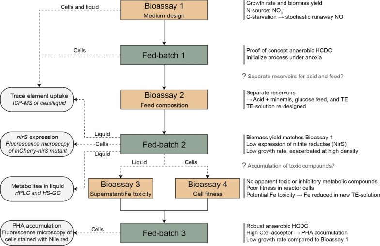
Results: Our fed-batch culture reached 20 g cell dry weight L-1, albeit with slower growth rates than observed in low cell density batch cultures. We explored reasons for slow growth, and the measured trace element uptake indicates it is not a limiting factor. Bioassays with spent medium excluded accumulation of inhibitory compounds at high cell density as the reason for the slow growth. The most plausible reason is that high metabolic activity led to CO2/H2CO3 accumulation, thus suppressing pH, leading to a paucity in HNO3-feeding until N2-sparging had removed sufficient CO2. The three free intermediates in the denitrification pathway (NO3- → NO2- → NO → N2O → N2) can all reach toxic concentrations if the electron flow is unbalanced, and this did occur if cells were glucose-limited. On the other hand, accumulation of polyhydroxyalkanoates occurred if the cells were NO3--limited. Carefully balancing glucose provision according to the HNO3 injected is thus crucial.

Conclusions: This work provides a proof of concept, while also identifying CO2/H2CO3 accumulation as a hurdle that must be overcome for further development and optimization of the method.

Keywords: Denitrification; High cell density cultivation; pH–stat.


Conflict of interest statement

Declarations. Ethics approval and consent to participate: Not applicable. Consent for publication: Not applicable. Competing interests: A patent application, describing the basic concept of HCDC by denitrification has been filed (European Patent App No. 22700061.9 and US Patent App. 18/270,538, 2024).

Figures

Fig. 1
Fig. 1
Principle of anaerobic high cell density culturing (HCDC) as fed-batch. A The culture is fed by peristaltic pumping of HNO3- and glucose-solutions, and by manual injection of trace element solutions. The feed rate is controlled by measured pH, ensuring that pH is kept within a narrow interval. The culture is sparged with N2 to remove CO2. B The respiratory reduction of HNO3 to N2 raises the pH, which is compensated by pumping a dose of HNO3. C Based on the titration curve of the growth medium, the pH interval translates into an HNO3 concentration interval. Our results indicated that the relationship between pH and HNO3 concentration could be obscured by high CO2 concentrations (lowering the pH), which would require a refined control algorithm taking monitored CO2 concentrations into account
Fig. 2
Fig. 2
Graphic summary outlining the bioassays and fed-batches. Bioassay 1 was used to determine the growth parameters and yield for anaerobic growth in the designed medium, which was then used in an initial attempt at high cell density cultivation in a fed-batch (Fed-batch 1; Strain: Pd1222). There were indications of toxicity when mixing glucose into the acid reservoir. To address this, we designed a bioassay to test the different feed compositions and combinations (Bioassay 2). Based on the results, we concluded that the acid, feed, and trace elements should be provided from three separate reservoirs. Cells and liquid samples from Bioassay 1 and Fed-batch 1 were analyzed by ICP-MS to determine the trace element content, which was used to re-design the trace element solution (TRES-2, Table 1) for Fed-batch 2. Fed-batch 2 (Strain: Pd1222_mC-nirS) had the expected biomass yield, but the growth rate was lower than expected based on Bioassay 1. During Fed-batch 2, cell-free liquid samples were analyzed by ICP-MS, HPLC, and HS-GC to determine the concentrations of trace metals and selected metabolites. Cells were analyzed by fluorescence microscopy to determine the expression of mCherry-nirS. At the end of Fed-batch 2, cells and reactor liquid were separated and used in Bioassay 3 and 4, respectively. Bioassay 3 aimed to test the presence of inhibitory or toxic compounds in the liquid by incubating with fresh cells. In addition, the toxicity of Fe(II) and Fe(III) under anoxic conditions was assessed. Bioassay 4 tested the fitness of the reactor cells by inoculation in a fresh medium. Bioassays 3 and 4, and the results from the liquid analysis, prompted us to re-design the trace element solution prior to Fed-batch 3 by reducing the Fe content. In Fed-batch 3 (Strain: Pd1222) we sustained a robust anaerobic HCDC, however, glucose excess resulted in PHA accumulation and the growth rate remained low
Fig. 3
Fig. 3
Gas kinetics in P. denitrificans growing under NO3 limitation, with (M2) and without (M1) NH4+. This show the results in representative single vials. Main: accumulation of N2 (µmol N vial−1), NO, and N2O (nmol N vial−1) during anaerobic growth by denitrification. Inserts: e-flow rates to N-oxides, Ve (µmol e vial−1 h−1), and apparent specific growth rate (µ, h−1) as estimated by nonlinear regression of rates against time
Fig. 4
Fig. 4
Stochastic accumulation of NO and N2O in single vials in response to C-starvation and injection of NO3. Runaway accumulation of NO occurred in two out of six glucose-limited cultures (2.9 mM glucose and 10 mM NO3) in Bioassay 1. This show measured N2, N2O, and NO (primary y-axis; µmol N vial−1), and the NO3 and glucose (secondary y-axis; mM) as estimated based on the measured N-gas production and the stoichiometry of growth (Table 2) in two single vials (M1 and M2)
Fig. 5
Fig. 5
Trace element analysis by ICP-MS after batch cultivation in M1 medium. A The initial trace element concentration in the medium and the concentration following aerobic (green) and anaerobic (orange) incubation of P. denitrificans. B The concentration of trace elements in cells grown aerobically (green) and anaerobically (orange). C Comparison of the content of trace elements in the biomass with the calculated uptake from the medium based on trace element concentration in the liquid. The average and standard deviation are based on 3 replicates
Fig. 6
Fig. 6
Glucose mixed with trace elements results in growth inhibition. P. denitrificans was inoculated in serum vials (50 mL medium M1; initial 1 vol% O2 in the headspace, 5 mM KNO3, and 10 mM glucose). After depletion of O2 and onset of denitrification, all vials got an additional 5 mM KNO3 and either glucose solution alone (3.125 M, 0.16 mL vial−1) or as a mix with trace elements. The figure shows O2-depletion (µmol vial−1, line) and subsequent N2 production (µmol N vial−1, circle) in the cultures (n = 2 replicate vials per treatment, standard deviations shown as vertical bars). The insert shows the estimated electron flow to NOx, Ve (µmol e vial−1 h−1) in the period after the injection. The result demonstrates that respiration was severely inhibited by the mixture of glucose and trace elements
Fig. 7
Fig. 7
Overview of the 330 h of anaerobic fed-batch cultivation of P. denitrificans with mCherry-nirS (Fed-batch 2). The bioreactor was operated as a pH–stat where denitrification-driven increase in pH (left number four and insert) was monitored and used to initiate the acid feed pump. The kfeed parameter (ratio between glucose- and HNO3-feed volume) was adjusted several times to avoid NO3-limitations and glucose excess. In addition, substrates were injected manually several times throughout the run, to explore limitations. The manual injections are shown in the top left. The N2 sparging flow rate was adjusted in response to increased cell density to limit CO2 accumulation in the reactor (upper right). Headspace samples were taken at intervals, and measured for NO, N2O, and CO2, with results shown in the three mid-right. N2O was below the detection limit (2 ppmv) in most samples (marked with a cross on the x-axis). The stirring speed (not shown) was increased from 250 to 350 rpm after 20 h and further increased to 500 rpm after 165 h. The lower right shows the offline measurements of NO2, NO3, glucose, and OD660. The lower left show pH and the cumulated input of HNO3 and glucose (mmol) during the first 20 h
Fig. 8
Fig. 8
Substrate consumption and biomass production during cycle 1 of Fed-batch 2. The top shows biomass calculated from cumulated consumption of NO3 (pYNO3-, assuming YNO3- = 13.68 g dw mol−1 NO3) and glucose (pYgluc, assuming Ygluc = 40.32 g dw mol−1 glucose), compared with biomass based on OD660 during a 190 h fed-batch cultivation. Open circles are the estimated dw based on measured OD, assuming a conversion factor of 0.36 g dw L−1 OD660−1, which is typical for P. denitrificans in small batches. In this fed-batch, however, the cells and medium darkened as the experiment progressed, apparently skewing the OD660 reads. A direct dry weight measurement at 188 h indicated a conversion factor of 0.26 g dw L−1 OD660−1. Corrected OD-based estimates, assuming that this conversion factor is valid throughout the experiment are also shown (closed circles). Transient periods of exponential growth are indicated as well as the first manual injection of trace elements (TRES-2) at 71 h. The growth rates were estimated by nonlinear regression of biomass against time. The bottom show measured concentrations of NO3 and NO2
Fig. 9
Fig. 9
Assessment of supernatant toxicity in Bioassay 3. Fresh cells were inoculated in bioreactor liquid (0, 5, 10, 20, 40, and 75 vol%) in Bioassay 3a. A Gas data for each of the treatments (n = 2). The top shows the O2 concentration (µmol vial−1) while the bottom shows the cumulative N2 concentration (µmol N2-N vial−1). B Initial electron flow rate (fmol e cell−1 h−1) estimated for each of the treatments in Bioassay 3a, corrected for individual sampling time for each vial, assuming a linear increase in rate during the time (1.7 h) from the first to the second headspace sampling. C Bioassay 3b investigated the toxicity of Fe2+ and Fe3+. The average N2 production rate 10–34 h after injection of Fe2+/Fe3+ was calculated for each Fe concentration and plotted against the concentrations. Since [S] > > Km both for glucose and nitrate, the inhibition coefficient (KI, i.e. the inhibitor concentration causing 50% inhibition) could be estimated by fitting V = Vmax / (1 + [I]/KI) to the data by least square ([I] is the concentration of the inhibitors Fe2+ or Fe3+). The estimated KI values are shown
Fig. 10
Fig. 10
Reactor cells in fresh medium (Bioassay 4). Vials with He-atmosphere and 50 mL mineral medium with glucose were inoculated with cells (8·1010 cells vial−1) from the bioreactor and monitored for N gas kinetics while stirred at 30°C. Four vials were provided with N2O as the sole electron acceptor (~ 1 vol% N2O in the headspace = 41 µmol vial−1), while four vials were provided with NO2 as the sole electron acceptor (2 mM KNO2 in the liquid). The large show the measured amount of NO, N2O, and N2 per vial (average, with standard deviation as vertical lines, n = 4), and the cell numbers per vial (calculated from the cumulated respiratory electron flow). The inserts show the cell-specific rates of N2-production (VN2, fmol N cell−1 h−1) and electron flow (Ve, fmol e cell−1 h−1). For the N2O-fed cultures (left), only VN2 is shown because Ve equals VN2 (1 electron per N). The dotted line represents Vmax in cells with µ = 0.19 h−1 assuming yield = 1.93 · 1013 cells mol−1 e to NO3- [27]
Fig. 11
Fig. 11
mCherry-NirS fluorescence in cells after 165 h. Images were obtained and analyzed as in Lycus et al. [28]. Fluorescence in each cell (n = 3769) was corrected for background and plotted as a histogram on the left y-axis (the number below shows the upper limit for each bar) and as the cumulative percentage in each bin on the right y-axis. The light grey is negative cells (≤ 10; average signal in negative controls: ~ 5). The insert shows one of the fluorescent images (phase contrast (grey) and mCherry (red) channels were merged in ImageJ), revealing very weak mCherry intensity in most of the cells
Fig. 12
Fig. 12
Growth rate and liquid analysis during the first cycle of Fed-batch 3. The top shows the total dry weight in the reactor, which was found by considering the liquid volume in the reactor during each OD660 measurement and assuming 0.36 g dw OD660−1 L−1. The insert shows the apparent specific growth rate throughout the cycle. The lower shows the offline measurements of NO2, NO3, and glucose
Fig. 13
Fig. 13
Reactor cells stained with Nile red. The cells were harvested at the end of the reactor run, stained with Nile red, and visualized by fluorescence microscopy (phase contrast (grey) and Nile red (red) channels were merged in ImageJ). The presence of PHA granules was visible in all cells

References

    1. UN Population Division. World population prospects 2022: Summary of results. UN DESA/POP/2022/TR/NO. 3. 2022.
    1. Westhoek H, Rood T, Van den Berg M, Janse J, Nijdam D, Reudink M, et al. The protein puzzle. Hague: PBL Netherlands Environmental Assessment Agency; 2011. p. 221.
    1. Suman G, Nupur M, Anuradha S, Pradeep B. Single cell protein production: a review. Int J Curr Microbiol App Sci. 2015;4(9):251–62.
    1. Matassa S, Boon N, Pikaar I, Verstraete W. Microbial protein: future sustainable food supply route with low environmental footprint. Microb Biotechnol. 2016;9(5):568–75. - PMC - PubMed
    1. Spalvins K, Zihare L, Blumberga D. Single cell protein production from waste biomass: comparison of various industrial by-products. Energy Proc. 2018;147:409–18.

MeSH terms

LinkOut - more resources