Case Reports
. 2024 Nov 25;2024(11):rjae753.
doi: 10.1093/jscr/rjae753. eCollection 2024 Nov.

Lung resection from wedge to pneumonectomy as surgical options for pulmonary mucormycosis

Affiliations
Case Reports

Lung resection from wedge to pneumonectomy as surgical options for pulmonary mucormycosis

Alexander Pohlman et al. J Surg Case Rep. .

Abstract

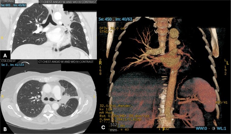
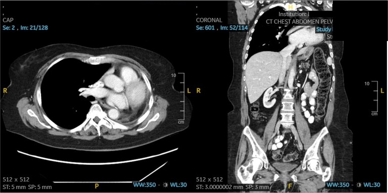
Pulmonary mucormycosis (PM) is a rare and life-threatening condition, most prevalent in immunocompromised patients. Early signs and symptoms are often nonspecific. A high index of suspicion in at risk patients should prompt early infectious work-up, including bronchoscopy, followed by aggressive antifungal therapy and early surgical resection when indicated. We demonstrate these core tenants of diagnosis and management of PM via two patient presentations, the first involving a kidney transplant recipient who presented with a mild cough, found to have a lung lesion with rapid growth over a few weeks; the second involving a patient with acute lymphoblastic leukemia who presented with hemoptysis and imaging revealing a 5 cm perihilar mass obliterating the left pulmonary artery. Both patients were managed with aggressive surgical therapy.

Keywords: immunosuppressed; leukemia; pulmonary mucormycosis; transplant.


Conflict of interest statement

None declared.

Figures

Figure 1
Figure 1
Patient 1 pre-operative images showing rapid progression of disease. (A) Initial CT scan with a 2.4 cm LUL pulmonary nodule. (B) Repeat CT scan 28 days later showing rapid growth and cavitation of the mass, now measuring 7.1 cm.
Figure 2
Figure 2
Patient 2 pre-operative images. (A) Coronal section of CT angiogram showing LUL mass abutting the aortic arch and invading the left main pulmonary artery and left mainstem bronchus. (B) Axial CTA showing complete occlusion of the left pulmonary artery. (C) 3D reconstruction of CTA showing complete occlusion of the left pulmonary artery and lack of opacification of any pulmonary artery branches and pulmonary oligemia.
Figure 3
Figure 3
Patient 2 post-operative CT scan showing complete resection of disease and expected post-operative anatomical changes, including leftward deviation of mediastinum and hemidiaphragmatic elevation.

References

    1. Hamilos G, Samonis G, Kontoyiannis DP. Pulmonary mucormycosis. Semin Respir Crit Care Med 2011;32:693–702. 10.1055/s-0031-1295717. - DOI - PubMed
    1. Muqeetadnan M, Rahman A, Amer S, et al. . Pulmonary mucormycosis: an emerging infection. Case Rep Pulmonol 2012;2012:120809. 10.1155/2012/120809. - DOI - PMC - PubMed
    1. Pavie J, Lafaurie M, Lacroix C, et al. . Successful treatment of pulmonary mucormycosis in an allogenic bone-marrow transplant recipient with combined medical and surgical therapy. Scand J Infect Dis 2004;36:767–9. 10.1080/00365540410021081a. - DOI - PubMed
    1. al-Abbadi MA, Russo K, Wilkinson EJ. Pulmonary mucormycosis diagnosed by bronchoalveolar lavage: a case report and review of the literature. Pediatr Pulmonol 1997;23:222–5. 10.1002/(SICI)1099-0496(199703)23:3<222::AID-PPUL9>3.0.CO;2-P. - DOI - PubMed
    1. Walsh TJ, Gamaletsou MN, McGinnis MR, et al. . Early clinical and laboratory diagnosis of invasive pulmonary, extrapulmonary, and disseminated mucormycosis (zygomycosis). Clin Infect Dis 2012;54:S55–60. 10.1093/cid/cir868. - DOI - PubMed

Publication types

LinkOut - more resources