

Pyogenic Liver Abscess and the Important Role of Point-of-Care Ultrasound (POCUS) in Daily Practice: A Report of Two Cases
- PMID: 39588419
- PMCID: PMC11588408
- DOI: 10.7759/cureus.72444
Pyogenic Liver Abscess and the Important Role of Point-of-Care Ultrasound (POCUS) in Daily Practice: A Report of Two Cases
Abstract
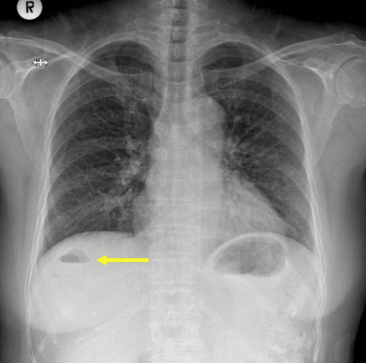
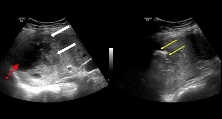
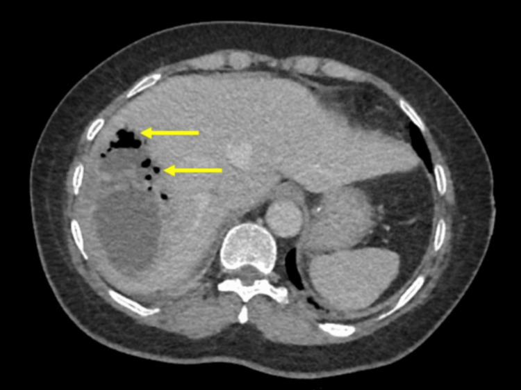
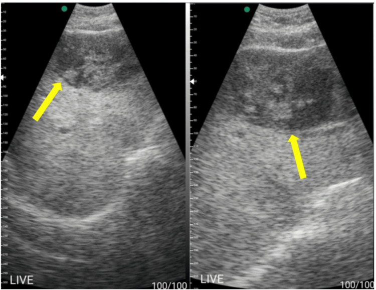
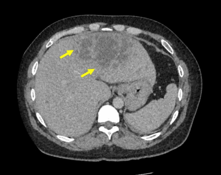
A pyogenic liver abscess (PLA) is uncommon and a potentially life-threatening condition. Clinical manifestations and laboratory investigations can be non-specific and the detection of PLAs requires imaging, which can often be delayed. Point-of-care ultrasound (POCUS) is now becoming more widely adopted and plays an important role in clinical practice. We report two cases of PLA, one of which was a gas-forming PLA that we encountered in a district general hospital where POCUS played an important role in the diagnosis and management. The diagnoses of PLAs were initially unsuspected due to a combination of non-specific symptom manifestations, initial negative imaging, and a subtle radiological clue that was missed due to a lack of awareness. Bedside POCUS examinations were done due to clinical deterioration in one patient and lack of inflammatory marker improvement in both patients. These cases highlight the important role of POCUS in the management of patients with liver abscesses.
Keywords: diagnosis; liver abscess; pocus; transabdominal ultrasound; ultrasound.
Copyright © 2024, Rubel et al.
Conflict of interest statement
Human subjects: Consent for treatment and open access publication was obtained or waived by all participants in this study. Medical and Health Research Ethic Committee, Ministry of Health, Brunei Darussalam issued approval Not applicable. Conflicts of interest: In compliance with the ICMJE uniform disclosure form, all authors declare the following: Payment/services info: All authors have declared that no financial support was received from any organization for the submitted work. Financial relationships: All authors have declared that they have no financial relationships at present or within the previous three years with any organizations that might have an interest in the submitted work. Other relationships: All authors have declared that there are no other relationships or activities that could appear to have influenced the submitted work.
Figures
References
Publication types
LinkOut - more resources
Full Text Sources