. 2024 Dec 11;52(22):13996-14012.
doi: 10.1093/nar/gkae1130.

Bacterial Rps3 counters oxidative and UV stress by recognizing and processing AP-sites on mRNA via a novel mechanism

Affiliations

Bacterial Rps3 counters oxidative and UV stress by recognizing and processing AP-sites on mRNA via a novel mechanism

Mohammad Afsar et al. Nucleic Acids Res. .

Abstract

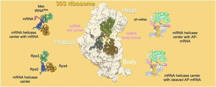
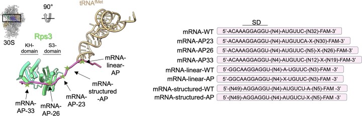
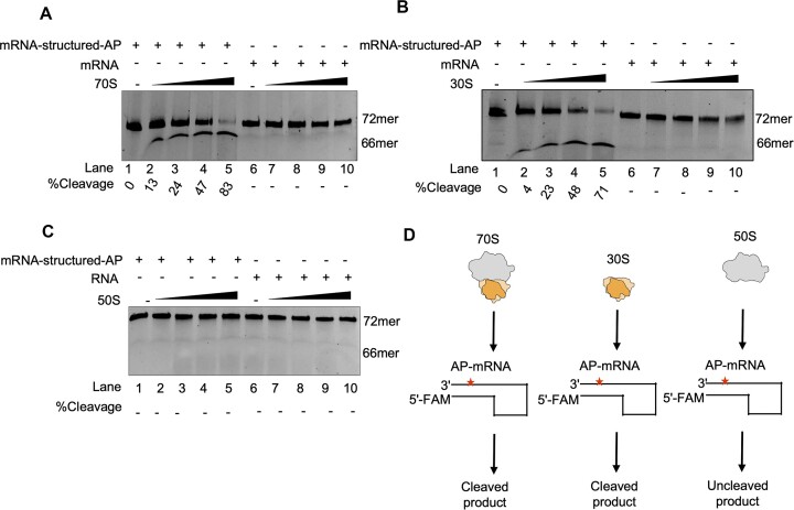
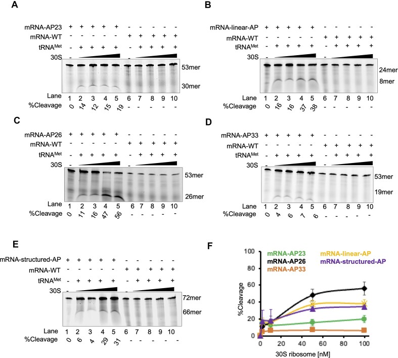
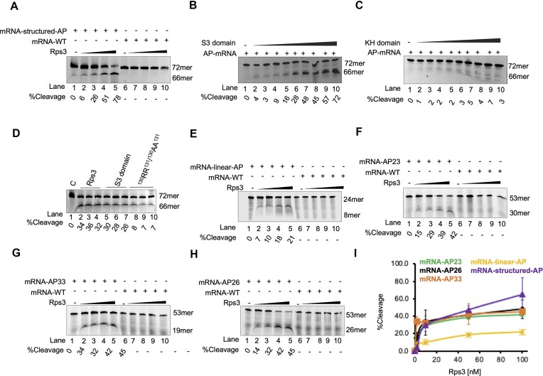
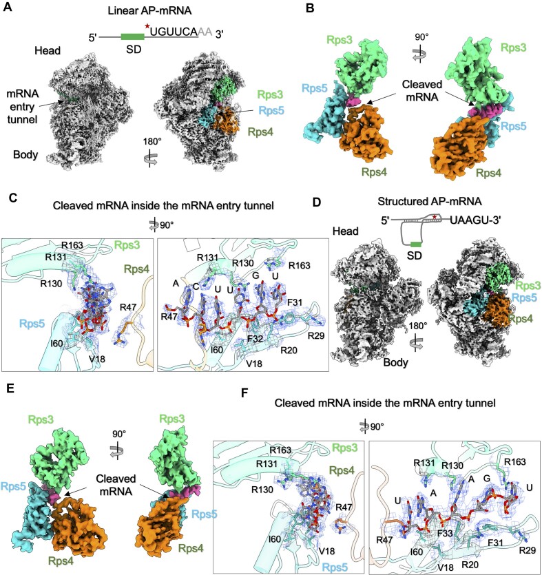
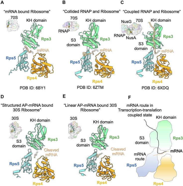
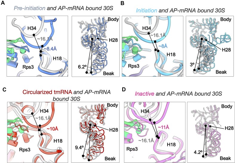
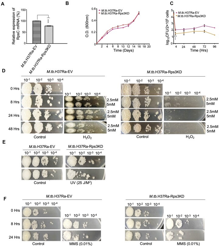
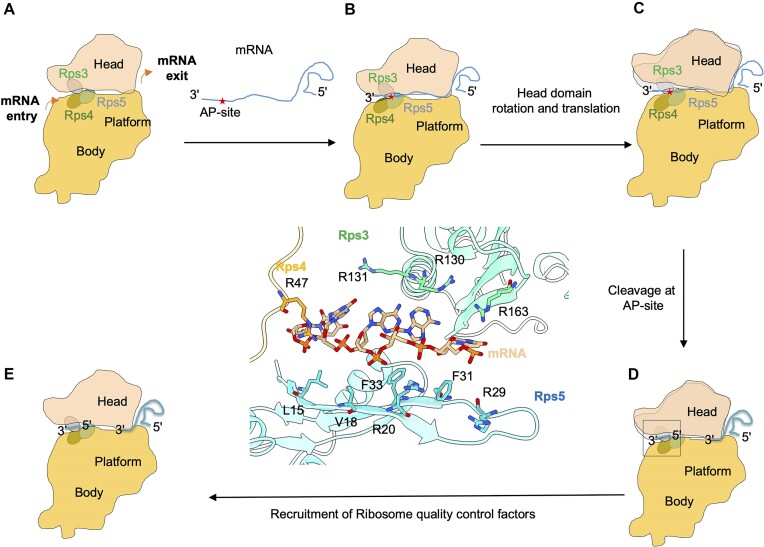
Lesions and stable secondary structures in mRNA severely impact the translation efficiency, causing ribosome stalling and collisions. Prokaryotic ribosomal proteins Rps3, Rps4 and Rps5, located in the mRNA entry tunnel, form the mRNA helicase center and unwind stable mRNA secondary structures during translation. However, the mechanism underlying the detection of lesions on translating mRNA is unclear. We used Cryo-EM, biochemical assays, and knockdown experiments to investigate the apurinic/apyrimidinic (AP) endoribonuclease activity of bacterial ribosomes on AP-site containing mRNA. Our biochemical assays show that Rps3, specifically the 130RR131 motif, is important for recognizing and performing the AP-endoribonuclease activity. Furthermore, structural analysis revealed cleaved mRNA product in the 30S ribosome entry tunnel. Additionally, knockdown studies in Mycobacterium tuberculosis confirmed the protective role of Rps3 against oxidative and UV stress. Overall, our results show that prokaryotic Rps3 recognizes and processes AP-sites on mRNA via a novel mechanism that is distinct from eukaryotes.


Figures

Graphical Abstract
Graphical Abstract
Figure 1.
Figure 1.
Preparation of apurinic/apyrimidinic (AP) site containing mRNA molecules. The positions and the sequence of AP site containing AP-mRNA molecules on the 30S ribosome with respect to initiator-tRNAMet and Rps3 protein in the mRNA channel (PDB ID: 6ZTJ).
Figure 2.
Figure 2.
Identification of AP-endoribonuclease activity of the ribosome on AP-mRNA. (A) AP-endoribonuclease activity exhibited by the 70S ribosome on mRNA-structured-AP. (B) AP-endoribonuclease activity of 30S ribosome on mRNA-structured-AP. (C) AP-endoribonuclease activity of 50S ribosome on mRNA-structured-AP. For gels in A–C, lanes 1 and 6 have no ribosome, while lanes 2–5 have increasing concentration of ribosome (0–100 nM) with mRNA-structured-AP. Lanes 7–10 contain undamaged mRNA molecule with increasing concentration of respective ribosomes. The reaction products (10 ul) were analysed on 8 M urea-15% polyacrylamide gels. (D) The biochemical assays involving the 70S ribosome and their subunits is summarized here. Data in panels A–C are representative of at least three technical replicates.
Figure 3.
Figure 3.
Characterization of AP-endoribonuclease activity of 30S ribosome on AP-mRNA. (A) AP-endoribonuclease activity of 30S:i-tRNAMet complex on mRNA-AP23. (B) AP-endoribonuclease activity of 30S:i-tRNAMet complex on mRNA-linear-AP. (C) AP-endoribonuclease activity of 30S:i-tRNAMet complex on mRNA-AP26. (D) AP-endoribonuclease activity of 30S:i-tRNAMet complex on mRNA-AP33. (E) AP-endoribonuclease activity of 30S:i-tRNAMet complex on mRNA-structured-AP. For gels in A–E, lanes 1 and 6 have no ribosome, while lanes 2–5 have increasing concentration of ribosome (0–100 nM) with mRNA-AP. Lanes 7–10 contain undamaged mRNA molecule with increasing concentration of respective ribosomes. The i-tRNAMet was added in each reaction in 1:2 molar ratio to 30S ribosomes. The reaction products (10 ul) were analyzed on 8 M urea-15% polyacrylamide gels. (F) Percent cleavage of 30S:i-tRNAMet complex on different mRNA-AP molecules are quantified and plotted (%cleavage versus nM concentration of ribosome). Data in panels A–E are representative of at least three replicates.
Figure 4.
Figure 4.
Biochemical characterization of the endoribonuclease activity of ribosomal protein S3 (RpS3). (A) AP-endoribonuclease activity of Rps3 on mRNA-structured-AP. (B) and (C) Role of S3 and KH domains in the observed endoribonuclease activity under standard reaction conditions. Lanes 2–10 have increasing concentration of S3 and KH domains, respectively (0–100 nM). (D) Role of ‘RRA’ motif in the endoribonuclease activity of RpS3. The wildtype (Lanes 2,3,4; 10, 50 and 100 nM, respectively), S3 domain (Lanes 5,6,7; 10, 50 100 nM, respectively) and site-directed mutants (Lanes 8,9,10; 10, 50 and 100 nM) were incubated with AP-mRNA (100 nM) under standard reaction conditions. The percentage activity of the wildtype RpS3, deletion mutants S3 domain and site-directed mutants (130RR131/130AA131) were quantified. (E) AP-endoribonuclease activity of Rps3 on mRNA-linear-AP. (F) AP-endoribonuclease activity of Rps3 on mRNA-AP23. (G) AP-endoribonuclease activity of Rps3 on mRNA-AP33. (H) AP-endoribonuclease activity of Rps3 on mRNA-AP26. For gels in A, E, F, G and H, lanes 1 and 6 have no Rps3, while lanes 2–5 have increasing concentration of Rps3 (0–100 nM) with mRNA-AP. Lanes 7–10 contain undamaged mRNA molecule with increasing concentration of respective Rps3. The reaction products (10 ul) were analyzed on 8 M urea-15% polyacrylamide gels. (I) Percent cleavage of Rps3 on different mRNA-AP molecules are quantified and plotted (%cleavage versus nM concentration of ribosome). Data in panels (A–H) are representative of at least three replicates.
Figure 5.
Figure 5.
The presence of cleaved mRNA product in entry tunnel and adaptation of partial inactive conformation. (A) High resolution Cryo-EM reconstruction of linear AP-mRNA molecule bound 30S ribosome. (B) The analysis of mRNA entry tunnel constituted by Rps3, Rps4 and Rps5 shows the presence of cleaved mRNA (5′-UGUUCA-3′) molecule. (C) Cleaved mRNA molecule in the mRNA entry tunnel is stabilized by the presence of positively charged residues of Rps3, Rps4 and Rps5. (D) Cryo-EM reconstruction of structured AP-mRNA molecule bound 30S ribosome. (E) Investigation of mRNA entry tunnel constituted by Rps3, Rps4 and Rps5 shows the presence of cleaved mRNA (5′-UAAGU-3′) molecule. (F) The cleaved mRNA molecule in the mRNA entry tunnel is stabilized by the presence of positively charged residues of Rps3, Rps4 and Rps5.
Figure 6.
Figure 6.
The path of the mRNA molecule and the state of ribosomes in various complexes. (A) The extended mRNA molecule in the mRNA entry tunnel interacts with the S3 domain of Rps3 protein. (B) The mRNA route in the collided state RNAP–ribosome complex shows the presence of mRNA near the S3 domain. (C) The density map of the transcription–translation-coupled complex shows density for the mRNA molecule extending from the S3 domain to the KH domain. (D) and (E), The mRNA route of post endoribonuclease activity shows its interaction with S3 domain of Rps3 protein. (F) A schematic summary of the observed mRNA routes in the entry tunnel in the different ribosome states is depicted here.
Figure 7.
Figure 7.
Head domain rotation and movement in the presence of AP-mRNA bound 30S ribosome. (A) Distance between the H18 (present in body) and H34 (present in head) is 8.4 Å in pre-initiation 30S ribosome complex (30S, IF-1, IF-3, mRNA, PDB ID: 5LMN) while this is 16.1 Å in AP-mRNA bound 30S ribosome. Beak region in head is also rotated by 6.2° from the H28. (B) H18 and H34 are present 8.0Å apart in initiation 30S ribosome complex (30S, IF-1, IF-2, mRNA, PDB ID: 6O7K), and beak region shows the rotation of 3° in comparison with AP-mRNA bound 30S ribosome. (C) Stalled ribosomes were rescued by tmRNA–SmpB complex. The superimposition of the tmRNA-SmpB bound 30S ribosome (PDB ID: 6Q95) with AP-mRNA bound 30S ribosome shows the rotation of head domain by 9.4° with 10 Å distance between the H18 and H34. (D) Furthermore, we determined the structural adaptation between the inactive conformation of 30S ribosome (PDB ID: 6W7M) and AP-mRNA bound 30S ribosome. This analysis shows that structured AP-mRNA 30S ribosome shows the rotation of head domain by 4.2° and distance between H18 and H34 is 11Å.
Figure 8.
Figure 8.
Role of RpS3 in recuing bacterium under oxidative stress. (A) mRNA transcripts was isolated from M.tb.H37Ra-Rps3KD and M.tb.H37Ra-EV, and fold change was measured by qRT-PCR. (B) Growth curve for the knock down and wild type strain. (C) PMA-differentiated THP-1 macrophages were infected with M.tb.H37Ra-Rps3KD and M.tb.H37Ra-EV at an MOI of 10. Intracellular CFUs were determined at the indicated post infection time points (0, 4, 24, 48, 72 and 96 h) by serial dilutions. Each data point represents mean ± standard deviation (error bars) from three independent experiments. Statistically significant differences are indicated by *(P ≤ 0.05), **(P ≤ 0.01) and ***(P ≤ 0.001). (DF), Cultures of OD 600 ∼0.1 were exposed to 2.5 or 5mM H2O2, 25 J/m2 UV light and 0.01% MMS, respectively. Each panel represents 10-fold serial dilutions of untreated (control) and treated cultures. Ten microliters of each dilution were spotted onto supplemented solid MB7H10 medium. The images are representative data from at least two independent experiments.
Figure 9.
Figure 9.
Recognition of AP-site containing mRNA molecule by 30S ribosome. (A) 30S ribosome is composed of head and body domains that are connect by neck region. The Rps3, Rps4 and Rps5 proteins are present in the mRNA entry tunnel of neck region. (B) AP-site in mRNA, first got recognized by mRNA helicase fold of ribosome in the entry tunnel. (C) The accompanied head domain rotation may facilitate the recognition of AP-stie in the mRNA molecule. (D) The Rps3 protein performs the endoribonuclease activity at the abasic site in mRNA molecule. (E) The trapped cleaved product in the mRNA entry and exit tunnels can be subsequently removed by RQC factors.

Similar articles

References

    1. Mauger D.M., Cabral B.J., Presnyak V., Su S.V., Reid D.W., Goodman B., Link K., Khatwani N., Reynders J., Moore M.J.et al. .. mRNA structure regulates protein expression through changes in functional half-life. Proc. Nat. Acad. Sci. USA. 2019; 116:24075–24083. - PMC - PubMed
    1. Wurtmann E.J., Wolin S.L.. RNA under attack: cellular handling of RNA damage. Crit. Rev. Biochem. Mol. Biol. 2009; 44:34–49. - PMC - PubMed
    1. Prashar T., De La Selle F., Hudak K.A.. Abasic RNA: its formation and potential role in cellular stress response. RNA Biol. 2023; 20:348–358. - PMC - PubMed
    1. Calabretta A., Kupfer P.A., Leumann C.J.. The effect of RNA base lesions on mRNA translation. Nucleic Acids Res. 2015; 43:4713–4720. - PMC - PubMed
    1. Reichle V.F., Petrov D.P., Weber V., Jung K., Kellner S.. NAIL-MS reveals the repair of 2-methylthiocytidine by AlkB in E. coli. Nat. Commun. 2019; 10:5600. - PMC - PubMed

MeSH terms

LinkOut - more resources