. 2025 Jan;41(1):e12911.
doi: 10.1002/kjm2.12911. Epub 2024 Nov 26.

ELAVL1-dependent SOAT2 exacerbated the pancreatitis-like cellular injury of AR42J cells induced by hyperstimulation with caerulein

Affiliations

ELAVL1-dependent SOAT2 exacerbated the pancreatitis-like cellular injury of AR42J cells induced by hyperstimulation with caerulein

Yu-Jing Sun et al. Kaohsiung J Med Sci. 2025 Jan.

Abstract

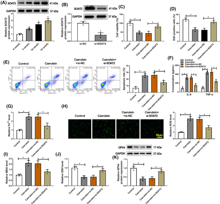
Pancreatitis is a severe inflammatory condition characterized by damage to the pancreas. Sterol o-acyltransferase 2 (SOAT2) has been reported to aggravate acute pancreatitis, however, the underlying mechanism remains to be elucidated. Rat pancreatic exocrine cells (AR42J) were treated with caerulein to induce pancreatitis-like cellular injury. Cell viability was determined using a cell counting kit-8 (CCK-8) assay, while cell proliferation was analyzed through a 5-Ethynyl-2'-deoxyuridine assay. Cell apoptosis was measured using flow cytometry, and enzyme-linked immunosorbent assays were performed to detect levels of pro-inflammatory cytokines IL-6 and TNF-α. Additionally, Fe2+ levels were analyzed using a colorimetric assay kit, reactive oxygen species (ROS) levels were assessed with a Cellular ROS Assay kit, and lipid peroxidation was measured using a malondialdehyde assay kit. Glutathione levels were analyzed with a detection assay. Protein and mRNA expression were evaluated through western blotting and quantitative real-time polymerase chain reaction, respectively. Furthermore, an RNA immunoprecipitation assay was conducted to investigate the association between ELAV-like RNA binding protein 1 (ELAVL1) and SOAT2. Actinomycin D assay was performed to explore the effect of ELAVL1 depletion on the transcript stability of SOAT2 mRNA. SOAT2 and ELAVL1 expression were upregulated in caerulein-exposed AR42J cells. Caerulein treatment induced pancreatitis-like cellular apoptosis, inflammatory response, ferroptosis, and cell proliferation inhibition. Silencing of SOAT2 protected against caerulein-induced AR42J cell injury. Moreover, ELAVL1 stabilized SOAT2 mRNA expression in AR42J cells. SOAT2 overexpression attenuated the effects induced by ELAVL1 silencing in caerulein-exposed AR42J cells. Additionally, ELAVL1 knockdown activated the NRF2/HO-1 pathway by downregulating SOAT2 expression in caerulein-exposed AR42J cells. SOAT2 silencing protected AR42J cells from caerulein-induced injury by inactivating the NRF2 pathway. In conclusion, ELAVL1-dependent SOAT2 exacerbated pancreatic exocrine cell injury by inactivating the NRF2/HO-1 pathway in pancreatitis. These findings provide new insights into the molecular mechanisms underlying pancreatitis and offer potential therapeutic targets for the treatment of this condition.

Keywords: ELAVL1; NRF2/HO‐1 pathway; SOAT2; pancreatitis.


Conflict of interest statement

The authors declare that they have no conflicts of interest.

Figures

FIGURE 1
FIGURE 1
Caerulein treatment induced pancreatitis‐like cellular apoptosis, inflammatory response, and ferroptosis. AR42J cells were treated with PBS or caerulein (5, 10, and 15 nmol/L). (A) Cell viability was assessed by the CCK‐8 assay. (B) Cell proliferation was analyzed by the EdU assay. (C) Cell apoptosis was assessed by flow cytometry. (D) ELISAs were performed to detect IL‐6 and TNF‐α levels. (E) Fe2+ colorimetric assay kit was used for Fe2+ level analysis. (F) ROS level was detected using a Cellular ROS Assay kit. (G) MDA level was analyzed using a lipid peroxidation MDA assay kit. (H) Glutathione detection assay was performed to analyze GSH level. (I) GPX4 protein expression was detected by western blotting assay. *p < 0.05.
FIGURE 2
FIGURE 2
SOAT2 silencing protected against caerulein‐induced AR42J cell injury. (A) The effects of caerulein treatment (0, 5, 10, and 15 nmol/L) on SOAT2 protein expression were assessed by western blotting assay. (B) The efficiency of SOAT2 knockdown was determined by western blotting assay. (C–K) AR42J cells were divided into control group, caerulein group, caerulein+si‐NC group, and caerulein+si‐SOAT2 group. (C) Cell viability was assessed by the CCK‐8 assay. (D) Cell proliferation was analyzed by the EdU assay. (E) Cell apoptosis was assessed by flow cytometry. (F) ELISAs were performed to detect IL‐6 and TNF‐α levels. (G) Fe2+ colorimetric assay kit was used for Fe2+ level analysis. (H) ROS level was detected using a Cellular ROS Assay kit. (I) MDA level was analyzed using a lipid peroxidation MDA assay kit. (J) Glutathione detection assay was performed to analyze GSH level. (K) GPX4 protein expression was detected by western blotting assay. *p < 0.05.
FIGURE 3
FIGURE 3
ELAVL1 stabilized SOAT2 mRNA expression in AR42J cells. The ENCORI online database was used to predict the RNA binding proteins of SOAT2. (B) The RIP assay was conducted to identify the association between ELAVL1 and SOAT2 in AR42J cells. (C) The efficiency of ELAVL1 knockdown was analyzed by western blotting assay in AR42J cells. (D and E) The effects of ELAVL1 silencing on SOAT2 expression were determined by quantitative real‐time polymerase chain reaction and western blotting assay. (F) Actinomycin D assay was performed to determine the effect of ELAVL1 depletion on the transcript half‐life of SOAT2 mRNA. (G) The effects of caerulein treatment (0, 5, 10, and 15 nmol/L) on ELAVL1 protein expression were assessed by western blotting assay. *p < 0.05.
FIGURE 4
FIGURE 4
SOAT2 overexpression attenuated ELAVL1 silencing‐induced effects in caerulein‐exposed AR42J cells. (A) The efficiency of SOAT2 overexpression was analyzed by western blotting assay in AR42J cells. (B–L) AR42J cells were divided into control group, caerulein group, caerulein+si‐NC group, caerulein+si‐ELAVL1 group, caerulein+si‐ELAVL1 + vector group, and caerulein+si‐ELAVL1 + SOAT2 group. (B) Cell viability was assessed by the CCK‐8 assay. (C) Cell proliferation was analyzed by the EdU assay. (D and E) Cell apoptosis was assessed by flow cytometry. (F) ELISAs were performed to detect IL‐6 and TNF‐α levels. (G) Fe2+ colorimetric assay kit was used for Fe2+ level analysis. (H and I) ROS level was detected using a Cellular ROS Assay kit. (J) MDA level was analyzed using a lipid peroxidation MDA assay kit. (K) Glutathione detection assay was performed to analyze GSH level. (L) GPX4 protein expression was detected by western blotting assay. *p < 0.05.
FIGURE 5
FIGURE 5
ELAVL1 knockdown activated the NRF2/HO‐1 pathway by downregulating SOAT2 expression in caerulein‐exposed AR42J cells. AR42J cells were divided into control group, caerulein group, caerulein+si‐NC group, caerulein+si‐ELAVL1 group, caerulein+si‐ELAVL1 + vector group, and caerulein+si‐ELAVL1 + SOAT2 group. (A–C) The protein expression of SOAT2, NRF2, and HO‐1 was analyzed by western blotting assay. *p < 0.05.
FIGURE 6
FIGURE 6
SOAT2 silencing protected AR42J cells from caerulein‐induced injury by upregulating NRF2 expression. AR42J cells were divided into five groups, including control, caerulein group, caerulein+si‐NC group, caerulein+si‐SOAT2 group, and caerulein+si‐SOAT2 + ML385 group. (A) Cell viability was assessed by the CCK‐8 assay. (B) Cell proliferation was analyzed by the EdU assay. (C and D) Cell apoptosis was assessed by flow cytometry. (E) ELISAs were performed to detect IL‐6 and TNF‐α levels. (F) Fe2+ colorimetric assay kit was used for Fe2+ level analysis. (G) ROS level was detected using a Cellular ROS Assay kit. (H) MDA level was analyzed using a lipid peroxidation MDA assay kit. (I) Glutathione detection assay was performed to analyze GSH level. (J) GPX4 protein expression was detected by western blotting assay. *p < 0.05.

References

    1. Hall JC, Crawford HC. The conspiracy of autophagy, stress and inflammation in acute pancreatitis. Curr Opin Gastroenterol. 2014;30(5):495–499. - PMC - PubMed
    1. Abdullah M, Firmansyah MA. Diagnostic approach and management of acute abdominal pain. Acta Med Indones. 2012;44(4):344–350. - PubMed
    1. Umar M, Noor E, Ali U, Khan I, Ahmed Z. Cocaine‐induced acute pancreatitis: a rare etiology. Cureus. 2020;12(7):e9029. - PMC - PubMed
    1. Lankisch PG, Apte M, Banks PA. Acute pancreatitis. Lancet. 2015;386(9988):85–96. - PubMed
    1. Teterin YS, Kulikov YD, Askerov AC, Yartsev PA. Intraluminal endoscopy in diagnosis and treatment of fluid collections in acute pancreatitis. Khirurgiia (Mosk). 2022;8:31–37. - PubMed

MeSH terms