. 2024 Nov 26;10(1):e174126.
doi: 10.1172/jci.insight.174126.

Role of cGAS/STING pathway in aging and sexual dimorphism in diabetic kidney disease

Affiliations

Role of cGAS/STING pathway in aging and sexual dimorphism in diabetic kidney disease

Sherif Khedr et al. JCI Insight. .

Abstract

Diabetic kidney disease (DKD) is the leading cause of chronic renal pathology. Understanding the molecular underpinnings of DKD is critical to designing tailored therapeutic approaches. Here, we focused on sex differences and the contribution of aging toward the progression of DKD. To explore these questions, we utilized young (12 weeks old) and aged (approximately 50 weeks old) type 2 diabetic nephropathy (T2DN) rats. We revealed that the cyclic GMP-AMP synthase (cGAS)/stimulator of interferon genes (STING) pathway was upregulated in T2DN rats compared with nondiabetic Wistar rats and in type 2 diabetic human kidneys. The activation of the cGAS/STING signaling pathway exhibited distinct protein expression profiles between male and female T2DN rats, with these differences becoming more pronounced with aging. RNA-Seq analysis of the kidney cortex in both male and female T2DN rats, at both younger and older ages, revealed several key molecules, highlighting crucial genes within the cGAS/STING pathway. Thus, our study delved deep into understanding the intricate sexual differences in the development and progression of DKD and we propose the cGAS/STING pathway as an essential contributor to disease development.

Keywords: Chronic kidney disease; Diabetes; Nephrology.


Conflict of interest statement

Conflict of interest: The authors have declared that no conflict of interest exists.

Figures

Figure 1
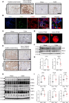
Figure 1. The expression pattern of cGAS/STING pathway molecules, TREX1 and mtTFA, in human and rat kidneys.
(A) Representative images of immunohistochemical staining of kidneys for STING in healthy and type 2 diabetic humans (left), nondiabetic (Wistar) rats, and rats with type 2 diabetic neuropathy (T2DN) (right). G, glomerulus. Scale bars: 150 μm. (B) Examination of CD68 and STING by immunofluorescence microscopy in diabetic human kidney. Scale bars: 50 μm (left) and 5 μm (right). (C) Immunohistochemical detection of CD68 in kidneys of old male Wistar and T2DN rats. Scale bar: 150 μm. (D) Representative 3D image from a human kidney stained for inactive and active STING molecules. Scale bar: 2 μm. (E) Immunohistochemical staining for mtTFA and TREX1 in renal cortical sections from healthy and diabetic human kidneys. Scale bars: 150 μm. (F) Western blot analysis of mtTFA and TREX1 in the kidneys of Wistar and T2DN old male rats. n = 6 rats in each group. β-Actin was used as a loading control. (G) Summary graphs of relative abundance for Western blots shown above. (H and I) Western blot analysis of the cGAS/STING pathway molecules (cGAS, STING, p-TBK1, TBK1, p-IRF3, and IRF3) in the kidneys of Wistar and T2DN old male rats. n = 6 rats in each group. Data presented as mean ± SEM. Statistical analysis was performed using unpaired, 2-tailed Student’s t test. *P < 0.05, **P < 0.01, ****P < 0.0001. NS, nonsignificant; a.u., arbitrary units.
Figure 2
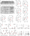
Figure 2. Differential levels of renal injury in T2DN rats of different sex and age groups.
(A) Renal cortical nephrin and KIM1 expression tested using Western blotting for both sexes in young and old T2DN rats. n = 6 rats for nephrin and 4 rats for KIM1, respectively. (B) Summary graphs of relative abundance of nephrin and KIM1. Each dot represents 1 rat. Data shown as mean ± SEM. Statistical analysis was performed using 2-way ANOVA. ***P < 0.001. NS, nonsignificant; a.u., arbitrary units. (C) Representative images of picrosirius red staining of old female and male T2DN rat kidneys. G, glomerulus. Scale bars: 150 μm. (D) Expression level of fibrosis-associated genes (Vim, Tgfb1, Col1a1, and Col3a1) obtained from RNA-Seq data represented as RPKM. n = 4 rats in each group. Statistical analysis was performed using unpaired and paired 2-tailed Student’s t tests. ****P < 0.0001 indicates a statistically significant difference within the same genes.
Figure 3
Figure 3. Expression analysis for TREX1, mtTFA, and cGAS/STING pathway molecules in T2DN rats of different sex and age groups.
(A) Western blot analysis of mtTFA and TREX1 expression levels for both sexes in young and old T2DN rats; β-actin was used as a loading control. n = 6 rats in each group. (B) Summary graphs of relative abundance of mtTFA and TREX1 shown in A. Each dot represents 1 rat. (C) Expression level of the cGAS/STING pathway proteins (cGAS, STING, p-TBK1, TBK1, p-IRF3, and IRF3) in the kidneys of both sexes in young and old T2DN rats. Each lane represents 1 rat. n = 6, except for p-IRF3 in which n = 4. (D) Summary graphs of relative abundance of cGAS/STING pathway proteins after normalization to β-actin loading control. Data shown as mean ± SEM. Statistical analysis was performed using a 2-way ANOVA test. *P < 0.05; **P < 0.01; ***P < 0.001; ****P < 0.0001. NS, nonsignificant; a.u., arbitrary units. (E) IRF3 adaptor protein activator (Mavs, Trif, and Sting) levels obtained from RNA-Seq data represented as RPKM. Statistical analysis was performed using unpaired and paired 2-tailed Student’s t tests. *P < 0.05, **P < 0.1, ****P < 0.0001. (F) Type I IFN signaling pathway molecule (Ifnar1, Ifnar2, Jak1, Tyk2, Irf9, Stat1, and Stat2) levels obtained from RNA-Seq data represented as RPKM. n = 4 rats in each group. Statistical analysis was performed using unpaired and paired 2-tailed Student’s t tests. *P < 0.05, **P < 0.01, ***P < 0.001, ****P < 0.0001, indicates statistically significant difference within the same genes. NS, nonsignificant.
Figure 4
Figure 4. Differential inflammatory profile in T2DN rats of different sex and age groups.
(A and F) Proinflammatory gene expression level. (B and G) Chemokine gene expression level. (C and H) Chemokine receptor gene expression level. (D and I) Adhesion molecule gene expression level. (E and J) Inflammatory cell marker gene expression level. Expression level obtained from RNA-Seq data represented as RPKM. Data are compared between old female and male T2DN rats (AE) and young and old male rats (FJ). n = 4 rats in each group. Statistical analysis was performed using unpaired and paired 2-tailed Student’s t tests. *P < 0.05; ****P < 0.0001, indicates statistically significant difference within the same genes. NS, nonsignificant.
Figure 5
Figure 5. Flow cytometric analysis of the leukocytic renal infiltration in different groups.
Representative images of immunohistochemical staining of STING (A) and CD68 (B) in T2DN rats of different sexes and ages. Scale bars: 150 μm. G, glomerulus. (C) Examination of CD68 and STING by immunofluorescence microscopy in the kidneys of old male T2DN rats. Scale bar: 10 μm. (D) Flow cytometric gating strategy for identification of leukocytes and their subsets from the kidney. (a) Two-parameter dot plots of forward versus side scatter was used to identify cells from debris. (b) The correlation between forward scatter area (FSC-A) and forward scatter height (FSC-H) identified single cells from doublets. (c) Live cells were gated as DAPI-negative. (d) From that, CD45+ total leukocytes were gated. Of the CD45+ cells, subpopulations were gated. (e) CD11b/c+ myeloid cells. (f) CD45R+ B lymphocytes. (g) CD3+ T lymphocytes. (h) Subpopulations of the CD3+ T lymphocytes of CD3+CD4+ T helper cells and CD3+CD8+ cytotoxic T cells. (E) Summary graphs of leukocytes and their subsets isolated from the kidney tissue of T2DN rats. Data are shown as mean ± SEM. Statistical analysis was performed using a 2-way ANOVA test. n = 4 rats in each group. *P < 0.05; **P < 0.01; ***P < 0.001.
Figure 6
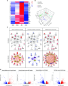
Figure 6. RNA-Seq analysis of cGAS/STING-specific genes.
(A) RPKM expression level of genes normalized to the z scale. (B) PCA plot with centroids (open circles) and different animal groups (filled circles) using RPKM expression values of genes. (C) Gene networks and upstream regulatory analysis of cGAS/STING-specific genes for different animal groups. Gene networks: significant gene fold changes (|fold change| ≥ 2 and FDR < 0.05) with respect to controls are represented in red (upregulation) and blue (downregulation) nodes, while the nonsignificant genes have white nodes. Upstream regulatory analysis: color scheme of nodes is the same as in gene networks. Yellow and purple nodes represent predicted activated or inhibited transcription factors, respectively. Orange lines indicate predicted activation, blue lines indicate predicted inhibition, yellow lines indicate that the predicted relationship is inconsistent with gene expression, while gray lines indicate no predicted effect. (D) Volcano plots of statistically significant differentially expressed genes identified from the RNA-Seq data among different groups. n = 4 rats in each group.
Figure 7
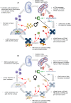
Figure 7. Summary of cGAS/STING activity.
The results of this study are summarized schematically for male (A) and female (B) T2DN rats. The old male renal tissue showed clear signs of increased cGAS/STING pathway activity, with an elevation in IFN causing a significant inflammatory cell recruitment in comparison with their age-matched counterpart sex.

References

    1. Iwasaki A, Medzhitov R. Regulation of adaptive immunity by the innate immune system. Science. 2010;327(5963):291–295. doi: 10.1126/science.1183021. - DOI - PMC - PubMed
    1. Barber GN. STING: infection, inflammation and cancer. Nat Rev Immunol. 2015;15(12):760–770. doi: 10.1038/nri3921. - DOI - PMC - PubMed
    1. Ahn J, et al. Intrinsic self-DNA triggers inflammatory disease dependent on STING. J Immunol. 2014;193(9):4634–4642. doi: 10.4049/jimmunol.1401337. - DOI - PMC - PubMed
    1. Ablasser A, Chen ZJ. cGAS in action: expanding roles in immunity and inflammation. Science. 2019;363(6431):eaat8657. doi: 10.1126/science.aat8657. - DOI - PubMed
    1. Crispin JC, et al. Gene-function studies in systemic lupus erythematosus. Nat Rev Rheumatol. 2013;9(8):476–484. doi: 10.1038/nrrheum.2013.78. - DOI - PubMed

MeSH terms