. 2024 Nov 26;135(3):e183576.
doi: 10.1172/JCI183576.

Ferumoxytol nanozymes effectively target chronic biofilm infections in apical periodontitis

Affiliations

Ferumoxytol nanozymes effectively target chronic biofilm infections in apical periodontitis

Alaa Babeer et al. J Clin Invest. .

Abstract

Bacterial biofilms are pervasive and recalcitrant to current antimicrobials, causing numerous infections. Iron oxide nanozymes, including an FDA-approved formulation, ferumoxytol (FMX), show potential against biofilm infections via catalytic activation of hydrogen peroxide (H2O2). However, clinical evidence regarding the efficacy and therapeutic mechanisms of FMX is lacking. Here, we investigate whether FMX nanozymes can treat chronic biofilm infections and compare their bioactivity to that of the gold standard sodium hypochlorite (NaOCl), a potent but caustic disinfectant. Clinical performance was assessed in patients with apical periodontitis, an intractable endodontic infection affecting half of the global adult population. Data show robust antibiofilm activity by a single application of FMX with H2O2 achieving results comparable to those seen with NaOCl without adverse effects. FMX binds efficiently to the bacterial pathogens Enterococcus faecalis and Fusobacterium nucleatum and remains catalytically active without being affected by dental tissues. This allows for effective eradication of endodontic biofilms via on-site free radical generation without inducing cytotoxicity. Unexpectedly, FMX promotes growth of stem cells of the apical papilla (SCAPs), with transcriptomic analyses revealing upregulation of proliferation-associated pathways and downregulation of cell cycle suppressor genes. Notably, FMX activates SCAP pluripotency and WNT/NOTCH signaling that induces its osteogenic capacity. Together, these results show that FMX nanozymes are clinically effective against severe chronic biofilm infection with pathogen targeting and unique stem cell-stimulatory properties, offering a regenerative approach to antimicrobial therapy.

Keywords: Bacterial infections; Drug therapy; Infectious disease; Nanotechnology.


Conflict of interest statement

Conflict of interest: The authors have declared that no conflict of interest exists.

Figures

Figure 1
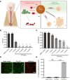
Figure 1. FMX nanozymes eliminate drug-resistant bacterial biofilms associated with severe endodontic infection through catalytic activation of H2O2.
(A) A schematic depiction of antibiofilm activity and stem cell–stimulatory effects of FMX (Created in BioRender. Babeer, A. (2025) https://BioRender.com/y71u571). (B) Dose-dependent antimicrobial activity of FMX/H2O2 against planktonic E. faecalis cells after 10 minutes (n = 6). (C) Dose-dependent efficacy of FMX/H2O2 against E. faecalis biofilm after 10 minutes (n = 6). Different letters indicate statistically significant differences. (D) Representative confocal images of E. faecalis biofilm. Live cells were stained with SYTO 9 (green), and dead cells were stained with propidium iodide (red). Scale bars: 20 μm. (E) Relative percentage of live and dead cells of the total biovolume (n = 7). The statistical analysis was performed using ANOVA, followed by Tukey’s test for multiple comparisons. All values are reported as mean ± SD, ****P < 0.0001; ns, not significant; n.d., non-detectable.
Figure 2
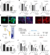
Figure 2. Bioactivity of FMX nanozymes on in vitro and ex vivo mixed-species biofilms.
(A) Antibiofilm effect of FMX/H2O2 on the total viable bacteria in the in vitro biofilm model (n = 6). (B) The effect of FMX/H2O2 on the viability of different bacterial species in in vitro mixed-species biofilms (n = 6). (C) Representative images of mixed-species biofilms. All samples were stained with SYTO 60 (green) or propidium iodide (red); GFP-labeled E. faecalis cells are shown in blue. Dashed squares highlight zoomed-in areas within the micrographs. Scale bars: 5 μm. (D) A schematic of the ex vivo biofilm model using extracted human tooth. BHIS, Brain heart infusion-supplemented. (E) Antibiofilm effect of FMX/H2O2 on the total number of viable bacteria in the mixed-species biofilm (n = 6). (F) The effect of FMX/H2O2 on the viability of different species in ex vivo mixed-species biofilms (n = 6). The statistical analysis was performed using ANOVA followed by Tukey’s test for multiple comparisons (A, B, E, and F, left panel) or Kruskal-Wallis test followed by Dunn’s test for multiple comparisons (F, middle and right panels). All quantitative values are reported as mean ± SD, *P < 0.05, **P < 0.01, ****P < 0.0001; ns, not significant; n.d., non-detectable.
Figure 3
Figure 3. FMX at antimicrobial dosages promotes cell proliferative capabilities without cytotoxicity in human stem cells of the apical papilla.
(A) Cell viability assay showed that topical exposure of FMX did not induce cell death in stem cells of the apical papilla (SCAPs). Scale bar: 25 μm. (B) Flow cytometry analysis showed that FMX did not induce cell apoptosis in SCAPs. (C) Cell proliferation assay showed that FMX treatment significantly increased Ki67+ cell percentage in SCAPs. Scale bar: 25 μm. Quantitative data are shown at the right of each panel. The statistical analysis was performed using ANOVA, followed by Dunnett’s test for multiple comparisons. All quantitative values are reported as mean ± SD, *P < 0.05.
Figure 4
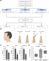
Figure 4. Clinical effectiveness of FMX/H2O2.
(A) A flowchart of the clinical study. (B) A simplified schematic diagram of the clinical treatment regimen and sample collection. Created in BioRender. Babeer, A. (2025) https://BioRender.com/o72b703 (C) The number of colony-forming units (CFU) before and after treatments. Values are reported as mean ± SD. (D) Antimicrobial effectiveness of different clinical protocols. Values are reported in box plots and were subjected to Mann-Whitney U test for pairwise comparisons. P values are represented within the graph. Red dashed line highlights the relationship of the median of log reduction of FMX/H2O2 relative to the median log reduction of NaOCl.
Figure 5
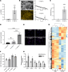
Figure 5. Surface retention, in situ catalysis, and regenerative potential of FMX nanozymes.
(A) Amount of FMX retained in treated biofilms after 10 minutes of treatment measured by ICP-OES (n = 6). (B) Left: A representative environmental scanning electron microscope image of the FMX-treated biofilm and the corresponding elemental mapping image showing iron ion (yellow) distribution. Scale bars: 10 μm. Right: EDS spectra of untreated and FMX-treated biofilms. (C) Catalytic activity of retained FMX in biofilms (n = 12). (D) Amount of FMX bound to bacterial cells after 10 minutes of treatment (n = 6). (E) Ki67 staining showed that 0.1 mg/mL or higher concentrations of FMX significantly increased SCAP proliferation after a 24-hour treatment. Scale bar: 25 μm. Original magnification × 20. (F) Quantitative analysis of the percentage of Ki67+ cells after treatment (n = 3). (G) Heatmap of transcriptomics analysis showing genes differentially regulated after 1 mg/mL FMX treatment for 24 hours. Gene expression is shown in normalized log2 counts per million. Differentially expressed genes were selected based on a 4-fold change. (H) qPCR assay confirmed that cell cycle genes were highly activated while cell cycle suppressor genes were greatly diminished after FMX treatment. Statistical analyses were performed using Welch’s 2-tailed t test (A and C), ANOVA followed by Tukey’s test (D), or ANOVA followed by Dunnett’s test (F and H) for multiple comparisons. All values are reported as mean ± SD, *P < 0.05, ***P < 0.001, ****P < 0.0001; ns, not significant.
Figure 6
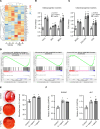
Figure 6. FMX promotes osteogenic differentiation in SCAPs.
(A) Heatmap from RNA-Seq showing differential regulation of stemness genes in SCAPs following treatment with FMX. (B and C) qPCR assays demonstrating significant increases in the levels of osteogenic and chondrogenic markers in FMX-treated SCAPs (n = 3). (D) Gene set enrichment analysis indicates that osteogenic pathways, including WNT and NOTCH signaling, are enriched in FMX-treated SCAPs. (E) Alizarin red staining reveals enhanced osteogenic capacity of SCAPs with FMX treatment, indicating increased mineralized nodule formation (n = 3). (F) Further qPCR analysis confirms significant upregulation of the osteogenic markers RUNX2 and ALP after FMX treatment (n = 3). Statistical analyses were performed using ANOVA followed by Dunnett’s test for multiple comparisons (vs. the control group). All values are presented as mean ± SD, *P < 0.05.

References

    1. Ciofu O, et al. Tolerance and resistance of microbial biofilms. Nat Rev Microbiol. 2022;20(10):621–635. doi: 10.1038/s41579-022-00682-4. - DOI - PubMed
    1. Lebeaux D, et al. Biofilm-related infections: bridging the gap between clinical management and fundamental aspects of recalcitrance toward antibiotics. Microbiol Mol Biol Rev. 2014;78(3):510–543. doi: 10.1128/MMBR.00013-14. - DOI - PMC - PubMed
    1. Tibúrcio-Machado CS, et al. The global prevalence of apical periodontitis: a systematic review and meta-analysis. Int Endod J. 2021;54(5):712–735. doi: 10.1111/iej.13467. - DOI - PubMed
    1. Niazi SA, Bakhsh A. Association between endodontic infection, its treatment and systemic health: a narrative review. Medicina (Kaunas) 2022;58(7):931. doi: 10.3390/medicina58070931. - DOI - PMC - PubMed
    1. Gomes BPFA, et al. Microbiota present in combined endodontic-periodontal diseases and its risks for endocarditis. Clin Oral Investig. 2023;27(8):4757–4771. doi: 10.1007/s00784-023-05104-0. - DOI - PubMed

Substances