

Development and evaluation of a deep learning model to reduce exomass-related metal artefacts in cone-beam CT: an ex vivo study using porcine mandibles
- PMID: 39589904
- PMCID: PMC11784918
- DOI: 10.1093/dmfr/twae062
Development and evaluation of a deep learning model to reduce exomass-related metal artefacts in cone-beam CT: an ex vivo study using porcine mandibles
Abstract
Objectives: To develop and evaluate a deep learning (DL) model to reduce metal artefacts originating from the exomass in cone-beam CT (CBCT) of the jaws.
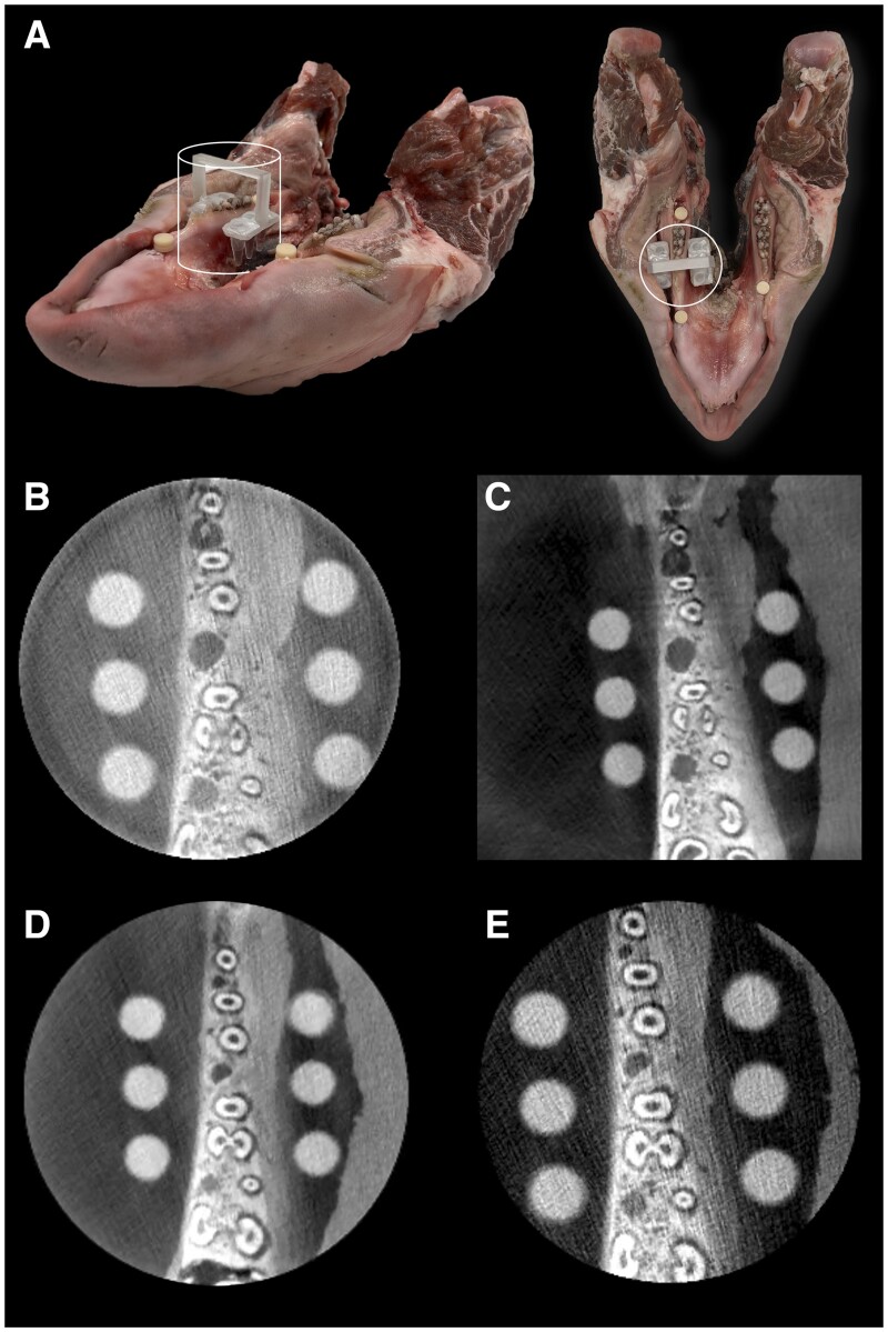
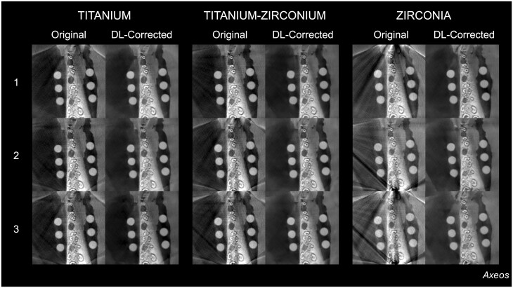
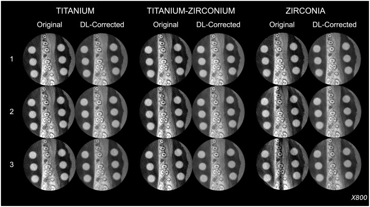
Methods: Five porcine mandibles, each featuring six tubes filled with a radiopaque solution, were scanned using four CBCT units before and after the incremental insertion of up to three titanium, titanium-zirconium, and zirconia dental implants in the exomass of a small field of view. A conditional denoising diffusion probabilistic model, using DL techniques, was employed to correct axial images from exomass-related metal artefacts across the CBCT units and implant scenarios. Three examiners independently scored the image quality of all datasets, including those without an implant (ground truth), with implants in the exomass (original), and DL-generated ones. Quantitative analysis compared contrast-to-noise ratio (CNR) to validate artefact reduction using repeated measures analysis of variance in a factorial design followed by Tukey test (α = .05).
Results: The visualisation of the hard tissues and overall image quality was reduced in the original and increased in the DL-generated images. The score variation observed in the original images was not observed in the DL-generated images, which generally scored higher than the original images. DL-generated images revealed significantly greater CNR than both the ground truth and their corresponding original images, regardless of the material and quantity of dental implants and the CBCT unit (P < .05). Original images revealed significantly lower CNR than the ground truth (P < .05).
Conclusions: The developed DL model using porcine mandibles demonstrated promising performance in correcting exomass-related metal artefacts in CBCT, serving as a proof-of-principle for future applications of this approach.
Keywords: artefacts; artificial intelligence; cone-beam CT; deep learning; dental implants.
© The Author(s) 2024. Published by Oxford University Press on behalf of the British Institute of Radiology and the International Association of Dentomaxillofacial Radiology.
Conflict of interest statement
None declared.
Figures


Similar articles
-
Evaluation of Exomass-Related Artefacts Caused by Dental Implants of Different Materials in Cone-Beam Computed Tomography Scans: An Ex Vivo Study.Clin Oral Implants Res. 2025 Apr;36(4):449-459. doi: 10.1111/clr.14394. Epub 2024 Dec 20. Clin Oral Implants Res. 2025. PMID: 39707609 Free PMC article.
-
CBCT image artefacts generated by implants located inside the field of view or in the exomass.Dentomaxillofac Radiol. 2022 Feb 1;51(2):20210092. doi: 10.1259/dmfr.20210092. Epub 2021 Jul 29. Dentomaxillofac Radiol. 2022. PMID: 34289314 Free PMC article.
-
Objective assessment of the combined effect of exomass-related- and motion artefacts in cone beam CT.Dentomaxillofac Radiol. 2021 Jan 1;50(1):20200255. doi: 10.1259/dmfr.20200255. Epub 2020 Aug 19. Dentomaxillofac Radiol. 2021. PMID: 32706986 Free PMC article.
-
Mapping artifacts generated in a tooth adjacent to titanium and zirconia implants located in the endomass and exomass in cone beam computed tomography: an ex vivo study.Oral Surg Oral Med Oral Pathol Oral Radiol. 2024 Jan;137(1):73-82. doi: 10.1016/j.oooo.2023.08.002. Epub 2023 Aug 8. Oral Surg Oral Med Oral Pathol Oral Radiol. 2024. PMID: 37838553
-
Are metal artefact reduction algorithms effective to correct cone beam CT artefacts arising from the exomass?Dentomaxillofac Radiol. 2019 Mar;48(3):20180290. doi: 10.1259/dmfr.20180290. Epub 2019 Jan 28. Dentomaxillofac Radiol. 2019. PMID: 30540919 Free PMC article.
Cited by
-
Enhancing Image Quality in Dental-Maxillofacial CBCT: The Impact of Iterative Reconstruction and AI on Noise Reduction-A Systematic Review.J Clin Med. 2025 Jun 13;14(12):4214. doi: 10.3390/jcm14124214. J Clin Med. 2025. PMID: 40565955 Free PMC article. Review.
References
-
- Yashayaeva A, MacDonald RL, Robar J, Cherpak A.. Evaluation of a metal artifact reduction algorithm for image reconstruction on a novel CBCT platform. J Appl Clin Med Phys. 2024;25(11):e14516. https://aapm.onlinelibrary.wiley.com/doi/10.1002/acm2.14516 - DOI - PMC - PubMed
-
- Wanderley VA, de Faria Vasconcelos K, Leite AF, et al.Impact of the blooming artefact on dental implant dimensions in 13 cone-beam computed tomography devices. Int J Implant Dent. 2021;7(1):67. https://journalimplantdent.springeropen.com/articles/10.1186/s40729-021-... - DOI - PMC - PubMed
-
- Sahrmann P, Kühl S, Dagassan‐Berndt D, Bornstein MM, Zitzmann NU.. Radiographic assessment of the peri‐implant site. Periodontol 2000. 2024;95(1):70-86. https://onlinelibrary.wiley.com/doi/10.1111/prd.12577 - DOI - PubMed
-
- Brüllmann D, Schulze RKW.. Spatial resolution in CBCT machines for dental/maxillofacial applications—what do we know today? Dentomaxillofac Radiol. 2015;44(1):20140204. http://www.birpublications.org/doi/10.1259/dmfr.20140204 - DOI - PMC - PubMed
Publication types
MeSH terms
Substances
Grants and funding
LinkOut - more resources
Full Text Sources
